touchONCOLOGY touchONCOLOGY
Breast Cancer
Read Time: 4 mins

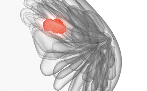
Diagnosis of Metastatic Breast Cancer— A Case Report Using Molecular Cancer Classification

Copy Link
Published Online: Sep 5th 2011 US Oncology & Hematology, 2011;7(2):116-8 DOI: https://doi.org/10.17925/OHR.2011.07.2.116
Authors: Yogesh Gandhi, Sunil Gandhi
Quick Links:
Abstract
Article
Article Information
Abstract:
Overview

An accurate cancer diagnosis is critical as it can direct the use of site-directed, and potentially more effective, treatment options for specific types of cancer. A differential or uncertain diagnosis could prevent cancer patients from receiving optimal treatment, thus affecting their overall prognosis. Advances in molecular technology have led to the development of molecular cancer classifiers that can direct or confirm the diagnosis of metastatic cancers which would otherwise be considered uncertain or unknown. This case report describes the role of molecular diagnostics in the evaluation of a patient with a large pancreatic mass and a history of breast cancer. Results from a 92-gene molecular profiling assay (CancerTYPE ID®) predicted that this new mass was breast cancer. This diagnosis allowed for effective treatment and complete response in this patient.

Keywords

Molecular cancer classification, CancerTYPE ID®, breast cancer

Article:

Despite the increasing sophistication of methods for detecting and diagnosing cancer, these methods fail to reveal a primary site of origin for a subset of patients with metastatic disease. Histopathology, the traditional cornerstone of cancer diagnosis, relies on cell morphology and tissue architecture, but can be subjective. Immunohistochemical analysis of specific tumor markers, in addition to histology and clinical findings, also aids in a differential cancer diagnosis, but can be subjective or misinterpreted as well, since many metastatic tumors do not retain the morphologic or phenotypic characteristics of their organ of origin.1,2

Despite comprehensive work-up, the tissue of origin remains uncertain or unknown in approximately 10–15 % of metastatic cases, and the possibility of misclassification remains a challenge. Failure to identify the primary origin of cancer of a metastasis despite extensive clinical work-up can lead to a diagnosis of carcinoma of unknown primary origin (CUP), or occult primary malignancy, and highlights the importance of diagnosis of the primary site.1,3,4 CUP accounts for approximately 3–5 % of all malignancies.1,3,4 A tumor’s primary anatomic site of origin usually dictates the optimal treatment, expected outcome, and overall prognosis for a cancer patient. The overall prognosis for patients with CUP is usually poor, with a median survival of three to nine months, even with newer treatment regimens.5–10 Multiple empiric chemotherapy regimens have been used for CUP patients, but there are few randomized data to support a specific regimen.11 The median survival in randomized studies is approximately seven months, which is both poor and significantly less than the expected survival for patients with breast and bowel malignancy following standard therapy.12 Survival improvement has been demonstrated in the CUP subset of patients if the primary site is identified, allowing for specific therapy to be initiated.2,13–17 Unfortunately, primary tumor detection remains a challenge in CUP: currently, the accuracy rate of a diagnostic work-up for CUP by histopathology, immunohistochemistry, and radiologic or other clinical methods is only 20–30 %.12,18,19 Additionally, extensive CUP work-up is expensive and time-consuming.20 Therefore, there is a need for more sophisticated tools to aid in correct primary diagnosis, particularly with the development in the last decade of targeted biologic therapies for specific tumor types, such as trastuzumab in breast cancer,21 erlotinib in lung cancer,22 and bevacizumab in renal cell,23 breast,24 and colorectal cancers,25 to name a few.In recent years, gene-expression-based techniques known as molecular cancer classification or gene profiling tests have been developed to assist in the diagnosis of malignancies.1,2,20,26–32 These assays are based on the hypothesis that, whereas the phenotype of a poorly differentiated tumor can differ from the tissue of origin, gene expression pattern remains more constant.2 One such molecular profiling assay is the CancerTYPE ID®, a 92-gene panel realtime polymerase chain reaction (RT-PCR) test. This assay identifies the most likely tumor origin based on the expression profiles of 87 informative genes and five reference genes analyzed by RT-PCR of RNA extracted from sections of formalin-fixed paraffin-embedded tissue. This RT-PCR-based test has been shown to have an accuracy of 85–87 % and over 99 % specificity in classifying 30 main tumor types and 54 subtypes.26,27

Case Report
A 63-year-old woman with a history of breast cancer presented during a routine visit with severe epigastric pain radiating to her back, anorexia, and weight loss (15lb over two months). A positron emission tomography– computed tomography scan showed a large pancreatic mass and diffuse retroperitoneal and pelvic lymphadenopathy (see Figure 1A). Six years previously, the patient had been diagnosed with breast carcinoma as detected by a routine mammogram. The tumor was 1.5 cm in size and was an ER-positive, PR-negative poorly differentiated carcinoma. The patient underwent lumpectomy and axillary lymph node dissection, which were negative. The patient had been treated with radiation therapy and adjuvant hormonal therapy with an aromatase inhibitor.

Given the patient’s history, there was doubt as to whether this new pancreatic mass was a primary pancreatic carcinoma with lymph node metastasis or a new metastasis from the breast carcinoma with metastasis to the pancreas and lymph nodes. Histopathology of the pancreatic lesion reported an adenocarcinoma consistent with pancreatic primary, and was negative for ER, PR, CK-20, CEA and p53, and positive for Her-2, pancytokeratin, CK-7 and mucicarmine. Notably, the tumor was negative for ER which would indicate a switch in receptor status if it was recurrent breast cancer. Her2/neu amplification was positive by immunohistochemistry. Given that adenocarcinomas other than breast cancer, such as gastric, pancreatic, or endometrial, have been reported to be positive for Her2/neu, this was not considered diagnostic of metastatic breast carcinoma. Although Ca-19-9 was normal, primary pancreatic cancer could not be ruled out as the reported sensitivity and specificity for pancreatic cancer is reported to be 80 % and 90 %, respectively. A definitive diagnosis was needed because therapy and prognosis would be significantly affected depending on whether it was metastatic pancreatic carcinoma or breast carcinoma, so we ordered the 92-gene assay for molecular cancer classification (Cancer TYPE ID). The 92-gene assay predicted a strong likelihood for breast carcinoma as the patient’s biopsy sample demonstrated significant similarity to breast cancer (P = 2.4 x 10⁻⁷). The patient was started on chemotherapy with paclitaxel and carboplatin plus trastuzumab, and achieved complete remission after only about three months of chemotherapy (see Figure 1B). This is a regimen known to be active in breast carcinoma but not in pancreatic carcinoma. Discussion
Metastasis to the pancreas is much less common than primary pancreatic carcinoma. Breast carcinoma commonly metastasizes to distant organs such as the lung, bones, liver, or brain, but metastases from breast carcinoma to the pancreas are relatively less common. The patient’s presentation of abdominal pain is one of the most common presenting symptoms for primary pancreatic carcinoma. More than 43,000 people develop exocrine pancreatic cancer each year in the US and, because of its aggressive character and the fact that most patients present with relatively advanced disease, most die from the disease. Median survival is eight to 12 months for patients with locally advanced unresectable disease and only three to six months for those who present with metastases.

Although metastatic breast cancer is unlikely to be cured, meaningful improvements in survival have been seen with the introduction of newer systemic therapies.33–5 Median overall survival approaches two years, with a range from a few months to many years.36 The patient discussed above was treated with combination therapy specific for metastatic breast carcinoma and not pancreatic carcinoma and still we achieved complete remission. This also confirms that her diagnosis by genetic assay was accurate. These results highlight the premise that accurate diagnosis of the primary site is critical for effective, site-specific cancer therapy.

Article Information:
Disclosure

The authors have no conflicts of interest to declare.

Correspondence

Sunil Gandhi, MD, FACP, 521 North Lecanto Highway, Lecanto, FL 34461. E: sunilggandhi@gmail.com

Received

2011-05-25T00:00:00

References

  1. Horlings, HM, van Laar RK, Kerst JM, et al., Gene expression profiling to identify the histogenic origin of metastatic adenocarcinomas of unknown primary, J Clin Oncol, 2008;26:4435–41.
  2. Bender RA, Erlander MG, Molecular classification of unknown primary cancer, Semin Oncol, 2009;36:38–43.
  3. Pavlidis N, Fizazi K, Cancer of unknown primary (CUP), Crit Rev Oncol Hematol, 2005;54:243–50.
  4. Varadhachary GR, Abbruzzese JL, Lenzi R, Diagnostic strategies for unknown primary cancer, Cancer, 2004;100:1776–85.
  5. National Cancer Institute, Carcinoma of Unknown Primary Treatment (PDQ®) Health Professional Version. Available at: www.cancer.gov.
  6. Greco FA, Burris HA 3rd, Litchy S, et al., Gemcitabine, carboplatin, and paclitaxel for patients with carcinoma of unknown primary site: a Minnie Pearl Cancer Research Network study, J Clin Oncol, 2002;20:1651–6.
  7. Pavlidis N, Briasoulis E, Hainsworth J, Greco FA, Diagnostic and therapeutic management of cancer of an unknown primary, Eur J Cancer, 2003;39:1990–2005.
  8. Hainsworth JD, Spigel DR, Farley C, et al., Minnie Pearl Cancer Research Network, Phase II trial of bevacizumab and erlotinib in carcinomas of unknown primary site: the Minnie Pearl Cancer Research Network, J Clin Oncol, 2007;25:1747–52.
  9. Schneider BJ, El-Rayes B, Muler JH, et al., Phase II trial of carboplatin, gemcitabine, and capecitabine in patients with carcinoma of unknown primary site, Cancer, 2007;110:770–5.
  10. Pimiento JM, Teso D, Malkan A, et al., Cancer of unknown primary origin: a decade of experience in a community-based hospital, Am J Surg, 2007;194:833–7.
  11. Briasoulis E, Tolis C, Bergh J, Pavlidis N, ESMO Minimum Clinical Recommendations for diagnosis, treatment and followup of cancers of unknown primary site (CUP), Ann Oncol, 2005;16(Suppl. 1):i75–6.
  12. Abbruzzese JL, Abbruzzese MC, Lenzi R, et al., Analysis of a diagnostic strategy for patients with suspected tumors of unknown origin, J Clin Oncol, 1995;13:2094–103.
  13. Varadhachary GR, Raber MN, Matamoros A, Abbruzzese JL, Carcinoma of unknown primary with a colon-cancer profile-changing paradigm and emerging definitions, Lancet Oncol, 2008;9:596–9.
  14. Pentheroudakis G, Pavlidis N, Serous papillary peritoneal carcinoma: unknown primary tumour, ovarian cancer counterpart or a distinct entity? A systematic review, Crit Rev Oncol Hematol, 2010;75:27–42.
  15. Pentheroudakis G, Lazaridis G, Pavlidis N, Axillary nodal metastases from carcinoma of unknown primary (CUPAx): a systematic review of published evidence, Breast Cancer Res Treat, 2010;119:1–11.
  16. Hainsworth JD, Fizazi K, Treatment for patients with unknown primary cancer and favorable prognostic factors, Semin Oncol, 2009;36:44–51.
  17. Varadhachary GR, Talantov D, Raber MN, et al., Molecular profiling of carcinoma of unknown primary and correlation with clinical evaluation, J Clin Oncol, 2008;26:4442–8.
  18. Ayoub JP, Hess KR, Abbruzzese MC, et al., Unknown primary tumors metastatic to liver, J Clin Oncol, 1998;16:2105–12.
  19. van de Wouw AJ, Jansen RL, Griffioen AW, Hillen HF, Clinical and immunohistochemical analysis of patients with unknown primary tumour: A search for prognostic factors in UPT, Anticancer Res, 2004;24:297–301.
  20. Monzon FA, Medeiros F, Lyons-Weiler M, Henner WD, Identification of tissue of origin in carcinoma of unknown primary with a microarray-based gene expression test, Diagnostic Pathology, 2010;5:3.
  21. Hudis CA, Trastuzumab – mechanism of action and use in clinical practice, N Engl J Med, 2007;357:39–51.
  22. Tsao MS, Sakurada A, Cutz JC, et al., Erlotinib in lung cancer: Molecular and clinical predictors of outcome, N Engl J Med, 2005;353:133–44.
  23. Escudier B, Pluzanska A, Koralewski P, et al., AVOREN Trial investigators, Bevacizumab plus interferon alfa-2a for treatment of metastatic renal cell carcinoma: A randomised, double-blind phase III trial, Lancet, 2007;370:2103–11.
  24. Miller K, Wang M, Gralow J, et al., Paclitaxel plus bevacizumab versus paclitaxel alone for metastatic breast cancer, N Engl J Med, 2007;357:2666–76.
  25. Hurwitz H, Fehrenbacher L, Novotny W, et al., Bevacizumab plus irinotecan, fluorouracil, and leucovorin for metastatic colorectal cancer, N Engl J Med, 2004;350:2335–42.
  26. . Ma XJ, Patel R, Wang X, et al., Molecular classification of human cancers using a 92-gene real-time quantitative polymerase chain reaction assay, Arch Pathol Lab Med, 2006;130:465–73.
  27. Erlander MG, Ma XJ, Kesty NC, et al., Performance and clinical evaluation of the 92-gene real-time PCR assay for tumor classification, J Mol Diagn, 2011;13:439–503.]
  28. Bloom G, Yang IV, Boulware D, et al., Multiplatform, multi-site, microarray-based human tumor classification, Am J Pathol, 2004;164:9–16.
  29. Dennis JL, Vass JK, Wit EC, et al., Identification from public data of molecular markers of adenocarcinoma characteristic of the site of origin, Cancer Res, 2002;62:5999–6005.
  30. Ramaswamy S, Tamayo P, Rifkin R, et al., Multiclass cancer diagnosis using tumor gene expression signatures, Proc Natl Acad Sci U S A, 2001;98:15149–54.
  31. . Su AI, Welsh JB, Sapinoso LM, et al., Molecular classification of human carcinomas by use of gene expression signatures, Cancer Res, 2001;61:7388–93.
  32. Dennis JL, Hvidsten TR, Wit EC, et al., Markers of adenocarcinoma characteristic of the site of origin: development of a diagnostic algorithm, Clin Cancer Res, 2005:11:3766–72.
  33. Chia SK, Speers CH, D’yachkova Y, et al., The impact of new chemotherapeutic and hormone agents on survival in a population-based cohort of women with metastatic breast cancer, Cancer, 2007;110:973.
  34. Gennari A, Conte P, Rosso R, et al., Survival of metastatic breast carcinoma patients over a 20-year period: a retrospective analysis based on individual patient data from six consecutive studies, Cancer, 2005;104:1742.
  35. Dafni U, Grimani I, Xyrafas A, et al., Fifteen-year trends in metastatic breast cancer survival in Greece, Breast Cancer Res Treat, 2010;119:621.
  36. Greenberg PA, Hortobagyi GN, Smith TL, et al., Long-term follow-up of patients with complete remission following combination chemotherapy for metastatic breast cancer, J Clin Oncol, 1996;14:2197.

Further Resources

Share this Article
Related Content In Breast Cancer
  • Copied to clipboard!
    accredited arrow-down-editablearrow-downarrow_leftarrow-right-bluearrow-right-dark-bluearrow-right-greenarrow-right-greyarrow-right-orangearrow-right-whitearrow-right-bluearrow-up-orangeavatarcalendarchevron-down consultant-pathologist-nurseconsultant-pathologistcrosscrossdownloademailexclaimationfeedbackfiltergraph-arrowinterviewslinkmdt_iconmenumore_dots nurse-consultantpadlock patient-advocate-pathologistpatient-consultantpatientperson pharmacist-nurseplay_buttonplay-colour-tmcplay-colourAsset 1podcastprinter scenerysearch share single-doctor social_facebooksocial_googleplussocial_instagramsocial_linkedin_altsocial_linkedin_altsocial_pinterestlogo-twitter-glyph-32social_youtubeshape-star (1)tick-bluetick-orangetick-red tick-whiteticktimetranscriptup-arrowwebinar Sponsored Department Location NEW TMM Corporate Services Icons-07NEW TMM Corporate Services Icons-08NEW TMM Corporate Services Icons-09NEW TMM Corporate Services Icons-10NEW TMM Corporate Services Icons-11NEW TMM Corporate Services Icons-12Salary £ TMM-Corp-Site-Icons-01TMM-Corp-Site-Icons-02TMM-Corp-Site-Icons-03TMM-Corp-Site-Icons-04TMM-Corp-Site-Icons-05TMM-Corp-Site-Icons-06TMM-Corp-Site-Icons-07TMM-Corp-Site-Icons-08TMM-Corp-Site-Icons-09TMM-Corp-Site-Icons-10TMM-Corp-Site-Icons-11TMM-Corp-Site-Icons-12TMM-Corp-Site-Icons-13TMM-Corp-Site-Icons-14TMM-Corp-Site-Icons-15TMM-Corp-Site-Icons-16TMM-Corp-Site-Icons-17TMM-Corp-Site-Icons-18TMM-Corp-Site-Icons-19TMM-Corp-Site-Icons-20TMM-Corp-Site-Icons-21TMM-Corp-Site-Icons-22TMM-Corp-Site-Icons-23TMM-Corp-Site-Icons-24TMM-Corp-Site-Icons-25TMM-Corp-Site-Icons-26TMM-Corp-Site-Icons-27TMM-Corp-Site-Icons-28TMM-Corp-Site-Icons-29TMM-Corp-Site-Icons-30TMM-Corp-Site-Icons-31TMM-Corp-Site-Icons-32TMM-Corp-Site-Icons-33TMM-Corp-Site-Icons-34TMM-Corp-Site-Icons-35TMM-Corp-Site-Icons-36TMM-Corp-Site-Icons-37TMM-Corp-Site-Icons-38TMM-Corp-Site-Icons-39TMM-Corp-Site-Icons-40TMM-Corp-Site-Icons-41TMM-Corp-Site-Icons-42TMM-Corp-Site-Icons-43TMM-Corp-Site-Icons-44TMM-Corp-Site-Icons-45TMM-Corp-Site-Icons-46TMM-Corp-Site-Icons-47TMM-Corp-Site-Icons-48TMM-Corp-Site-Icons-49TMM-Corp-Site-Icons-50TMM-Corp-Site-Icons-51TMM-Corp-Site-Icons-52TMM-Corp-Site-Icons-53TMM-Corp-Site-Icons-54TMM-Corp-Site-Icons-55TMM-Corp-Site-Icons-56TMM-Corp-Site-Icons-57TMM-Corp-Site-Icons-58TMM-Corp-Site-Icons-59TMM-Corp-Site-Icons-60TMM-Corp-Site-Icons-61TMM-Corp-Site-Icons-62TMM-Corp-Site-Icons-63TMM-Corp-Site-Icons-64TMM-Corp-Site-Icons-65TMM-Corp-Site-Icons-66TMM-Corp-Site-Icons-67TMM-Corp-Site-Icons-68TMM-Corp-Site-Icons-69TMM-Corp-Site-Icons-70TMM-Corp-Site-Icons-71TMM-Corp-Site-Icons-72