One of the main achievements in the field of radiation oncology for breast cancer has been preservation of the breast using breast conservation therapy (BCT), classically defined as local excision of the primary tumor to achieve negative margins followed by whole breast (WB) radiation therapy (RT). Historically, all patients with early-stage breast cancer (eBC) were treated with some form of mastectomy, requiring en bloc removal of the breast, the underlying musculature, and axillary lymph nodes. Multiple prospective, randomized studies have now demonstrated that BCT has equivalent long-term outcomes to mastectomy for eBC.1–3 Furthermore, the Early Breast Cancer Trialists Collaborative Group meta-analysis of the randomized studies, with over 7,300 patients, has demonstrated not only a significant decrease in ipsilateral breast tumor recurrence (IBTR) with the use of radiation, but also that radiation therapy can influence survival, changing the paradigm that radiation affects local control only.4 This meta-analysis has conclusively shown that adjuvant WBRT for eBC provides a modest improvement in overall survival at 15 years.
The vast majority of patients treated in these trials were treated with WBRT to doses of 45–50Gy, delivered once daily in 25–28 fractions, often followed by a cone-down to boost the tumor bed. This proven conventional fractionation is as effective as mastectomy with relatively minimal associated long-term morbidity, and has gained acceptance internationally as the standard treatment for eBC. However, there are several limitations associated with conventional fractionation that sometimes restrict its utility. The six- to seven-week course of daily treatments raises concerns in terms of access of care, particularly for patients who live in locations far from radiation facilities. The prolonged course increases medical costs and can be a major inconvenience for some patients. Lastly, it may be unnecessary to radiate the uninvolved portions of the breast and normal surrounding tissue. Recently, two new strategies that address some of the limitations of conventionally fractionated WB radiation therapy (cfWBRT) have gained popularity. Accelerated partial breast irradiation (APBI) treats the lumpectomy cavity plus a margin to definitive doses, typically delivered in less than five days, often on a twice-daily schedule. Accelerated hypofractionated WBRT (hWBRT) treats the WB with higher daily doses, and therefore over a shorter overall treatment period, to total doses that are biologically equivalent to those delivered by cfWBRT. This article will review these two strategies, the current literature supporting their use, the indications/patient selection criteria for each modality, and limitations compared with cfWBRT.
Accelerated Partial Breast Irradiation Clinical Demand
As evidence supporting the use of BCT has accumulated, with long-term follow-up from multiple randomized trials and several large metaanalyses, BCT has become the standard of care for eBC. While cfWBRT has gained worldwide acceptance for its efficacy, its utilization has not been uniform across the US and there continues to be marked variation in the use of BCT by geographic region.5–7 For example, it has been documented that up to one-third of patients undergoing breast-conserving surgery do not receive post-operative radiotherapy despite its well-established benefits.4,6,8,9 While foregoing radiation may be reasonable in patients over 70 years of age owing to the relatively small absolute benefit in terms of local control, all other subgroups of patients appear to benefit from adjuvant radiotherapy and, to date, no prospective studies with long-term follow-up have been published that consistently identify patient subgroups in which radiation can be withheld without increasing local relapse rates.10 The decision to omit radiotherapy is often multifactorial, resulting from concerns related to length of treatment, convenience, cost, access to care, geographic variations, ethnicity-based differences, and physician/patient preferences. Indeed, it has been shown that the distance of the patient from the nearest radiotherapy center is inversely proportional to the likelihood of receiving RT following breast-conserving surgery.8,11 It is quite possible that a shorter radiotherapy regimen would increase the probability of patients receiving post-operative RT.
Scientific Rationale
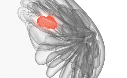
APBI typically involves treating the lumpectomy cavity plus a 1–2cm margin, thereby sparing a significant amount of normal, unaffected breast tissue from high-dose radiation. When treating only a portion of the breast, a theoretical concern is that occult foci of cancer located remotely from the lumpectomy cavity will be left untreated, thus increasing the risk of IBTR. However, both clinical and pathologic data suggest that the benefit of radiotherapy results from the eradication of microscopic disease immediately adjacent to the lumpectomy cavity.12 Data from several prospective and retrospective trials indicate that most IBTRs occur within or immediately adjacent to the lumpectomy bed.1,13,14 In fact, fewer than 4% of recurrences are found in an area of the breast distant from the original tumor.15–17 Therefore, the benefit of post-operative radiotherapy may still be attainable while treating a smaller volume of tissue. By treating a partial breast volume to larger daily fractions to achieve a biologically equivalent total dose, radiation can be delivered over a much shorter time period, with the added benefit of less normal, unaffected breast tissue receiving high doses of radiation.
Current Status
Most of the existing published data on APBI come from retrospective, single-institution, or single-arm prospective series. While there are three published randomized prospective studies comparing APBI with cfWBRT, only one of these trials showed equivalent local control with the use of APBI (see the section on interstitial brachytherapy techniques below).18–20 Notably, the patients treated in this study were highly selected, low-risk patients. The other two studies both reported increased IBTR for the patients treated with APBI. Although the existing data are deficient in terms of randomized study design and long-term follow-up regarding efficacy and toxicity, the available clinical data collectively suggest that APBI may be a safe and effective therapy in appropriately selected patients.
There are several techniques for delivering APBI, including interstitial brachytherapy, single-lumen or multiple-lumen balloon brachytherapy, 3D conformal radiation therapy (3D-CRT), and intraoperative radiotherapy (IORT). Owing to the recent development and marketing of new devices manufactured to deliver APBI with relative ease, APBI is being increasingly widely utilized, despite the limited phase III data supporting its use. The Radiation Therapy Oncology Group (RTOG) 0413/National Surgical Adjuvant Breast and Bowel Project (NSABP) B-39 phase III trial is an ongoing prospective, randomized study comparing APBI with cfWBRT, and has been one of the fastest accruing trials for breast cancer owing to its appeal to both physicians and patients. Patients are randomized to cfWBRT versus APBI, with the caveat that the choice of APBI technique (i.e. interstitial, lumen-based, or externalbeam- based) is left to the discretion of the treating physician. While mature results are not expected for many more years, APBI is being increasingly used in large academic centers, small community-based hospitals, and private practice settings, both on and off protocol. In order to help guide patient selection and promote best practices for APBI while awaiting results from the RTOG 0413/NSABP B-39 trial, the American Society for Therapeutic Radiology and Oncology (ASTRO) recently published consensus guidelines for appropriate patient selection for APBI.21 The proposed criteria classify patients into one of three groups: suitable, cautionary, or unsuitable (see Table 1). The suitable group, based on data from phase II studies supporting the use of APBI, includes low-risk patients for whom WBRT would be unlikely to confer a survival benefit. The cautionary and unsuitable groups represent patients for whom minimal data exist supporting APBI and in whom cfWBRT has a proven survival benefit.Accelerated Partial Breast Irradiation Techniques Interstitial Brachytherapy
The earliest APBI technique utilized interstitial brachytherapy catheters. This technique therefore has the longest average published patient follow-up (5.4 years) and the most patient-years of follow-up.21 Available data support that this technique provides acceptable tumor control and toxicity in properly selected patients. This relatively complex technique involves the insertion of approximately 10–20 afterloading catheters intraoperatively into the tumor bed so that the lumpectomy cavity plus a 1–2cm margin can be encompassed by the prescribed radiation dose. The catheters are open at the skin and a radioactive isotope is inserted into each catheter post-operatively with either low-dose-rate (LDR) or high-dose-rate (HDR) sources. LDR requires an inpatient stay during which 0.4–2Gy/hour is delivered over the course of two to five days. HDR can be performed on an outpatient basis and typically involves twicedaily treatment over four to five days, with each treatment typically lasting less than 15 minutes. While this technique has been shown to be highly efficacious in experienced hands, there is a significant learning curve for the 3D placement of these catheters.
The largest published experience with the longest follow-up using interstitial brachytherapy comes from the William Beaumont Hospital group. Vicini et al. reported on 199 patients with eBC treated with either LDR (50Gy) or HDR (32Gy in eight fractions or 34Gy in 10 fractions). With a median follow-up of 65 months for surviving patients, five IBTRs were noted, giving a five-year actuarial IBTR rate of 1%. Three of these failures occurred outside the irradiated volume.22 A matched-pair analysis was performed comparing the APBI patients with comparable patients who had undergone WBI. No significant differences were identified in any of the end-points evaluated.
RTOG 95-17 was a multi-institutional phase II trial that enrolled 100 carefully selected women with eBC for APBI. Ninety-nine patients were eligible and were treated with either HDR or LDR interstitial brachytherapy. With a median follow-up of seven years, the estimated five-year locoregional failure rate was 5% and the in-breast failure rate was 4%, comparable to rates seen in historic cfWBRT controls.23The only randomized trial to show efficacy of APBI compared with cfWBRT was performed at the National Institute of Oncology in Hungary, where 258 selected low-risk patients were randomized after breastconserving surgery to either standard cfWBRT or APBI. The partial breast technique consisted of HDR multicatheter brachytherapy (36.4Gy in 5.2Gy fractions) in the majority of patients. With a median follow-up of 66 months for all patients, the actuarial IBTR rate for APBI and cfWBI did not differ significantly (4.7 and 3.4%, respectively); the same was true of the other end-points evaluated. In fact, the rate of excellent to good cosmetic results was significantly better in the APBI arm compared with the cfWBI group (77.6 and 62.9%, respectively; p=0.009).20
Single-lumen-catheter Balloon Brachytherapy
With early APBI interstitial brachytherapy data suggesting partial breast radiotherapy as a potentially safe alternative to cfWBI for selected patients, several devices were developed to reduce the technical expertise required for interstitial delivery. The MammoSite (Hologic, Bedford, MA) balloon catheter device allows for a simpler, less operator-dependent delivery of APBI, with the cited advantage of reproducible dosimetry and potentially improved comfort for patients. The device consists of a silicone balloon connected to a catheter to pass an HDR iridium-192 (Ir-192) source after a mixture of saline and contrast has been used to inflate the balloon.
Typically, the single-lumen catheter is placed into the lumpectomy cavity intraoperatively after definitive surgery, the balloon is inflated, and a computed tomography (CT) scan is performed to document adequate device positioning to enable treatment planning. Post-operatively, an HDR Ir-192 source is remotely afterloaded in the radiation department, with a typical dose of 3.4Gy delivered twice daily over five days. Since being introduced in 2002, the MammoSite device has been rapidly adopted, with more patients treated with MammoSite than any other APBI technique.21 Recently, updated results were published from the American Society of Breast Surgeons (ASBS) MammoSite Registry trial. While this ‘trial’ was not randomized, MammoSite users from 97 institutions entered patient information at any time before, during, or after treatment for future analysis regarding parameters of MammoSite delivery, local relapse, and cosmesis. A total of 1,449 breasts in 1,440 patients with eBC treated with the MammoSite device were entered into this database. With a median follow-up of 54 months, the five-year actuarial IBTR rate was 3.80% and the actuarial axillary recurrence rate was 0.80%, with 90.6% of the patients reporting good to excellent cosmetic results. Complications included seroma (28%) and fat necrosis (2.3%).24 Keisch et al. recently analyzed data from the subset of patients with ductal carcinoma in situ (DCIS) in the ASBS MammoSite brachytherapy trial. Although there is concern that DCIS has an increased propensity for multifocality and therefore may be less amenable to APBI, the authors reported acceptable rates of local control in the 194 DCIS patients treated with MammoSite brachytherapy, with a four-year actuarial IBTR rate of 2.45%.25,26
Multiple-catheter Intracavitary Brachytherapy
While the single-lumen MammoSite is a simpler APBI technique compared with multicatheter brachytherapy, there are inherent limitations in dose shaping using a single lumen, particularly with lesions that are close to the skin or chest wall, where an increased risk of fat necrosis and severe skin toxicity is reported. To address this issue, devices have been developed that have multiple-lumen catheters, which allow for improved dosimetry by providing more source-placement options to improve dose conformation.
The first, the Strut-Adjusted Volume Implant (SAVI, Cianna Medical, Aliso Viejo, CA), consists of a central strut surrounded by six to 10 peripheral struts (see Figure 1), thus allowing for multiple source-placement options to avoid high doses to the skin and chest wall.27 Early clinical experience suggests that excellent dosimetry is achievable, even for patients who would have otherwise been ineligible for other brachytherapy owing to the proximity of the cavity to the skin or chest wall.28 The second device, the Contura (SenoRx, Inc., Irvine, CA), consists of a central catheter lumen plus four additional surrounding lumens, each containing multiple dwell positions. Similar to SAVI, early data suggest that the Contura device allows for adequate coverage of the treatment volume while limiting doses to the skin and chest wall.29–32 There is also now a multilumen MammoSite balloon brachytherapy applicator with four lumens to allow for dose optimization.
3D Conformal Radiation Therapy
More recently, 3D-CRT has been used to target the lumpectomy cavity plus margin using external-beam radiation therapy (EBRT) delivered with a conventional linear accelerator (see Figure 2). The advantages of this technique include the non-invasive delivery method, the widespread availability of linear accelerators compared with brachytherapy methods, and the familiarity of EBRT delivery techniques for all practicing radiation oncologists. One disadvantage of 3D-CRT is the multiple-beam configuration, which causes a significant distribution of low-dose radiation over a much larger volume, including the lungs, the ribs, and the contralateral breast owing to the multiple, often co-planar, beams that are required to converge in the high-dose region of the target volume; this has raised concerns regarding increased long-term toxicity. Furthermore, a larger margin around the tumor bed is required to take into account patient motion/set-up errors. Interestingly, the majority of patients in RTOG 0413 were treated with 3D-CRT, despite the fact that this technique has the least amount of clinical data on efficacy and toxicity and the shortest follow-up compared with the other APBI techniques. Thus, the RTOG 0413/NSABP B-39 trial will provide important information regarding 3D-CRT; while we await mature data from this important trial, 3D-CRT APBI should be utilized on-protocol only.
RTOG 0319 was a prospective, single-arm phase II trial designed to assess the reproducibility and technical feasibility of using 3D CRT, with secondary end-points of efficacy and toxicity, with the anticipation of being able to allow the technique to be used in RTOG 0413. This trial utilized 38.5Gy in 10 fractions delivered twice daily (biologically equivalent to 34Gy for brachytherapy). Initial efficacy results at a median follow-up of 4.5 years for 52 evaluable patients revealed an IBTR rate of 6% and an ipsilateral nodal failure rate of 2%, with 4% grade III toxicities.33 It is unclear how the toxicity and cosmetic outcome of 3D-CRT compares with that of WBI or other APBI techniques. Wernicke et al. reported the preliminary results of a prospective single-institution study using ‘mini-tangents’ in a prone position to deliver 30Gy in five fractions over 10 days. Ninety-two percent of patients reported good to excellent cosmesis at 28-month follow-up.34 By contrast, Jagsi et al. delivered IMRT with deep-inspiration breath-hold to 34 patients in a prospective study in which 38.5Gy in 10 fractions twice daily was used. They reported early closure of the trial at a median of 2.5 years owing to the high incidence (>20%) of compromised cosmesis with the use of breath-holding.35 Additional concerning toxicity data using 3D-CRT APBI come from the Tufts group, where 64 patients were treated according to the 3D-CRT technique specified in RTOG 0413. After a median follow-up of only 15 months, grade 3–4 toxicity was noted in 8.3% of patients, and 18.4% of patients reported either ‘fair’ (11.7%) or ‘poor’ (6.7%) cosmetic outcome.35,36
Intraoperative Radiotherapy
IORT is a technique whereby electrons or low-energy X-rays are generated by a mobile treatment machine and a single fraction of approximately 20Gy is delivered intraoperatively to the tumor bed. The advantages of intraoperative targeting include the reduction of the possibility of a geographic miss, the convenience of one treatment, and significant reductions in cost. However, a major disadvantage is the lack of final pathology (i.e. margin status), which is critical in guiding decisions regarding APBI.
The European Institute of Oncology has extensively used single fractionated electron APBI, treating nearly 600 patients with 21Gy IORT. With a median follow-up of 20 months, 1% of patients had IBTR (same quadrant n=3, other quadrants n=3), with breast fibrosis (3.2%) being the primary toxicity.37 Recently, the international phase III Targeted Intraoperative Radiotherapy for Breast Cancer A (TARGIT-A) trial reported a four-year IBTR rate of 1.2% with IORT compared with 0.95% with EBRT, with no significant difference in toxicity.38 While 14% of the IORT patients received additional EBRT after APBI owing to adverse pathologic features, these results are encouraging in that IORT was comparable to cfWBRT in selected patients.
Hypofractionated Whole Breast Irradiation Rationale for Fractionated Radiotherapy
The rationale for using conventional fractionation (small daily fractions to a high total dose) is based on theoretic radiobiologic modeling of the relative sensitivity to changes in the dose of normal cells to that of cancer cells. Typically, most tumor types have relatively low sensitivity to changes in fraction size, whereas normal, late-reacting tissue has higher sensitivity. Since theoretically the goal of radiation is to maximize the tumor cell kill while minimizing the cell kill of normal tissue, the rationale for using low doses of conventional fractionation (1.8–2.0Gy, see Figure 3A) was to limit the damage to normal late-reacting tissue while maximizing cell kill for tumor cells. While the exact sensitivity of breast tumor cells was not known until recently, it was presumed that breast tumors, like most other tumors, have low sensitivity compared with late-reacting tissue. Recently, in a landmark trial of hypofractionated radiation therapy for breast cancer, a radiobiologic surrogate for sensitivity (termed α/β ratio) was calculated for breast tumors based on coefficients estimated from the Cox multivariate model. Surprisingly, the surrogate was found to be similar to the normal, late-reacting breast tissue,39 suggesting that breast tumors and normal tissue have similar sensitivity to dose fraction size. Therefore, there may be little or even no therapeutic advantage of using a smaller fraction size for breast cancer to reduce cell kill in normal tissue relative to tumor cells (see Figure 3B).
Clinical Data for Hypofractionated Whole Breast Radiation Therapy
With the support of radiobiologic models and the increased demand for shorter radiotherapy regimens, several preliminary reports using daily fractions of 2.5–2.7Gy over approximately three weeks have demonstrated low rates of local recurrence and acceptable toxicity.40–42 Based on these experiences, larger randomized phase III trials were initiated comparing hWBRT versus cfWBRT.
The strongest phase III data supporting the use of hWBRT randomized patients to 50Gy in 25 fractions versus 42.5Gy in 16 fractions for nodenegative tumors measuring <5cm (no DCIS) after breast-conserving surgery. With a median follow-up of 12 years, there was no observed difference between the groups in the risk for IBTR or cosmetic outcome. Of note, this trial did not allow boost irradiation, which has since become routinely used for eBC.43 hWBRT has not yet been studied in a prospective fashion for DCIS, although limited retrospective data suggest that hWBRT may be safely used for in situ disease.44
Starting in the late 1990s, the UK Standardization of Breast Radiotherapy Trialists (START) group initiated two parallel phase III randomized trials comparing standard fractionation with hypofractionated regimens in pT1–3 pN0–1 patients. Early results from both trials supported the feasibility of hypofractionated regimens. In the START-A trial, 50Gy in 25 fractions was compared with 41.6Gy in 13 fractions and 39Gy in 13 fractions. Approximately 60% of the patients in each treatment arm received a 10Gy boost to the tumor bed in five fractions. With a median follow-up of 5.1 years, the five-year IBTR rates were not statistically different for the three arms. Patients who received 41.6Gy had similar rates of late adverse effects to the control group, whereas the 39Gy cohort had slightly fewer late sequelae based on photographic and patient self-assessment.39In the START-B trial, the randomization was 40Gy in 15 fractions versus 50Gy in 25 fractions, with more than 40% of women receiving a 10Gy boost to the tumor bed. After a median follow-up of six years, there was no significant difference in IBTR (2.2 and 3.3%, respectively). Again, a slightly lower rate of late adverse effects was noted in the hypofractionated group.45 Given the theoretically increased risk for late effects with larger fraction sizes, long-term follow-up of more than 10–15 years will be required to ensure the safety of these particular regimens.
Based on the early success of the hypofractionated regimens outlined above, the START investigators have initiated the FAST trial, which will randomize women to conventional fractionation, 30Gy in five fractions, or 28.5Gy in five fractions, given once per week using 3D-CRT or IMRT. A variety of other hWBRT techniques and fractionation schedules are being explored,41,46–49 such as concurrent boost treatment (where the boost dose is delivered at the same time as the WBRT, thus shortening the overall duration of treatment) and combinations of IORT followed by a hypofractionated external-beam course post-operatively.50,51
Recently, a task force authorized by ASTRO performed a systematic literature review of available data regarding hypofractionated radiation for breast cancer and have generated a consensus statement. The panel concluded that for patients with early-stage breast cancer meeting the criteria specified in Table 2, the data support the use of hWBRT. There were several patient groups for whom no consensus was reached, due to lack of sufficient data (i.e. patients < 50 years of age, receipt of chemotherapy, and >7% dose heterogeneity in the breast). No consensus was reached regarding the use of a boost in conjunction with hWBRT, since boost was not routinely delivered in the vast majority of patients treated in these trials. Lastly, the task force recommended that the heart be excluded from the primary treatment fields when hypofractionation is used.52
Conclusions
The benefits of cfWBRT as a component of BCT for eBC are well documented and include substantially improved local control, a modest survival benefit, and minimal toxicity. APBI and hWBRT are under intense investigation as possible alternatives to cfWBRT that would allow for expedited completion of radiotherapy. There are maturing phase III data suggesting that hWBRT is a safe and effective alternative to cfWBRT for eBC. While preliminary data suggest that APBI is feasible and safe for appropriately selected, low-risk patients, results from randomized phase III trials are eagerly awaited. Until mature data are available to support APBI, providers should approach its implementation cautiously.


