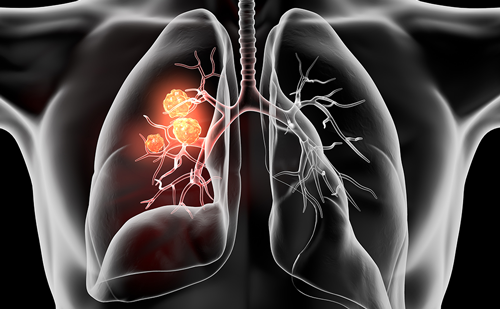
By way of orientation, I-ELCAP type screening is a clinical activity; a positive test CT initiates a process that leads to early diagnosis. Such diagnostic activity, as it is pursued in an individual, is squarely in the domain of clinicians. In this context, we suggest that it is the responsibility of the individual clinician to decide, from the evidence at hand, whether or not to recommend screening to individuals at risk for lung cancer. The clinician should not be deterred by those who remain pessimistic concerning CT screening, or by those health policy authorities who presume to be qualified to discourage high-risk members of the population from seeking early diagnosis and potentially curative treatment of lung cancer. In a matter as important as this, clinicians should not defer to non-clinical authorities. By definition, candidates for screening must be asymptomatic and without clinical signs to suggest lung cancer. Members of the high-risk population are identified at present according to age, exposure to cigarette smoke (current, former, or passive smoker), occupational hazards, co-existing lung disease, and family history of lung cancer.
Research efforts to refine the risk profile so that it reliably identifies an individual’s unique profile are underway and will inform the screening tests of the future, which will be discussed. In the meantime, lowradiation- dose chest CT has been shown to be the most sensitive test available. Obtaining the CT is not what we consider to be screening. The CT initiates the ‘regimen of screening’: a management algorithm initially derived empirically and regularly revised from the data of ongoing research. When judiciously followed, this regimen leads to early diagnoses and minimizes unnecessary tests and invasive interventions, namely biopsies and surgeries.2 We believe that it is not appropriate to initiate a screening program without coupling the CT to a regimen of screening of the I-ELCAP type. Also essential is a team of interested specialists to collaborate and monitor compliance with the management algorithm and the quality of its diagnostic components. Regular meetings of the team, which consists of pulmonologists, chest radiologists (trained in diagnostic and needle biopsy), cytologists, pathologists, thoracic surgeons, and medical and radiation oncologists, are essential to insure the timeliness and efficiency of the diagnostic pursuit. Successful programs have been active in community hospitals and occupational settings; an academic medical center is not essential.For programs that satisfy these criteria, we would also suggest that there is no reason to delay screening, i.e. early diagnosis, for lung cancer. However, screening for lung cancer is not recommended in the US or in most other countries. There is major controversy in the US about its implementation, first stimulated by I-ELCAP’s original report. Those responsible for the National Lung Cancer Screening Trial (NLST), sponsored by the National Cancer Institute, and other critics, insist that the results of that study, anticipated in 2009–2010, must be awaited before healthcare policy decisions can be made.3 Remarkably, that single study—a randomized, controlled trial (RCT), the most expensive ever conducted—is presumed by its advocates to be so powerful that the results will definitively set (or reset) healthcare policy on screening for lung cancer for the foreseeable future in the US. Our main concern, which was expressed to the trial’s planners and is unchanged now, is that the trial has the capacity to produce equivocal or falsely negative results in the same way that predecessors of the RCT genre performed in the evaluation of mammography.4 In our view, to wait for NLST results before starting to screen is to fail to cure many curable cases of lung cancer.
Two lines of criticism of I-ELCAP type screening have been monotonously repeated and are grossly misleading. First, the frequency of ‘falsepositives’— defined as lung nodules detected by CT that require additional evaluation but are not cancers—has been cited by some to be as high as 40%.5 As a consequence, unnecessary tests and biopsies are carried out with their attendant costs and morbidity. However, the fact is that in I-ELCAP screening of subjects 40 years or older with 10 or more years of cigarette smoking, a positive test is seen in 13% of subjects. This acceptable figure results from data-driven refinements of the definition of a positive test according to lung nodule size and radiographic characteristics for the baseline (prevalence or first test) and repeat screenings. Additional evaluation for those 13%, in the main, has been by repeat CTs after three months to assess the growth of the nodule before consideration of biopsy. It is evident that, since new or growing nodules on repeat annual screening CTs are the focus, ‘false-positives’ are less frequent than at baseline. Second, seemingly by rote, and with intention to suggest that CT screening for lung cancer is sure to fail, critics recite the familiar litany of biases: lead time, length time, and overdiagnosis. To these we have answered that the very objectives of screening for lung cancer, or any cancer, are to provide lead time and to diagnose latent, curable cancer.6 There is no bias introduced, unless one uses short-term survival rates (five years) to compare screening with nonscreening results to indicate the effectiveness and the success of screening. I-ELCAP does not do that.
We report long-term, 10-year rates, which in lung cancer equate with curability rates, and only compare survival rates of people screened who are treated compared with those who are not treated. Overdiagnosis bias refers to the inclusion in study results of lung cancers that are so slow to grow that death routinely results from another cause. We have responded to criticism regarding overdiagnosis by pointing out that, in I-ELCAP, all of the resected clinical stage I cancers (375) were reviewed by an expert panel of lung pathologists and all were found to be genuine, locally invasive cancers.7 All of the clinical stage I cases that were not resected1 were dead within five years. In addition, the incidence of lung cancer after the early rounds of screening is the same in the I-ELCAP screening group as in an unscreened population of similar age and cigarette smoke exposure. These findings speak strongly against overdiagnosis influencing the results of the I-ELCAP study in any important way. To address legitimate concerns about unnecessary invasive procedures, it is worth noting again that the I-ELCAP protocol recommends biopsy of a nodule only after growth at a malignant rate was ascertained on follow-up CT. Growth is defined as an increase in volume of a solid nodule, increase in size of the solid component of a part-solid nodule, the addition of a solid component, or overall increase in size of a non-solid nodule (ground glass).CT-guided fine- or core-needle biopsy is the preferred technique. Realtime on-site cytology interpretation provides immediate information about the adequacy of the specimen or need for additional samples. When needle biopsy is not possible, a bronchoscopic or image-assisted thoracosopy procedure is carried out. Limited thoracotomy for early diagnosis is rarely required. For critics5,8 to suggest that I-ELCAP screening may “do more harm than good” (the harm consisting of morbidity or mortality from unnecessary biopsies and surgeries) is to be grossly uninformed of the tenets on which the I-ELCAP study design rests. Furthermore, the critical role of the screening—coupling the CT test to explicit means of achieving early diagnosis—causes minimal adverse effects while maximizing favorable outcomes. Once the diagnosis of lung cancer is made, the diagnostic mission of screening is complete. The screening subject, now a patient with a life-threatening disease, needs to make a decision with the assistance of his or her physician. The focus here is on those diagnosed with clinical stage I disease. The overwhelming majority of these individuals, when provided with accurate information, will choose surgical intervention at an early date. Some, for a variety of reasons, will permanently refuse treatment of any sort. Others will defer for a time (late treatment) or select a non-surgical treatment for non-medical reasons. The outcomes will, of course, be influenced by these choices. The follow-up of all cases of cancer diagnosed in I-ELCAP is required for at least 10 years. For clinical stage I cancers, analysis of the time of symptoms and of metastasis, as well as the time and cause of death, provides prognostic information and insights about the natural history of early-stage disease as a whole. More importantly, in addition to initial size and cell type, future biomolecular research may produce new information about the etiology, novel diagnostics, and treatment of cancer subtypes.
While treatment is not an integral part of screening, it is essential for cure, and I-ELCAP considers its type and timing important information for the evaluation of efforts to achieve early treatment after early diagnosis in order to cure all potentially curable screen-detected cancers. There is now an unprecedented opportunity for the evaluation of various treatment modalities in stage I cancers. For treatment research (intervention), unlike diagnostic research (screening), the RCT is well suited and appropriately applied to assess the relative merits of different treatments, for example the comparison of lobectomy and lesser resections of lung (e.g. segmentectomy or wedge). Lung-sparing surgery is already becoming more important as the thoracic surgeon is presented with increasing numbers of small, screen-detected lung cancers (stage I). Cured of their initial primary disease years earlier, patients properly continue under screening and some may develop a second primary lung cancer. This scenario has already prompted the planning of a co-operative study to reconsider the question of whether wedge resection is sufficient to cure these small early lesions. The availability of even smaller (less than 2cm) stage IA lung cancers has provided an essentially new domain for research. The role and relative merits of non-surgical treatment modalities available—which include stereotactic radiation, high-frequency radioablation, cryotherapy, and the newer molecular targeted agents, such as angiogenesis, growth factor inhibitors and new vaccines—may be defined in well-designed trials. Products of future research will be tested in comparative trials until an agent proves curative and replaces surgery as a standard of care for the treatment of stage IA lung cancer. Future research is also expected to produce the means to:
• identify robust new risk factors that address the clinically relevant questions of who should be screened, at what age to begin, at what interval to screen, and when to stop;
• definitively diagnose early lung cancer without biopsy; and
• ultimately predict its development and the opportunity for primary lung cancer prevention.
It has been said that functional genomic and proteomic research will launch the next era of cancer molecular medicine. Molecular biologic techniques have been applied to lung cancer diagnostics since the 1990s. The linkage of the techniques of polymerase chain reaction, bioinformatics, high-throughput analysis, and molecular hybridization has led to the development of arrays of genes specific to an organ (e.g. breast, lung), body process (e.g. apoptosis, angiogenesis) and gene function (e.g. oncogenes, suppressor genes). Commercially available ‘chips’ are being sought to assist researchers and clinicians in the diagnosis and treatment of lung cancer. Potential screening techniques using micro-array analysis of DNA of sputum, buccal smears, and serum are under investigation. Although promising conceptually, this remains the province of the research laboratory.
Analysis of proteins in relevant samples in search of functional pathways (proteomics) is another area of research that has been accelerated by the advances in molecular and cell biology and bioinformatics. Proteomic analysis of blood, sputum, or other vital sources may prove useful for lung cancer screening in the future.
It has been recognized that changes in cellular metabolism resulting from cancer lead to alterations in the pattern of volatile organic compounds present in expired air. Analysis of samples from lung cancer patients is another method under investigation as a possible non-invasive and inexpensive screening test. Imaging technology, given its recent history and its accelerating capacity to demonstrate function, as well as microstructure to the cellular level, is expected to compete favorably with these other products of research, at least for the next decade.
All of these lines of investigation create great expectations for the future. However, the clinician who fantasizes that the ideal approach to lung cancer prevention will become a reality in his or her lifetime is quickly sobered when he or she reflects that, even if all cigarette smoke exposure were to stop today, there would remain a reservoir of millions of at-risk individuals, many of whom will die of the disease under the present pessimistic healthcare policies concerning screening. Right now, the best way to increase the curability of lung cancer is by screening, as demonstrated through evidence presented by I-ELCAP. Screening may reduce lung cancer deaths by up to 75%, saving 123,500 lives in the US each year. Clinicians will recognise that two compelling indicators—the rationale for CT screening and the evidence of its effectiveness— demonstrate that now is the time to implement CT screening for individuals at risk for lung cancer.