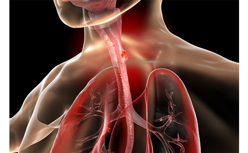
The on-board imager (OBI) has been in routine clinical use at the Karolinska University Hospital since June 2004. The OBI consists of a diagnostic X-ray tube and a kV flat panel imager, which are both mounted on robotic arms and designed for three main functions – orthogonal radiographs for three-dimensional (3-D) patient set-up, CB kVCT and realtime tumour tracking, and fluoroscopy. The availability of high-quality tomographic images and automatic tools for online 3-D image registration will lead to the introduction of new clinical applications and protocols. The most beneficial application is high-precision hypofractioned treatment of lung and liver metastases with online tumour position correction (see Figure 1).
Radiographic Online Set-up Verification of Prostate Patients Implanted with Gold Markers
The OBI has been used for the online set-up correction of patients using internal gold markers since June 2004. Displacements of these markers can be monitored radiographically during the treatment course and the displacements of the markers act as a surrogate for prostate motion. For this purpose, onboard kV-kV seems to be an ideal system in terms of image quality. Prostate patients are treated in the supine position and are given three gold markers (diameter = 0.9mm; length = 3mm) implanted under transrectal ultrasound control. Individually fabricated vacuum cushions are produced to immobilise the legs and to ensure stable positioning of the patient during the application of treatment fields. In the treatment room the patient is positioned according to the laser marks on the skin. Before each treatment, fraction orthogonal kV-kV images from the anteriorposterior (AP) and lateral directions are acquired using the OBI. Digitally reconstructed radiographs (DRRs) were obtained from planning CT data, and used as reference images, and the marker positions were contoured and displayed. The automatic 2-D matching procedure is based on bony structures used to match the two orthogonal images with reference images (DRRs) followed by a subsequent final manual alignment of the fiducial markers. The correction is applied on these gold markers rather than the bony structures, in order to correct the actual target position (see Figure 2).
The set-up deviations and required correction are displayed as couch displacements in X, Y, and Z directions, and couch rotation. Each displacement can be selected individually; the rotation of the couch was not considered. The position displacements can be applied using the remote table control. The total positioning errors, combining both set-up and internal organ motion errors after initial positioning according to the laser marks, have been quantified and analysed. The largest position error is due to the internal prostate motion (see Figure 2). The uncorrected results for 12 patients with prostate cancer are shown in Table 1, where moverall is the overall mean systematic deviation, σset-up is the random set-up error, and Σset-up is the set up systematic error. However, correcting AP, lateral, superior-inferior, pitch, and roll will not completely correct anatomical deformation. The majority of the patients treated using OBI were patients with prostate and gynaecological tumours. Other target volumes have also been positioned with OBI such as lung, head, and neck cancers, and brain tumours. The additional workload due to online registration is approximately two minutes added to the time slot booked for the patient. CB kV CT
The CBCT function of OBI was installed in March 2005 in the author’s department. It focuses on localising tumours based on internal anatomy and not just on the conventional external marks or tattoos. The CBCT system provides the capacity for soft-tissue imaging in the treatment position and realtime radiographic monitoring during treatment delivery.
Using the 3-D manual image registration, a match between the planning scan and verification CBCT was achieved and verified using the split view. This gave a good correlation between the two data sets. Currently, the author is using manual online registration tools for clinical set-up correction protocol. The image quality of the CBCT scans is slightly poorer than that of conventional CT scans and it also includes some artifacts from the tabletop. An example of a successful online registration of lung cancer can be seen in Figure 3, off-line registration of the prostate is shown in Figure 4a, and the head and neck in Figure 4b. The dose to the patient during CT acquisition is approximately 45mGy for full-fan geometry. The image quality is better for full-fan mode (the imager is centred with X-ray axis) than half-fan mode (the imager shifted a side to cover larger patient volume) due to more projections covering the whole field of view (FOV). The use of CBCT functions with an on-board imaging system in adjusting and verifying the position of the tumour and soft tissues before irradiation is a work in progress.
Future Work
Adaptive radiotherapy (ART) is the next step from IGRT and will be a valuable new tool for evaluating online and off-line treatment strategies to account for set-up uncertainty and anatomical changes. The adaptive framework should support different adaptation scenarios, including deformable target, tumour shrinkage, and organ atrisk modelling, re-planning and compensating for residual dose errors. In this treatment process multiple CT scans are acquired for each or few treatment fractions and are used as feedback to form a new planning target volume and modify the treatment plan accordingly. The clinical implementation of ART is now possible in some centres. The off-line adaptive planning approach used the daily scanned CT images during the first week of treatment delivery to shape an average over the planning target volume (PTV), the clinical target volume (CTV), and the critical surrounding organs in the new modified treatment plan. This technique has been implemented in different institutions especially for re-planning the prostate cancer where the target is contoured on each CBCT scan. The online adaptive automatic re-planning and target deformation will be the main issues to be solved in the very near future.
Conclusions
The linear accelerator with on-board imager seems to be very useful for the set-up of patients and for verification of the irradiation field just before irradiation. These systems will provide high-quality radiotherapy by delivering higher doses to tumours and fewer doses to normal tissue. The 3-D treatment plan verification using online correction based on soft tissue and target position has been used clinically and adds an approximate additional five minutes to the treatment time slot due to acquisition, reconstruction, and 3-D registration.
The radiographic mode with kV-kV images using the on-board imager systems offers a means for the repositioning of patients. Gold markers implanted into the prostate can be safely used for daily organ tracking using online set-up verification by OBI. The patient position is corrected according to the position of the markers detected in the localisation images, acquired prior to set-up, and also changes in work flow and responsibilities for the therapist. The workloads generated by set-up correction in relation to their benefits have to be considered. The initial experience of CBCT is very encouraging with the image quality adequate for internal soft target localisation.
Clinical Introduction of Image-guided Radiotherapy and Cone Beam Computed Tomography
Abstract:
Overview
Image-guided radiotherapy (IGRT) can be used to measure and correct target and critical structure positional errors immediately prior to or during treatment delivery. Single-slice megavoltage computed tomography (MVCT) using 50MV for the racetrack microtron (RTME) was first utilised at Karolinska University Hospital in 1987. There were some technical and clinical limitations with this system.
Some of the most recently available methods of target localisation are transabdominal ultrasound, implanted markers with in-room MV or kilovoltage (kV) X-rays, optical surface tracking systems, implantable electro-magnetic markers, inroom CT, such as kVCT on rail, kV or cone beam (CB) MVCT and helical MVCT.
The image-guided systems provide an increasing amount of information about daily target localisation and set-up errors during the course of fractionated treatment. This information can be compared online with the treatment planning system and utilised for the set-up adjustment of the treatment fraction. The verification of the accurate treatment position in conjunction with detailed anatomical information before every fraction is therefore essential for the outcome of the treatment.