Mechanisms of Inflammation-induced Cancer
Approximately 20% to 30% of the world’s cancer burden can be traced to infectious agents that are thought to act through the production of chronic infections and subsequent chronic inflammation. For example, adenocarcinoma of the stomach is characterized by a series of sequential events comprising Heliobacter pylori infection, resultant long- standing persistent inflammation, atrophic gastritis, dysplasia, and carcinoma.The molecular mechanisms of inflammation-induced cancer relate to the action of phagocytic inflammatory cells (neutrophils and macrophages) that release a variety of chemical agents designed to kill pathogens. These agents include superoxide, hydrogen peroxide, singlet oxygen, and nitric oxide, which can further react to form the highly reactive peroxynitrite. Some of these reactive oxygen and nitrogen species can directly interact with DNA in host bystander cells or react with other epithelial cellular components, such as phospholipids, initiating a free radical chain reaction – the end result being cell injury and cell death. Lost epithelial cells must regenerate quickly by DNA replication and cell division in order to maintain their barrier function.
This increased DNA synthesis, which is also stimulated locally via cytokines released from inflammatory cells, places epithelial cells at high risk for acquiring oxidative/nitrosative DNA damage and subsequent mutations.
Evidence Suggesting a Link Between Inflammation and Prostate Cancer
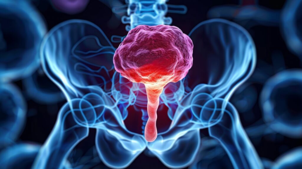
A growing body of work from studies of families with hereditary prostate cancer, as well as data from the fields of genetic epidemiology, histopathology, and molecular pathology, has begun to suggest that a link may exist between chronic inflammation and prostate cancer. For instance, case-control epidemiological studies have found an increased relative risk of prostate cancer in men with a prior history of certain sexually transmitted infections (STIs) or prostatitis. Furthermore, genetic epidemiological data have implicated germline variants of several genes directly involved in the response to infection (e.g. RNAseL or macrophage scavenger receptor 1 (MSR1)) in modulating prostate cancer risk. In addition, the overwhelming majority (more than 90%) of prostate cancers contain CpG island hyper- methylation within the upstream promoter of glutathione S-transferase pi gene (GSTP1), resulting in the inactivation of a gene involved in defenses against oxidant and electrophilic damage. Finally, the vast proportion of prostate tissues that are obtained by needle biopsy, transurethral resection, radical prostatectomy, or cystoprostatectomy have been noted to contain multiple foci of chronic and/or acute inflammation.
Model of Prostate Carcinogenesis
Over the past years, a new model of prostate carcinogenesis has emerged wherein phagocyte-derived oxidant stress increases the initiation rate of prostate cancer in areas of proliferative inflammatory atrophy (PIA) (see Figure 1). Pathologists have long recognized several patterns of focal epithelial atrophy in the periphery of the prostate – an area where prostate cancers typically arise. Many of these lesions are associated with inflammatory infiltrates consisting, at times, of lympho- cytes, macrophages, and neutrophils. Interestingly, as opposed to being quiescent,the atrophic epithelial cells in these foci are generally quite proliferative, with the number of cells undergoing cell division being increased from three- to 80-fold over that seen in matched adjacent normal appearing epithelium.To highlight the common association of inflammation and proliferation, the term ‘proliferative inflammatory atrophy’ as an umbrella term The Case for the Role of Chronic Inflammation in the Development of Prostate Cancer 112 B USINESS BRIEFING: US ONCOLOGY REVIEW 2006 Palapattu.qxp 16/12/05 11:16 am Page 112 for most of these lesions has been put forth. From a topographical standpoint, the direct merging with morphological transitions of areas of PIA with high-grade prostatic intra-epithelial neoplasia (PIN) – a putative prostate cancer precursor lesion – has been frequently observed. PIA lesions in the setting of early carcinoma, with occasional direct merging of atrophic epithelium with carcinoma, have also been observed. Additionally, regions of PIA have been shown to possess elevated levels of GSTP1, GST alpha (GSTA1), and cyclooxgenase-2 (COX-2) in many,albeit not all,epithelial cells,suggesting that these cells may be responding to increased oxidant/nitrosative stress.
In terms of somatic DNA alterations, the authors have recently reported that, while normal epithelium or benign prostatic hypertrophy (BPH) epithelium from cancer patients does not contain methylated GSTP1 alleles, approximately 6% of PIA lesions harbor epithelial cells with methylated GSTP1. Others have also described molecular changes in PIA, including chromosome-8 numeric abnormalities. For example, one group has recently reported that the median percentage of nuclei with more than three chromosome-8 centromeric probe signals as:
• 1.3% normal;
• 2.1% simple atrophy;
• 2.8% high-grade PIN (HGPIN);
• 4% post-atrophic hyperplasia (PAH); and
• 6% in carcinoma.
It should be noted that simple atrophy and PAH are both considered forms of PIA in the current classification schema. Similarly, another group has observed mutated p53 alleles in PAH, PIN, and carcinoma, but not in normal prostate. PIA lesions, therefore, share a number of histopathologic and genetic traits with both pre-malignant and malignant lesions of the prostate. The authors’ current working model describes PIA as a response to micro- environmental stress experienced by normal prostate epithelial cells (see Figure 1). Individual cells within given PIA lesions that are unable to adequately defend themselves against oxidative/nitrosative genome damage (perhaps due to silencing of GSTP1 via promoter CpG island hypermethylation) may subsequently progress to PIN or prostate cancer. In this model it is hypothesized that many, although not all, high-grade PIN lesions may develop by first proceeding through a period of atrophy. At other times, it is hypothesized that atrophy can directly proceed to carcinoma without a prior phase of high-grade PIN.
Conclusion
Novel insights into the development of human prostate cancer that implicate the process of chronic inflammation in prostate carcinogenesis have recently emerged. Examination of the development of prostate cancer in this context may yield innovative approaches to the prevention, detection, and treatment of this burdensome disease. ■


