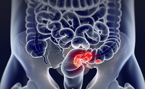
Screening for colorectal cancer is worthwhile;1–3 however, the ideal method has not yet been established. Adherence by an asymptomatic patient population to a colorectal cancer screening programme is known to be lower than 50%.4 This lack of patients complying with the existing screening tests is due to pre-, per- and post-procedural inconveniences causing the patients to interrupt their normal daily activity.5
Screening for colorectal cancer is worthwhile;1–3 however, the ideal method has not yet been established. Adherence by an asymptomatic patient population to a colorectal cancer screening programme is known to be lower than 50%.4 This lack of patients complying with the existing screening tests is due to pre-, per- and post-procedural inconveniences causing the patients to interrupt their normal daily activity.5
From its introduction by David Vining in 1994,6 virtual colonoscopy or computed tomography (CT) colonography was almost immediately been proposed as a viable alternative to the existing tests that screen for colorectal cancer.7 In fact, several single-centre trials obtained very good sensitivity and specificity of lesion detection.8,9 Based on these qualities, CT colonography is accepted as a standard examination for patients after incomplete optical colonoscopy or for those patients who cannot be examined with optical colonoscopy.10 Furthermore, research has been performed to alleviate the preparation.11
Despite the high expectations and invaluable improvements in technique, CT colonography has not yet been established as a widely accepted test for screening for colorectal cancer. This may be due to there being two large multi-centre trials in the last year with very disappointing results of lesion detection. Both the Cotton12 and Rockey trial13 failed to fulfill high expectations. In the former study, sensitivity for polyp detection on a per patient basis was 39% and 55% for lesions 6–9mm and lesions >1cm, respectively, in 615 patients. In the latter study these results were 51% and 59% for lesions 6–9mm and lesions >1cm, respectively, in 614 patients. The reasons for the failure of these studies were numerous.14–17 Although recently published, both studies started in 2000 resulting in suboptimal scanning and visualisation technique, and neither used the optimal method to prepare the patients. In many cases there was no evaluation of the preparation and colonic distention, and the radiologists received no feedback from the endoscopists during the trial. Some of the radiologists were not experienced (<10 examinations performed), underestimating the considerable learning curve of CT colonography, or the examinations were interpreted during routine work. There was no consistency in the method of reading the data sets.
These shortcomings are related to the technique of CT colonography, underscoring the importance of applying a rigorous method of preparation, examination technique and image interpretation when performing CT colonography.
How to Perform CT Colonography?
CT colonography consists of three important parts:
• the preparation preferably with faecal tagging;
• the examination with the colonic distention and the scanning parameters; and
• reading of the data sets on a good workstation by an experienced reader.
It has become clear that state-of the-art application of CT colonography is mandatory to obtain good results of lesion detection.
Preparation
As for other full structural examinations of the colon, it is mandatory to prepare the colon prior to CT colonography otherwise faecal residue hampers visualisation of the mucosal surface of the colon. There are two options:
• the regular preparation, which can also be used for optical colonoscopy; or
• the preparation with faecal tagging.
The Regular Preparation
This preparation combines a low residue or clear liquid diet with oral sodium phosphate (Phosphosoda – a single dose of 45ml) and two bisacodyl tablets.18 Despite an extensive cathartic cleansing, residual stool frequently mimics a polypoid lesion (false positive) or inversely a polyp is interpreted as faecal residue (false negative). Furthermore, residual fluid may cause drowned segments resulting in incomplete visualisation of the colonic surface.19 This is less the case with oral sodium phosphate than with polyethylene glycol, which is not considered a suitable cathartic for CT colonography anymore.20
Faecal Tagging
Faecal tagging is the peroral ingestion of contrast material (barium and/or iodinated contrast) prior to CT colonography to label or tag faecal residue remaining in the colon after the preparation. The faecal residue appears hyperdense or white on the CT images. As a consequence, polyps are recognised as they are not labelled or as they appear as negative filling defects in tagged fluid (see Figure 1). This avoids the false positive and negative diagnosis occurring with a regular preparation.
Faecal tagging can be performed in combination with a full cathartic cleansing. This has been achieved successfully by Pickhardt et al.21 In the largest trial on CT colonography ever performed, they obtained tremendous results of polyp detection in 1,233 patients who were at average risk for colorectal cancer (see below).
Not only does faecal tagging reduce the lack of accuracy in polyp detection, it offers the occasion to reduce the preparation prior to CT colonography. In fact, as the faecal residue is labelled in the colon, it is possible to leave more of it in the colon, thus reducing the preparation. In a first study, Lefere et al.22 combined 750ml of a 2.1% w/v barium suspension with a reduced cleansing and obtained a sensitivity of 92% and 100% for lesions 6–9mm and >1cm, respectively. In a recent study of 180 patients they obtained similar results (6–9mm: 88%; >1cm: 100%) using only a total of 60ml of a 40% w/v barium suspension to achieve efficient tagging.23,24 Furthermore reduction of the cathartic cleansing improves patient compliance.
The Examination
To adequately perform CT colonography the following conditions need to be fulfilled:
• adequate colonic distention; and
• optimal scanning parameters.
Colonic Distention
Adequate colonic distention is achieved with:
• smooth muscle relaxation;
• inflation of the colon with CO2; and
• scanning the patient in supine and prone position (dual positioning).
Although not mandatory, it is now generally accepted in Europe that improved distention of the colon and improved patient comfort is obtained with 20mg of intravenous scopolamine buthylbromide.25 It is preferable to inflate the colon with CO2.
Before the use of CO2 to inflate the colon, patients frequently suffered from post-procedural discomfort such as abdominal fullness, bloating and cramps due to the inflation of the colon with air. In fact, the air had to be expelled after the exam. The use of scopolamine buthylbromide made this a difficult task. The introduction of CO2 resolved these post-procedural inconveniences. Compared with room air, the absorption of CO2 in the human body is much faster.26 So even before the patients leave the CT suite the inflated CO2 is completely reabsorbed. When used with an automated CO2 injector, colonic inflation can be achieved in a rapid and comfortable way for the patient. Finally, to further improve colonic distention, the patient is scanned in both supine and prone position. This technique of dual positioning allows for distending parts of the colon that are collapsed because of a dependent position in the supine position, which becomes non-dependent in prone and vice versa.27
Scanning Parameters
The use of multi-detector scanners (16 and 64 slices) has resulted in thinner slice thickness (collimation) and a faster examination (less than 15 seconds). Using a thinner slice collimation should improve the accuracy of the technique.
As a screening procedure should be harmless, the radiation dose generated by CT could be a blocking issue when screening a population at average risk of colorectal cancer. However, since the introduction of the multi-row detector scanners, efforts to reduce the radiation dose have been performed. Iannaccone et al.28 obtained a sensitivity of 83.3% and 100% for lesions 6–9mm and >1cm respectively, using an ultra-low dose of 140kV and 10mAs. With the new 64-slice scanners the identical ultra-low dose can be used without deterioration of the image quality (see Figure 2).
Reading the Data Sets
The Workstation
After scanning the patient, the obtained images are transmitted to a dedicated workstation. This workstation is provided with dedicated software allowing performance of a virtual ‘fly-through’ of the colon, quick comparison between two- and three-dimensional, and supine and prone images. Several vendors are making tremendous efforts to constantly improve their workstation concerning ergonomy and tools enabling ease of reading and reporting for the radiologist.
Experience of the Reader
It should be remembered that CT colonography is not an easy technique. There is a considerable learning curve in becoming used to this new visualisation technique of the colon. This period of learning to interpret the examination is particularly important because the radiologist will be confronted with the ground truth provided by the optical endoscopy. Too many misses will unavoidably cause a disbelief in the technique, so to overcome this painful period it is mandatory for the radiologist to make a good commitment with the gastroenterologist and to start with at least 50 examinations being controlled by optical colonoscopy.17,29 In the meantime the radiologist has to refine his skills by participating in dedicated workshops on CT colonography enabling training on several data sets. Taking these remarks into consideration it will be possible to achieve good results of lesion detection as was done by Pickhardt et al.21
Compared with the Cotton and Rockey trial, Pickhardt et al. used a much more rigorous method based on a preparation with faecal tagging, adequate examination technique with good colonic distention and thin slice collimation, the exclusive participation of experienced centres and the use of a very good workstation. They obtained impressive results of polyp detection in 1,233 patients who were at average risk of colorectal cancer, constituting the largest trial on CT colonography to date. In this study the endoscopists were unaware of the results of CT colonography. Using a method of segmental unblinding revealing the results of CT colonography per segment virtual colonoscopy obtained better results than optical colonoscopy for lesions >8mm and >1cm. CT colonography detected 85.7%, 92.6% and 92.2% of lesions >6mm, >8mm and >1cm, respectively, while optical colonoscopy detected 90%, 89.5% and 88.2% of lesions >6mm, >8mm and >1cm, respectively.
With regard to flat adenomas, in the same study, encouraging results were obtained. Optical colonoscopy retrieved 55 lesions with a flat appearance, 29 being an adenoma and 26 a hyperplastic lesion. Overall sensitivity for the flat lesions was 79.6%, with a sensitivity of 76.7% and 82.2% for the hyperplastic lesions and adenomas respectively.30 This could be an indication that flat adenomas are not a significant drawback for CT colonography when adequately performed.
Future Developments
Two important developments could dramatically change the future for CT colonography:
• computer-aided diagnosis (CAD); and
• laxative-free CT colonography.
Computer-aided Diagnosis
With computer-aided diagnosis (CAD), the radiologist makes a diagnosis based on the findings of a software program performing automated image analysis by detecting shapes in the colon.31 As with mammography and detection of lung nodules CAD is particularly helpful when screening for disease with a low prevalence, as is the case with colorectal cancer. CAD draws the radiologist’s attention to abnormalities or ‘polyp candidates’ in the colon. The radiologist has to decide which of these are true positives and which are false positives. By doing so, CAD could act as a second reader or help to perform polyp detection after consensus reading (double reading), and detect lesions that otherwise would have been missed by the radiologist. In that way CAD could help avoiding bad results as in the Cotton and Rockey trials. It would also consistently help the radiologist during his learning curve. In the small studies performed so far promising results with sensitivities of 70% to 100% for polyp detection on a per lesion basis were obtained.32 Research is under way to combine CAD with faecal tagging.33
Laxative-free CT Colonography
It is generally accepted that the poor adherence of an asymptomatic patient population to undergo a full structural colonic examination is directly related to the discomfort caused by the cathartic preparation.34 Both radiologists and gastroenterologists admit that if ever CT colonography could be performed without cathartic cleansing, this would obviate any discussion of whether virtual or optical colonoscopy should be used for screening for colorectal cancer.35 It would also dramatically improve the success of a colon cancer screening programme. This method of performing CT colonography has successfully been adapted by Iannaccone et al.36 They successfully examined 203 patients with laxative-free CT colonography. A low residue diet was combined with faecal tagging over two days. They obtained very good results of polyp detection – 86% for lesions >6mm (79 lesions), 95.5% for lesions >8mm (45 lesions), 100% for lesions >1cm (24 lesions).
Conclusion
CT colonography is a promising technique to detect tumoural lesions in the colon. It is indicated when optical colonoscopy is incomplete or contra-indicated. Using a meticulous technique, performed by an experienced reader it is a very feasible examination and could be used for screening for colorectal cancer on a local basis. However, it is not yet ready for widespread application to screen for colorectal cancer amongst a large radiological community. Laxative-free CT colonography and CAD are promising developments. ■


