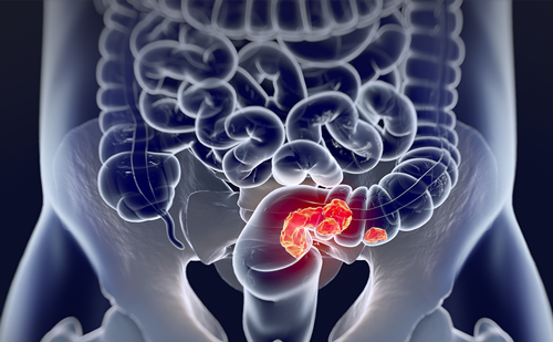
Historically, these patients have been considered to have a poorer prognosis than those subsequently found to have developed metachronous diseases.1,5 A further 40–50% will develop liver metastases, usually within the first three years of follow-up after successful resection of the primary tumour.1,4,5 Surgery remains the only treatment that offers the prospect of cure for CRLM. Until recently, only far fewer than 20% of these patients were considered suitable for attempted curative resection, the remaining patients being offered palliative and symptomatic treatment.6
This article focuses on a variety of recent strategies that have been designed to increase the pool of those patients presenting with synchronous metastatic disease for whom curative treatment may be possible. These include improved pre-operative staging techniques, new standards for surgical resection, novel surgical strategies, the application of modern systemic chemotherapy in a ‘neoadjuvant’ setting, an emerging role for ablative therapies and an emphasis on the collaborative, multidisciplinary management of this disease.
Pre-operative Staging – The Key to Selection of Candidates for Curative Treatment
Individual imaging techniques used in pre-operative staging have different strengths and weaknesses. One clear aspect of these rapidly advancing technologies has been the earlier detection of low-volume metastatic disease. From a pathological perspective, all metastases (those found at the time of initial presentation, and those subsequently found after an apparently ‘curative’ resection of the primary tumour) are synchronous to the time of diagnosis of primary colorectal cancer. It is only our ability to detect low-volume disease that is subsequently ‘metachronously’ diagnosed when the disease becomes clinically or radiologically overt that is evolving. Consensus is now emerging on the optimal choice of technique and the sequence in which they should be employed.7–10
Recent advances in computed tomography (CT) technology, such as helical CT and multidetector-row helical CT, have improved the performance of CT in terms of speed of acquisition, resolution and ability to image the liver during various phases of contrast enhancement with greater precision.7,10 Intravenous iodinated contrast media should be used routinely. These techniques help to characterise liver lesions based on their enhancement patterns during the various phases of contrast circulation in the liver.10 CT has limitations, including the need for a high radiation dose and low sensitivity for the detection and characterisation of lesions smaller than 1cm.
Magnetic resonance imaging (MRI) is a highly effective imaging modality for detecting and characterising liver lesions, as it provides high lesion-toliver contrast. Gadolinium, the most commonly used MRI contrast agent, behaves similarly to the iodinated contrast agents used in CT.7,10 Liver-specific contrast media, such as super paramagnetic iron oxide, further improve the contrast between liver and metastases.10,11 However, it is limited by low sensitivity for detecting extrahepatic disease in the peritoneum and chest.
Positron emission tomography CT has emerged as an important diagnostic tool in the evaluation of metastatic colorectal cancer. This modality is highly sensitive. However, any focal area of hypermetabolism (including inflammation and abscesses) can give false-positive results. Other disadvantages include high cost, poor lesion localisation and limited sensitivity for lesions smaller than 1cm.10,12 Surgery – The Old and New Standards for Resection
Many substantial prospective and retrospective studies of surgical resection of CRLM consistently show five-year survival rates following liver resection of 30–50%, depending on selection criteria.13 Those patients who survive for five years can usually be considered cured of the disease.
In the past, the decision to resect CRLM was relatively straightforward. On the basis of old studies that established certain adverse clinicopathological factors, liver resection was attempted only in patients who had one to three unilobar metastases, preferably presenting at least 12 months after resection of the primary tumour, whose disease was resectable with at least a 1cm margin of healthy liver tissue and who had no hilar lymphadenopathy or extrahepatic disease.14
More recent experience has demonstrated that patients outside the above traditionally accepted factors can experience long-term survival following liver resection.15-16 Thus, a shift has occurred in the criteria used for assessing resectability, from morphological criteria to new criteria based on whether a macroscopically and microscopically complete (R0) resection of the liver can be achieved. Instead of resectability being defined by what is removed, resectability should now be determined by what will remain.
Specifically, CRLM should be determined as resectable if:
- the disease can be completely resected;
- two adjacent liver segments can be spared with adequate vascular inflow and outflow and biliary drainage; and
- the volume of the liver remaining after resection, i.e. the future liver remnant (FLR), will be adequate.11 Clearly, the FLR limit for safe resection varies from patient to patient, but in those with an otherwise normal liver the safe FLR volume is 20%.17
These new standards clearly challenge the ‘1cm rule’, which required that liver resection be attempted only if a margin of at least 1cm could be achieved. In fact, various studies show that the width of the surgical margin has no effect on survival, as long as the margin is microscopically clear.18,19
New Surgical Strategies to Improve Resectability
In conjunction with neoadjuvant therapy, new surgical strategies have been increasingly employed in patients with unresectable CRLM to improve resectability. Portal vein embolisation induces atrophy of the liver to be resected and hypertrophy of the liver that will remain (i.e. increases the FLR). Similarly, two-stage hepatectomies involve delayed re-hepatectomy after hypertrophy of the residual liver and may be used for large bilateral lesions in which a one-stage resection of all of the involved segments would lead to liver failure.20,21 Extrahepatic colorectal metastases may be resected with curative intent, such as direct diaphragmatic invasion, adrenal metastases and lung metastases that are few in number and readily resectable.1 Recent data suggest that if pulmonary colorectal metastases are resectable, 35% of patients are alive at five years.22
Neoadjuvant Chemotherapy and ‘Rescue Surgery’
Modern chemotherapeutic regimens combining fluorouracil (5-FU), folinic acid and oxaliplatin and/or irinotecan are associated with high response rates of up to 50% and can allow 10–30% of patients with disease that is initially considered unresectable to be successfully brought to liver surgery.20,23
The largest study reported so far20 was a consecutive series of 1,439 patients with CRLM managed over an 11-year period. Of these patients, 1,104 (77%) were initially considered unresectable and were treated with chemotherapy, mainly in the form of 5-FU and folinic acid combined with oxaliplatin (70%), irinotecan (7%) or both (4%). Of these 1,104 patients with unresectable CRLM, 138 (12.5%) had a good response to chemotherapy, enabling potentially curative liver surgery to be performed in 93% of these cases. Survival was 33 and 23% at five and 10 years, respectively, with a median survival of 39 months, although this was significantly lower than that of patients resected primarily within the same period at the same institution (48 and 30% at five and 10 years, respectively). These data suggest that the ability to achieve secondary liver resection of initially inoperable CRLM is directly proportional to the degree of response to the chemotherapy regimen.24
Phase II and III studies evaluating novel biological agents, such as the monoclonal antibodies directed against vascular endothelial growth factor (bevacizumab) and epidermal growth factor receptor (cetuximab and panetumumab), suggest even greater response rates (and possibly higher secondary CRLM resection rates) compared with conventional chemotherapy alone. Therefore, even more patients with initially unresectable CRLM may respond to treatment with combinations of systemic treatments in the future.25
What Role for Ablative Therapies?
Much of the current interest in radiofrequency ablation (RFA) derives from its low morbidity and mortality.26 A recent meta-analysis of 95 published series reported a complication rate of 8.9%,27 with the most common complications being intra-abdominal bleeding, sepsis and biliary tree injury. Mortality rates range from 0 to 0.5%. An apparent disadvantage of RFA is a high rate of local recurrence, ranging from 1.8 to 12%, with a surgical approach to as high as 40% with percutaneous placement. Undoubtedly, this relates to the types of lesions being treated by RFA. Ablative therapies are often used for the treatment of metastases that are often too close to major vascular structures to be considered resectable with a clear margin. Just as a surgical margin would be likely to be compromised, blood flow will conduct away heat, leading to incomplete ablation and recurrence.2 The efficacy of RFA in unresectable CRLM has been established by several large cohort studies, with median survivals of 28.9–36 months being achieved.26 There are currently no prospective randomised, controlled trials to show an advantage for RFA over chemotherapy alone in unresectable CRLM, but this deficiency is being addressed by a trial of chemotherapy plus local ablation versus chemotherapy alone (CLOCC trial). A significant improvement in the chemotherapy plus RFA group would provide strong evidence of its value, but it is difficult to foresee a successful trial comparing RFA with surgical resection, as the results of the latter are good and operative mortality is low. However, RFA may have a future role in combination with surgery as part of the effort to expand the definition of resectability.20
Management Strategies for Patients Presenting with Synchronously Detectable Colorectal Liver Metastases
In a number of cases, this can be relatively straightforward.28–30 Patients who present with a technically ‘easily’ resectable primary tumour (right, transverse, left and sigmoid colon) and peripherally placed low-volume liver disease (segments 2, 3, 4B, 5, 6 and subcapsular lesions in segments 4A, 7 and 8) are amenable to synchronous resection of both primary and metastatic liver disease at the same procedure, without increased morbidity or mortality.31 Those patients (a decreasing minority) who present with large bowel obstruction and synchronous CRLM should have immediate definitive treatment for their life-threatening colonic emergency (endoscopic stenting, resection with either a stoma or immediate reconstruction).
Most surgical oncologists would recommend that when resection of the primary tumour may be more demanding (T2–T3 rectal carcinoma), when the treatment of the primary requires neoadjuvant treatment (chemoradiotherapy) to downstage the primary tumour (T3–T4 rectal carcinoma) before surgery or when the liver disease (albeit technically resectable) is of such an extent that at least a hemi-hepatectomy or more is required, a planned sequential staged procedure carries lower peri-operative risk.30,31
However, when proposing a staged sequential treatment strategy, the surgeon must remain concerned about the risk of tumour progression during treatment.32–35 Clearly, for patients who present with a relatively asymptomatic primary tumour (not bleeding and non-obstructive) in the presence of unresectable liver metastases, it would be reasonable to propose a course of systemic chemotherapy and to base subsequent treatment strategies on the degree of response.32 Those patients whose response to chemotherapy is sufficient that their CRLM disease is now potentially resectable can be considered for surgery with curative potential.32-34 For those patients whose initially unresectable disease continues to progress while on chemotherapy (which is in the order of 6–10%),20,35 consideration can be given to further lines of chemotherapy, but overall the outlook remains poor and futile surgery can be avoided. If colon cancer patients (as opposed to rectal cancer patients) with initially unresectable liver disease respond so well that an R0 resection can be achieved using a relatively minor liver resection, a synchronous liver-bowel procedure will then be an option.32 The real debate relates to those patients who respond sufficiently for their liver disease to be amenable to potentially curative surgery, albeit by an extended hepatectomy (possibly facilitated by either two-stage surgery or portal vein embolisation), and those patients who will still require major pelvic surgery to excise their primary rectal tumour. The fundamental question is now whether having achieved a window of therapeutic opportunity to deal with the liver disease the liver disease takes precedence. In this, the work of Mentha et al. is illuminating and helpful.33,34 The authors propose that we should resect the liver disease first and, if successful in eradicating the liver metastases, subsequently deal with the primary bowel tumour. They support their argument with their own experience, in which this strategy has been successful in achieving potentially curative surgery for both primary and secondary disease in 16 of 20 such patients (a success rate of 80%).33 We now wait with interest to see whether others can reproduce these very interesting results.
Conclusions
Despite increasing awareness of symptoms of colorectal cancer among both the public and clinicians, patients still present at an advanced stage of their disease with CRLM or more distant spread. Modern chemotherapy regimens offer increasing numbers of patients with CRLM that is initially thought to be unresectable the chance of being brought to potentially curative liver surgery. The current area of controversy in this field is now the timing of such surgery and the strategic decisions around which operation (bowel first, liver first or synchronous combined surgery) is now the first procedure of choice.