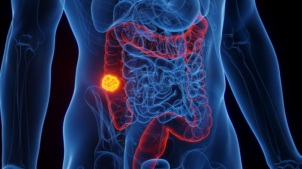
Colorectal cancer (CRC) is the third most common cause of cancerrelated deaths worldwide.1,2 Upon diagnosis, 19% of CRCs are metastatic, and while the overall five-year survival rate for patients with early CRC is around 60%, the rate drops to less than 10% in advanced disease.1
The multidisciplinary approach to CRC treatment is currently based on radical surgery and fluoropyrimidine-based chemotherapy. Indeed, for many years 5-fluorouracil (FU) has been the backbone of therapy since it demonstrated an improvement in overall survival in both the adjuvant setting and metastatic disease.1,2 In the last 15 years newer chemotherapeutic agents such as irinotecan (IRI) and oxaliplatin (l-OHP), used as monotherapy or in combination with FU, have had a significant impact on treatment strategies for patients with CRC, and these new regimens have improved patient outcomes.3 Indeed, it is very well-established that regimens combining FU/leucovorin with IRI or l-OHP are equally effective in terms of response rate and overall survival4 and represent the standard first-line treatment for advanced CRC.5 In addition, the combination of l-OHP with FU/leucovorin was demonstrated as being superior to bi-monthly FU/leucovorin as adjuvant therapy6 and is considered a standard first-line therapy in stage III CRC.2
In the last few years, the scenario has been further complicated by the advent of new molecular-targeted agents.3 Indeed, the monoclonal anti-vascular endothelial growth factor (VEGF) antibody bevacizumab and the monoclonal anti-epidermal growth factor receptor 1 (EGFR1) antibodies cetuximab and panitumumab have become available for the clinical management of advanced CRC. These novel drugs demonstrated a single-agent antitumour activity and, more importantly, exhibited a synergistic activity with IRI/FU or l-OHP/FU regimens, reducing drug resistance and improving the efficacy of combination chemotherapy.7–10
However, in spite of this progress, a significant percentage of patients still relapse after adjuvant chemotherapy and current treatments are largely ineffective against metastatic diseases. Thus, new therapeutic strategies are needed to improve the outcome of patients with CRC.
Several new studies are ongoing with the aim of finding the best combinations of cytotoxic and targeted therapies and to validate novel biomarkers able to predict sensitivity/resistance to specific anticancer agents. In this article we review some of the strategies aimed at improving the efficacy of drug therapies in human CRC.
The Combination of 5-fluorouracil and Irinotecan in Human Colorectal Carcinoma
Doublet chemotherapy (i.e. fluoropyrimidines combined with IRI or l-OHP) represents the standard of care for the treatment of metastatic CRC, since it has been demonstrated to increase the time to progression and overall survival rates. However, while most of the schedules combining l-OHP and FU differ only marginally, the combinations of IRI and FU are characterised by major differences in terms of doses and schedules.3–5,11,12 Indeed, regardless of the specific combination regimen, FU/IRI-based chemotherapy achieves a response rate of 30–50% and an overall survival period of 14–20 months in advanced CRC.3,5 Interestingly, three regimens of IRI and FU (FU, IRI and leucovorin [IFL]; folinic acid, FU and IRI [FOLFIRI]; and AIO+IRI) have been studied in phase III trials,11,12 and several other different schedules have also been proposed.3,13 These phase III clinical trials clearly demonstrated that the addition of IRI to FU/leucovorin, administered either as bolus or as a continuous infusion, resulted in a doubling of the tumour response rate, prolonged median survival and a substantially reduced relative risk of death compared with FU/leucovorin alone.11,12 Interestingly, in the study by Saltz et al., IRI was combined with bolus FU/leucovorin in a weekly schedule (IFL),11 whereas in the Douillard schedule, IRI was combined with infusional FU/leucovorin (FOLFIRI).12 It is noteworthy that the regimens with IRI and infusional FU were associated with reduced toxicity compared with the IFL regimen alone.14 More recently, a phase III study suggested that the up-front use of a triplet schedule with IRI, l-OHP and FU/leucovorin (FOLFOXIRI) significantly improved the outcome in terms of response rate and survival compared with a standard doublet of IRI and FU/leucovorin, but was characterised by relevant toxicities.15
Our group has been involved in studying the interaction between the IRI active metabolite SN-38 and FU in human colon carcinoma HT-29 cells (cell line with non-functional p53)16 in order to obtain in vitro evidence for optimising chemotherapeutic schedules. We observed that the sequential exposure of colon carcinoma cells to the two agents produces a supra-additive effect, with maximal cytotoxic activity when cells are pre-exposed to SN-38 before FU. Concomitant exposure to both drugs produces only additive effects. Interestingly, this synergism depends on the extent of cell exposure to FU and the interval between administration of the two drugs. Indeed, it is possible to potentiate this synergism of action by prolonging the exposure of tumour cells to FU and by administering the two agents sequentially with a minimal interval between them. Furthermore, the sequential exposure to SN-38 before continuous infusion of FU elicits the maximal increase in apoptotic cell death.17 These results are consistent with the evidence from Poele and Joel, who demonstrated that the cytotoxic activity of IRI depends on the schedule and the p53 status of tumour cells.18 Indeed, these authors observed significantly higher levels of apoptosis for prolonged exposure to SN-38 in p53-deficient human CRC cells and for shorter exposure to higher concentrations in p53 wild-type CRC cells.18
Based on this pre-clinical evidence and considering the low toxicity profile of infusional FU,19 we evaluated a modified IRI/FU schedule with IRI administered on day one followed by a four- or five-day infusion of FU starting on day two. This schedule was designed with the aims of using the sequence that, in pre-clinical studies, demonstrated the higher synergism of action (i.e. SN-38 followed by FU); avoiding any simultaneous exposure of tumour cells to the two agents; and combining short-term exposure to SN-38 with prolonged exposure to FU, a condition that elicited the highest rates of apoptosis in vitro.17 We tested this alternative FU/IRI-based regimen as a first-line treatment in a phase I trial in 25 patients with advanced CRC, evaluating three dose levels of IRI and two of FU in an every-three-weeks schedule. Compared with the commonly used two-drug regimens,3 our schedule was demonstrated as feasible and did not increase haematological and gastrointestinal toxicities. The maximum tolerated dose was not reached since two dose-limiting toxicities at the same dose level were not observed. However, at the highest dose level the theoretical weekly dose intensities of IRI and FU were very similar to the dose intensities of IRI and FU in FOLFIRI and IFL regimens,11,12 suggesting that these dose levels may deserve to be used in phase II studies. The schedule obtained a response rate higher than 50%, a disease control rate of 80% and a time to progression of seven months. Similar results have been achieved in another phase I dose-escalating trial of IRI and continuous-infusion FU as a first-line treatment of metastatic CRC. Interestingly, the combination demonstrated significant clinical activity, obtaining an overall response rate of 55%, a clinical response benefit of 82% and a time to progression of eight months.20 These results clearly suggest that the schedule of administration of FU and IRI is critical in achieving the maximal supra-additive cytotoxic activity and that the lack of a full synergism in some traditional schedules of IRI and FU may depend on the use of bolus FU and/or the need to optimise the sequence of administration of the two agents. It seems possible to improve the clinical activity of the FU/IRI combination by avoiding simultaneous administration of the two drugs and/or by prolonging the infusion of FU.
Integration of Traditional Chemotherapeutic Regimens with Molecular-targeted Agents
In the last few years advances in the understanding of cancer biology have led to the development of agents that target tumour-specific pathways and act by a cytostatic mechanism. Three of these agents (i.e. the anti-VEGF monoclonal antibody bevacizumab and the anti- EGFR1 monoclonal antibodies cetuximab and panitumumab) are already having a significant impact on treatment strategies for metastatic CRC and are under evaluation in the adjuvant setting.3 The supra-additive effect of these biological agents depends either on the blockage of tumour-specific molecular mechanisms responsible for cancer and endothelial cell proliferation, or on the inhibition of survival anti-apoptotic mechanisms that trigger the resistance of tumour cells to traditional chemotherapeutic agents (see Figure 1).21
The EGFR1-signalling pathway is thought to play a pivotal role in the growth and progression of various human malignancies, including CRC.22 EGFR1 belongs to the HER family of receptors and binds several ligands that induce receptor homo- or hetero-dimerisation and downstream signals (see Figure 1A). Various studies have demonstrated that EGFR1 signalling is dysregulated in CRC and other tumour types23 and that the over-expression of EGFR1 correlates with disease progression, poor prognosis and reduced sensitivity to chemotherapy.24 Therefore, EGFR1 is considered a molecular target in CRC therapy and, interestingly, anti-EGFR1 antibodies exert a single-agent activity and are synergistic with chemotherapeutic agents.25,26
The combination of cetuximab or panitumumab with IRI- or l-OHPbased chemotherapy was proved to increase response rate and prolong progression-free survival in the first-line treatment of patients with metastatic CRC.27 Furthermore, the inhibition of the EGFR1 pathway results in the restoration of the sensitivity to IRI or l-OHP by restoring mechanisms of chemoresistance.7,27
Recently, several groups have shown that activating mutations in the K-RAS protein, which partially transduces the activation signal from EGFR1, abrogates the therapeutic effect of anti-EGFR1 therapy.28–30 These results have restricted the use of anti-EGFR1 to the set of patients with wild-type K-RAS tumours. However, selection of patients on the basis of K-RAS status is not perfect. While the test for nonresponse is highly specific (nearly 95% of patients with somatic mutations of K-RAS fail to respond to anti-EGFR1 therapy), 40–60% of patients with wild-type K-RAS fail to respond to the treatment.31 This suggests that there are other important molecular determinants of response that have yet to be determined. The identification of these predictive biomarkers of the response to anti-EGFR1 agents represents a major strategy for tailoring treatments on the molecular profile of each tumour in order to improve patient outcomes and reduce toxicities.
Angiogenesis, the formation of new blood vessels from pre-existing vessels, allows tumours to absorb nutrients and oxygen for their further growth, and helps cancer cells to access the systemic circulation and establish metastases. The switch to an angiogenic phenotype is caused by an increased production of pro-angiogenic factors, including VEGF and basic and acidic fibroblast growth factor (FGF), and a decrease in angiogenic inhibitors.32 VEGF is a specific mitogen for the endothelial cell and acts as a survival factor through the inhibition of apoptosis, also playing an important role in mobilising endothelial cell precursors to sites of angiogenesis (see Figure 1B).33 VEGF is upregulated in most human tumours, including CRC.33 Bevacizumab is an anti-VEGF humanised monoclonal antibody and is the most advanced agent of its class in clinical development. Several studies have examined bevacizumab in combination with chemotherapy in first- and second-line settings in patients with metastatic CRC. Indeed, the addition of bevacizumab to FU/l-OHP- or FU/IRI-based chemotherapy results in a significantly longer survival time, greater overall response rate, longer duration of response and longer progression-free survival in metastatic CRC.34,35 It is noteworthy that, at present, there are no molecular determinants predictive of the response to anti-angiogenic therapies.
Drug Resistance in Colorectal Carcinoma
Drug resistance is a multifactorial phenomenon involving multiple inter-related or independent pathways, including changes in cellular responses, such as increased cell ability to repair DNA damage or tolerate stress conditions and acquiring mechanisms for escaping apoptosis.36 Most heat shock proteins (HSPs) have strong cytoprotective effects and have been characterised as molecular chaperones, proteins with the property of modifying the structures and interactions of other proteins.37 The levels of HSPs are elevated in many cancers, and HSP over-expression results in a poor prognosis in terms of survival and response to therapy in specific cancer types.38 Elevated HSP expression in malignant cells plays a key role in protection from the spontaneous apoptosis associated with malignancy, as well as the apoptotic cell death induced upon drug therapy, mechanisms that may underlie the role of HSPs in tumour progression and drug resistance.38,39 At a molecular level, the cytoprotective activity of HSPs can be explained in several ways: as molecular chaperones they can more efficiently repair the damaged proteins resulting from cytotoxic drug administration, protect cancer cells from apoptosis40 and enhance DNA repair.41 HSPs are considered potential targets for anticancer therapy.
Due to our specific scientific interest in the functional characterisation of the mitochondrial HSP TNF-receptor-associated protein 1 (TRAP1), especially its role in chemo-resistance, we conclude our discussion by presenting a recently identified role of the TRAP1/HSP90 pathway in cell survival and drug resistance and its potential role as antiapoptotic mechanism in CRC.42 A major advance in the knowledge of mitochondrial HSP TRAP1 function was demonstrated by recent results suggesting that TRAP1 and HSP90 are components of a mitochondrial survival pathway that is selectively activated in tumour cells and is responsible for antagonising the pro-apoptotic activity of cyclophilin D (CypD), thus favouring mitochondria integrity and cell survival.42 The disruption of TRAP1/HSP90 interaction, using the mitochondriadirected ATP-binding peptidomimetic shepherdin, induces CypD release and the consequent opening of the mitochondrial pore (see Figure 2). This finding correlates with several other observations that suggest that HSP90 is involved in favouring resistance to antiblastic agents in human tumours. It has been proposed that, because of its restricted repertoire of client proteins, mainly kinases and signalling molecules, HSP90 occupies a critical role in cellular homeostasis.43,44 HSP90 chaperones are, indeed, required for the activity of several key regulators of apoptosis and through these associations may confer survival advantages to tumour cells.43 A derivative of geldanamycin, 17AAG, has been proposed for clinical use in cancer patients due to its ability to block the HSP90 pathway by binding the regulatory pocket in the N-terminal domain of HSP90.45 17AAG has shown a significant antitumour activity in vitro and in animal models, and thus has entered clinical testing in cancer patients.46 Early evidence of therapeutic activity has been described in human solid tumours by using 17AAG alone or in combination with paclitaxel.47 However, recent reports suggest that 17AAG is unable to accumulate in mitochondria:42,48 hence, HSP90 inhibition as well as cytochrome-c release cannot occur and therefore apoptosis is not activated. The advantage of using shepherdin to block adenosine triphosphate (ATP)-binding and ATPase activities over the widely used pharmacological ATPase inhibitors 17AAG, geldanamycin and radicicol42,48,49 depends on the specificity of effects. In fact, shepherdin is directed to mitochondria due to the antennapedia sequence, where TRAP1 and HSP90 are active in protecting against apoptosis (see Figure 2).
Our group has previously demonstrated that Saos-2 osteosarcoma cells, chronically adapted to grow in mild oxidising conditions, upregulate several genes, including TRAP1.50 Several recent findings obtained by our group on the protective role played by TRAP1 against apoptosis and in the induction of a drug-resistant phenotype suggest that this chaperone seems to be involved in favouring a condition of resistance to apoptosis and antiblastic agents in human tumours and can be regarded as a suitable target for future therapeutic approaches to improve patient outcomes.51,52 Saos-2 osteosarcoma cells, expressing constitutively high TRAP1 levels, are more resistant to H2O2-induced DNA damage and cisplatin-triggered apoptosis, do not release the apoptosis-inducing factor from mitochondria upon cisplatin treatment and exhibit reduced activation of caspase 3.51 In addition, we observed that TRAP1 is over-expressed in 60–70% of human CRCs and in drug-resistant CRC cells. Interestingly, TRAP1 is responsible for a multidrug-resistant phenotype in human colon carcinoma cells, since HT-29 CRC cells transfected with the TRAP1 gene exhibit a phenotype resistant to FU, l-OHP and IRI.52 Accordingly, the transfection of a dominant negative N-terminal deletion mutant of TRAP1 and the inhibition of TRAP1 activity by shepherdin increase the sensitivity to apoptosis induced by l-OHP and IRI in wild-type HT-29 colon cancer cells and in cells resistant to one or other of the agents.52 Taken together, these results suggest that TRAP1 may be regarded as a novel molecular target for overcoming drug resistance in human CRC.
Conclusions
Major progress has been achieved in the clinical management of CRC patients. The results have been obtained by either the advent of novel anticancer agents or the optimisation of drug regimens. Indeed, a significant number of studies have been performed to improve the clinical activity of traditional chemotherapeutic schedules and, at present, such regimens achieve response rates of 50–60% and an overall survival rate of approximately 20–22 months in advanced disease. By contrast, the understanding of the interactions between molecular-targeted agents and chemotherapeutic drugs is far from being complete. While initial studies revealed promising results, major goals of pre-clinical and clinical research in this field are the optimisation of the combination of these biological agents with traditional chemotherapeutics and the validation of novel molecular determinants of sensitivity/resistance to specific drugs.
At a cellular and molecular level, CRC is a remarkably heterogeneous disease and several pathways have been implicated that might be potential targets for anticancer therapeutics. Given the complexity of this disease, further increases in long-term survival might be achieved by translating recent insights at the molecular and cellular level to personalise individual strategies for treatment and optimise early detection of drug resistance. Our studies of TRAP1 and other predictive biomarkers will provide proof-of-principle of target modulation, could be used for patient selection and will be essential in the development of novel molecular-targeted therapies for CRC. ■