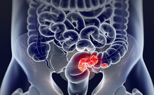
Gastrointestinal stromal tumors (GIST) are the most common mesenchymal tumors of the gastrointestinal (GI) tract. Although they have been recognized as a distinct entity for several decades, it has only been over the past 10–15 years that these tumors have been truly studied and further defined by rigorous diagnostic and molecular criteria. Indeed, one might argue that it was a landmark study involving the successful treatment of unresectable GIST that launched a new era of targeted therapy in clinical oncology. In the ensuing years we have further refined our knowledge of GIST, developed rigorous diagnostic techniques, surgical interventions and prognostic criteria. Considerable strides have also been made in adjuvant therapy of successfully resected localized tumors and in the setting of widespread disease. In this article, we review the recent progress that has been made in all of these domains.
Section 1—Epidemiology—Clinical and Pathologic Considerations
Elena Tsvetkova, E Celia Marginean, and Shailendra Verma
Epidemiology
In the past, the diagnosis of GIST has been fraught with difficulty and they have often been misdiagnosed as leiomyoma, leiomysarcoma, neurofibroma, or benign/borderline malignant potential tumors. Consequently, information on the true incidence or prevalence is scarce and has been largely dependent on retrospective or population-based studies. The identification of the specific expression of the KIT protein (CD117) as a reliable phenotypic marker of GIST has led to a re-appraisal and re-evaluation of pathologic diagnosis and has provided more concrete information. International epidemiologic data derived from Sweden and The Netherlands suggests an annual incidence of 14.5 and approximately 12.3 per million, respectively.1,2 A population-based study by Tran et al.,3 utilizing the Surveillance, Epidemiology and End Results (SEER) registry, revealed an age-adjusted yearly incidence of approximately 6 per million. The incidence was noted to steadily increase with age: from 0.06/100,000 (95 % confidence interval [CI] 0.01–0.09) in the 20–29 age group, to 2.29/100,000 (95 % CI 1.99–2.62) in those greater than 80 years. In this study, the mean age at diagnosis was 63 years, men were more commonly affected (54 %), and age adjusted incidence was highest among African Americans. As has been documented by others, the review by Tran confirmed that a GIST might arise from any part of the GI tract. Fifty-one percent of cases were in the stomach, 36 % small intestine, 7 % colon, 5 % rectum, and 1 % arose in the esophagus. Rarely, a GIST has been noted to arise in the retroperitoneum or omentum (see Figure 1).4
Clinical Presentations
The retrospective review of GIST diagnosed in western Sweden by Nilsson found that 90 % were detected clinically due to the presence of symptoms, 21 % were incidental findings at surgery, and 10 % were asymptomatic,detected at autopsy.1 A study from Japan suggested that the incidence of asymptomatic GIST may be much higher than hitherto suspected and it has been proposed that only a few of the very small incidental tumors may actually develop into larger ones with malignant potential.5 GIST may be associated with nonspecific symptoms such as abdominal discomfort, bloating, and early satiety. When the tumor is ulcerated or large enough to elicit symptoms, patients may present with overt GI bleeding (40 %), abdominal pain (40 %), or an abdominal mass (20 %).6 Over 25 % ofpatients may present with bowel obstruction. Bowel perforation, in the setting of GIST, occurs infrequently. In the case of esophageal GIST, dysphagia may represent the first site-specific symptom. Interestingly, paraneoplastic syndromes involving hypoglycemia7 or hypothyroidism have been described.8
Unusual Clinical Presentations
GIST Syndromes
In adults, GIST has been associated with neurofibromatosis type 1 (NF1)9 and the Carney triad10/syndrome11 (gastric epithelioid GIST, extra-adrenal paraganglioma, and pulmonary chondroma).
- NF1-associated GIST has a propensity for multicentricity within the GI tract, spindle cell morphology and do not harbor KIT or plateletderived growth factor receptor alpha (PDGFRA) gene mutations. NF1- associated GIST is typically positive for the CD117 antigen.9
- Carney triad syndrome-associated GIST is predominantly of epithelioid morphology, tends to occur in the antrum of the stomach, lacks conventional KIT and PDGFRA gene mutations, and tend to run an indolent course.9,12,13
- GIST-paraganglioma syndrome is extremely rare and is similar to the Carney triad, but lacking pulmonary chondroma with an equal gender distribution and is also referred to as the Carney Stratakis syndrome.11
Familial GIST—As of 2008, approximately 24 kindred with heritable mutations in KIT or PDGFRA have been identified. Penetrance in these kindred is high, with most affected members developing one or more GISTs by middle age. The tumors are usually small, lack mitotic activity and arise in a background of interstitial cells of Cajal (ICC) hyperplasia and follow a benign course.12
Pediatric GIST—These are rare (1–2 % of all GIST) and fall into one of two subgroups: with mutations (either KIT or PDGFRA) or without mutations, the latter being more frequent. The patients are almost exclusively young females, developing gastric epithelioid GIST, which are KIT positive by immunohistochemistry (IHC). Unlike adult GIST, these tumors can spread to lymph nodes.14–16
Multiple GIST—Notwithstanding the above described GIST syndromes, sporadic, synchronous, and metachronous tumors have been observed in patients without identifiable germ line risk factors, suggesting that other genes that predispose to the development of GIST have yet to be discovered.12,17
Diagnosis
Preoperative pathologic diagnosis usually is not required if imaging is consistent with a GIST, the tumor is resectable without significant morbidity, and the patient is an operative candidate. Typically on computed tomography (CT) scan, a GIST appears as a smoothly contoured mass that enhances brightly with intravenous contrast (see Figure 2A/B). Very large tumors may appear more complex due to necrosis, hemorrhage, and degenerating components. In cases where operative morbidity is high or the diagnosis is unclear, biopsy may be warranted. If biopsy is required, endoscopic ultrasound guided fine needle aspiration (EUSFNA) biopsy is an acceptable option and can be a safe and reliable way to obtain the diagnosis and is associated with a lower risk for seeding or potential spread of disease. In a study of 65 patients, EUS-FNA had a sensitivity of 82 % and a specificity of 100 %.18 Another method of biopsy that can be used is endoscopic ultrasound-guided trucut biopsy. This type of biopsy also allows for high agreement between biopsy and surgical pathology specimen with respect to yield of diagnosis and CD117 status.19 Percutaneous biopsy carries the risk for tumor capsule rupture with peritoneal spread of disease20 and thus is avoided if at all possible. However, in selected cases, careful, guided percutaneous biopsy by expert radiologists may be attempted when a neoadjuvant approach is being considered or if there is diagnostic uncertainty and there is no ability to perform an EUS-guided biopsy.
Pathology and Differential Diagnosis of GIST
GIST appears to originate from the ICC or their stem cell-like precursors.21,22 ICCs are pacemaker-like cells located between the layers of the muscularis propria throughout the GI tract, which regulate GI motility and autonomic nerve function. ICC or their stem cell-like precursors can differentiate into smooth muscle cells if KIT gene signaling is disrupted.23
The morphologic spectrum of GIST includes spindle cell tumors, with thin, elongated nuclei forming long, interlacing fascicles (see Figure 3A/B), and epithelioid tumors, with polygonal cells in a syncytial pattern, without visible cell borders and large, round nuclei (see Figure 3 C/D). Based on morphology,24–26 GIST can be subtyped into the following categories:
- Spindle cell variants: sclerosing, palisading-vacuolated, hypercellular, and sarcomatous spindle cell; and
- Epithelioid cell variants: sclerosing, discohesive, and hypercellular epithelioid.
Based on nuclear atypia, presence of necrosis, hemorrhage and mitotic activity, the tumors can appear very bland and cytologically benign (see Figure 4 A/B), or frankly malignant (see Figure 4 C/D).
Immunohistochemistry of GIST
KIT (CD117)—In recent years, IHC staining for KIT expression (CD117) has become integral to the diagnosis of GIST, nearly 90 % of which harbor activating mutations in the KIT receptor tyrosine kinase gene.27 Approximately 95 % of GIST are positive for the CD117 antigen by IHC staining.12 The staining pattern may be cytoplasmic, membranous, and/or paranuclear (Golgi pattern) (see Figure 5 A/B/C). Although KIT positivity is a major defining feature of GIST, it is no longer an absolute requirement; KIT expression is a constitutional feature and not a consequence of the mutation. Moreover, a GIST having a weak or negative KIT expression tends to be KIT wild-type or PDGFRA mutants. In rare cases, tumors cease to express the KIT protein because of clonal evolution after tyrosine kinase therapy.
CD117 is not specific for GIST. In fact, weak reactivity can occur with other mesenchymal neoplasms. Accordingly, CD117 immunostaining of tumors should be interpreted cautiously in the context of other immunomarkers and the anatomic location and morphology of the tumor in order to differentiate GIST from other mesenchymal, neural, and neuroendocrine neoplasms.12
DOG1 (“discovered on GIST 1”)—This is a protein of unknown function that is expressed strongly on GIST and is rarely expressed on other soft tissue tumors.28 The staining pattern of DOG1 varies from cytoplasmic to membranous, with usually strong, diffuse intensity (see Figure 6). The results from several series have shown a high overall sensitivity and specificity for DOG1 in the detection of GIST. Overall, about 6 % of GIST exhibit a DOG1+/KIT-immunoprofile.28–30 DOG1 antibodies are more sensitive than KIT antibodies in detecting tumors of gastric origin, tumors with epithelioid morphology, and tumors harboring PDGFRA mutation.28–31
Protein Kinase C Theta (PKC-θ)—This is a novel PKC isotype involved in T-cell activation, and is highly and specifically expressed in GIST showing a diffuse cytoplasmic staining pattern. Although PKC-θ has a lower specificity than DOG-1, it can be a useful biomarker, especially in CD117/ DOG1-negative GIST.32–34 Of note, PKC-θ is also reported to be expressed in schwannomas, malignant peripheral nerve sheath tumors, and Ewing’s sarcoma/primitive neuroectodermal tumors.35
CD34—Is expressed in 100 % of esophageal GIST, 65 % of colonic GIST, 96 % of rectal GIST, and 65 % of GIST in non-GI locations, such as the mesentery and omentum.36 A smooth muscle marker, h-caldesmon, is expressed in >70 % of GIST and smooth muscle antigen (SMA) in 20–30 %, whereas desmin positive staining is observed in <5 % (usually epithelioid gastric, KIT-negative tumors).
Neural markers such as S100 are observed in <1 % of GIST.
In summary, an IHC panel including KIT (CD117), DOG1, S100, desmin,SMA, and CD34 are useful to confirm the diagnosis of GIST and help distinguish GIST from other mesenchymal tumors. c-KIT, PKC-θ, and DOG1 antigens are the most sensitive and specific immunomarkers for confirming extraintestinal GIST.
Differential Diagnosis of GIST
The tumors that enter the differential diagnosis of GIST vary widely according to the histologic pattern, and can be grouped into tumors with spindled or epithelioid morphology, either cytologically bland or frankly malignant.
Spindle Cell Tumors, Cytologically Bland
Included in these are the following:
- Abdominal/mesenteric fibromatosis is the most common primary tumor of the mesentery. This entity is usually CD117 negative andshows strong nuclear positivity with β catenin.37,38
- Leiomyomas commonly occur in the esophagus and rectum and are true neoplasms of smooth muscle origin. By IHC, they are positive for smooth muscle markers (desmin, SMA, muscle specific actin [MSA]) and negative for CD117 and DOG1.
- Schwannomas are rare benign mesenchymal tumors, common in the stomach, colon, and rectum. By IHC they commonly express glyofibrillar acid protein (GFAP) and S100 but are negative for CD117 and DOG1. They also usually do not express the NF2 gene alterations commonly seen in typical soft tissue schwannomas.
- Inflammatory fibroid polyps are most common in the submucosa of the gastric antrum and are rare in the small intestine, colon, and esophagus. These are hypocellular tumors, with loose edematous stroma, “onion skin” arrangement of spindle cells around vessels, and numerous inflammatory cells, especially eosinophils. By IHC they express CD34, focal positivity for SMA, but usually lack expression of CD117 and DOG1.
- Inflammatory myofibroblastic tumors (IMT) are uncommon neoplasms of intermediate biologic potential composed of proliferating myofibroblasts with a plasma cell-predominant inflammatory infiltrate. About 25 % can recur locally, with very rare cases showing malignant transformation or metastases. By IHC, the myofibroblasts express vimentin, smooth muscle actin, focally CD34 and cytokeratins and 50 % of IMT express anaplastic lymphoma kinase (ALK), which may be a favorable prognostic indicator.39
- Solitary fibrous tumors are rare, usually pleural-based tumors, which can also occur in the retroperitoneum and may be a diagnostic challenge because of overlapping clinicopathologic features with GIST. Histologically, they have cellular alternating with hypocellular areas, have ropy collagen fibers, and typical hemangiopericytoma-like vessels. By IHC, 80–90 % express CD34, focal SMA, and are negative for both CD117 and S100.40
- Mesenteric sclerosing fibrotic lesions, including sclerosing mesenteritis, sclerosing peritonitis, and retroperitoneal fibrosis, are characterized by dense fibrosis of mesenteric fat, with proliferation of fibroblasts, peripheral inflammation with lymphoid aggregates, and occasional calcifications. By IHC, only vimentin is positive, while β catenin, CD34, CD117, and DOG1 are all negative.37
Spindle Cell Tumors, Cytologically Malignant
Included in this group are the following:
- Malignant melanoma—This is the most common solid tumor that metastasizes to the GI tract, especially the small intestine. Amelanotic melanoma can be misdiagnosed as a GIST, carcinoma, or sarcoma. A potential pitfall in its diagnosis is the fact that >75 % of melanomas can express CD117. However, most melanomas express S100, HMB45, Melan A, and tyrosinase, but lack DOG1.
- Sarcomatoid carcinoma—Either primary or metastatic to the GI tract, can be diagnosed if the epithelial origin is confirmed by keratin stains (low- or high-molecular keratins) and/or the presence of epithelial dysplasia at the edge of the tumor.
- Leiomyosarcoma—Is an uncommon, high-grade tumor, showing infiltrative margins, high mitotic rate, and marked nuclear pleomorphism. By IHC, like their benign counterpart, they express smooth muscle markers (desmin, SMA, MSA, h-caldesomon) but lack CD117 and DOG1.
- Malignant peripheral nerve sheath tumor (MPNST) – These are extremely rare tumors that arise from a peripheral nerve or exhibit nerve sheath differentiation. They are highly cellular tumors, with a high mitotic rate, necrosis and marked nuclear pleomorphism. Unlike schwannomas, they show very focal S100 positivity, and are negative for CD117, DOG1, and HMB45.41
Other rare mesenchymal tumors, such as clear cell sarcoma, follicular dendritic cell sarcoma, endometrial stroma sarcoma (ESS), angiosarcoma, undifferentiated sarcoma, PEComas, synovial sarcoma and dedifferentiated liposarcomas with spindle cell morphology should be considered occasionally, and the clinical history, imaging and IHC panel can help in resolving this differential diagnosis.
Epithelioid Cell Tumors, Cytologically Bland
This group includes the following:
- Glomus tumors—These tumors have been well described in the gastric antrum, are small, multinodular tumors that usually behave in a benign fashion, but rarely can metastasize. The round tumor cells, with well-delineated cell borders, are strongly positive for SMA and negative for chromogranin A, neuronspecific enolase (NSE), carcinoembryonic antigen (CEA), epithelial membrane antigen (EMA), CD117, and DOG1.42,43
- Epithelioid schwannomas—These are rare benign lesions of the colon or rectum, strongly positive for S100; negative for CD117 and DOG1.
- Paragangliomas—Usually of the duodenum, can be epithelioid, ganaglion-like, or spindle cell, with a nested pattern, show neuroendocrine expression with synaptophysin, chromogranin A, and lack CD117 and DOG1. They comprise part of the Carney syndrome11,44 (gastric epithelioid GIST, extra-adrenal paraganglioma, and pulmonary chondroma).
Epithelioid Cell Tumors, Cytologically Malignant
The main considerations in this group include malignant melanoma, poorly differentiated carcinoma, epithelioid leiomyosarcoma, and neuroendocrine tumors, which can be differentiated by clinical history, imaging, and IHC.
Molecular Classification of GIST
Approximately 85 % of GISTs contain oncogenic mutually exclusive mutations in one of two receptor tyrosine kinases: KIT or PDGFRA.12 Constitutive activation of either of these receptor tyrosine kinases plays a central role in the pathogenesis of GIST.21 The proper identification of GIST with genotyping is very important because of the availability of specific, molecular-targeted therapy with KIT/PDGFRA tyrosine kinase inhibitors (TKIs), such as imatinib mesylate, or, in the case of imatinib-resistant GIST, sunitinib maleate.45–47
GIST may fall into one or more of the following subgroups (Table 1):
KIT-mutant GIST. Approximately 80 % of all GISTs contain a mutation in the KIT receptor tyrosine kinase that results in constitutive activation of the protein.12 The KIT gene is located on chromosome 4q12-13, in the vicinity of genes encoding the receptor tyrosine kinases PDGFRA, and vascular endothelial growth factor receptor 2 (VEGFR2).48 Mutations in five different KIT exons have been observed in GIST: exon 11 (67 %), exon 9 (10 %), and exons 8,13, and 17 (3 %).12,46,49 KIT mutation variants have distinct anatomic distributions: exon 8 (small intestine), exon 9 (small intestine, colon), and exons 11, 13, and 17 (all sites).12 It is important to note that mutations in exon 11 are most commonly observed in gastric GIST. KIT-mutant tumors usually express CD117 PKC-θ and DOG1 by IHC.28,34,50
PDGFRA-mutant GIST. Approximately 5 % to 8 % of GIST harbor a mutation in PDGFRA, a close homolog of KIT with similar extracellular and cytoplasmic domains.51 PDGFRA is activated at the PDGFRA juxtamembrane domain (encoded by exon 12), the ATP-binding domain (encoded by exon 14) or the activation loop (encoded by exon 18).49 PDGFRA-mutant GIST may differ from KIT-mutant GIST in a number of ways, including a marked predilection for the stomach, epithelioid morphology, myxoid stroma, nuclear pleomorphism, variable expression of CD117, and generally lower potential for malignancy.50,52,53 As with KITmutant GIST, PDGFRA-mutant tumors express PKC-θ and DOG1.28,34,54
Wild-type GIST. Wild-type GIST comprise approximately 12 % to 15 % of all GIST. In these tumors no detectable mutations have been identified in either KIT or PDGFRA, although KIT is still usually phosphorylated, suggesting that KIT is still activated. For these tumors, there is noparticular association with anatomic location or clinical outcome.12 However, recent studies have revealed that wild-type GISTs are a heterogeneous group and display various oncogenic mutations (see Table 1). For example, the BRAF V600E substitution that is seen in papillary thyroid carcinoma and melanoma is present in up to 7–15 % of wild-type GIST.55 Defects in the succinate dehydrogenase (SDH) complex of respiratory chain complex II, which comprises four subunits (SDHA, SDHB, SDHC, and SDHD), have recently been identified in wildtype GIST.56 Germline mutations in SDHB, SDHC, or SDHD increase the risk not only of the development of GIST, but also of the development of paragangliomas (Carney Stratakis syndrome).
KIT-negative GIST. Approximately 5 % of GISTs do not express CD117 by IHC; in these instances, IHC may lack sufficient sensitivity to detect small amounts of mutant kinase.12 Approximately 30 % of these tumors harbor PDGFRA gene mutations while more than half have KIT mutations.12
Although mutation status is not included in risk stratification schemes, certain mutations have been linked with survival and treatment response. In a recent population-based series of 1505 GISTs by Joensuu et al., it was observed that patients with a PDGFRA mutation had a more favorable relapse-free survival (RFS) than those with KIT mutations (hazard ratio [HR] 0.34; p=0.004). In this study, only one of 35 GIST witha KIT exon 11 mutation recurred and patients with deletions of only one codon of KIT exon 11 had a better RFS than those with other deletiontypes. Those with a KIT exon 11 substitution mutation also had a better RFS.57 In general, KIT exon 11 mutations are most sensitive to imatinib and have the best prognosis with a regular dose of 400 mg/day; KIT exon 9 mutations might require a higher dose of imatinib (800 mg/day) and generally have a worse prognosis, whereas PDGFRA exon 18 mutation leading to Asp842Val is imatinib resistant and may likely benefit most from sunitinib.58
Section II—Management of Early and Advanced GIST
Elena Tsvetkova, Carolyn Nessim, and Shailendra Verma
Surgical Management of Resectable Disease
The mainstay of potentially curative treatment for resectable GIST is surgery with clear resection margins. Since lymphatic spread of GIST is uncommon, lymphadenectomy is not necessary and is not routinely performed.59,60 Consequently, there are limited data on the influence of lymph node involvement on outcome. In a study by Aparicio et al., only one out of 59 patients undergoing curative resection had a positive lymph node.60 In another reported series by Agaimy et al., involving 210 patients,only two patients had positive nodal disease.61 Interestingly, in contrast to elderly patients, lymphatic spread was more commonly found in younger patients.61 However, significance of nodal involvement in GIST remains unclear. Tumor pseudocapsule rupture—either spontaneous or at the time of surgery—has been identified as an independent adverse prognostic factor carrying a poor prognosis (p=0.0014)62–64 and thus caution must be exerted at the time of surgery particularly if resection is to be performed laparoscopically. A systematic review and meta-analysis of open versus laparoscopic resection of gastric GIST showed no difference in operative time, adverse events, estimated blood loss, overall survival, and recurrence rates.65 This study supported laparoscopic resection stating that it was safe and effective and was associated with a significantly shorter length of hospital stay. This technique is thus feasible in well-selected patients.
As has been aforementioned, the major breakthrough in the management of GIST came about with the development of TKIs—small molecules such as imatinib and others that followed suit, which have revolutionized the management of this disease and have prompted investigation of similar approaches in the wider field of oncology. The major studies investigating imatinib are well known and are summarized in Table 2.
Neoadjuvant Therapy
In cases of borderline resectable or potentially resectable disease, which requires extensive organ disruption, initial therapy with the TKI, imatinib, is a rational approach. This allows for downstaging of the tumor for possible resection. Figure 7A/B shows an example of a large borderline resectable GIST that becomes resectable after several months of neo-adjuvant imatinib treatment. There is limited published data on neo-adjuvant therapy in GIST: most of the information has been based on the high response rate to imatinib in the setting of locally advanced or metastatic disease and on small retrospective study reports.66–68 In a study by Andtbacka, 46 patients who underwent potentially curative surgery after initial treatment with imatinib were assessed retrospectively for surgical resection rate and outcome.66 Of them, 11 with locally advanced disease were treated for a median 11.9 months, followed by complete resection, and were alive at a median of 19.5 months (10 were disease free). Of the remaining 35 patients with recurrent metastatic GIST, 11 underwent complete resection and were alive at a median of 30.7 months: six out of 11 had recurrent disease at a median of 15.1 months. Patients with a partial radiologic response to imatinib had significantly higher rates of complete resection than patients with progressive disease (91 % versus 4 %; <0.001).
A phase II trial by the US Intergroup prospectively investigated the effect of imatinib 600 mg daily for 8–12 weeks prior to curative surgery in 63 patients with primary or resectable metastatic KIT-positive GIST.69 Of the 52 patients included in the final analysis, 30 had primary GIST (group A) and 22 had recurrent metastatic disease (group B). Stable disease and partial response (as per Response Evaluation Criteria in Solid Tumors [RECIST] criteria) were observed in 83 % and 7 % of patients in group A; 91 % and 4.5 %, respectively, in group B. Two-year progression-free survival (PFS) was 83 % in group A and 77 % in group B. No major surgical complications were reported. After a median of 5.1 years of follow up, the estimated 5-year PFS and disease-specific survival (DSS) were 57 % and 77 % ingroup A, respectively, and 30 % and 68 % in group B, respectively.70,71
The largest series thus far of patients treated with neoadjuvant imatinib is from the European Organisation for Research and Treatment of Cancer-Soft Tissue and Bone Sarcoma Group (EORTC-STBCG).72 In this series, overall survival (OS), disease-free survival (DFS), and DSS in 161 patients with locally advanced, nonmetastatic GIST who received neoadjuvant imatinib, were analysed. The primary tumor was in the stomach (55 %), rectum (20 %), duodenum (10 %), ileum/jejunum/other (11 %), and esophagus (3 %). Sixty-five percent of patients had an exon 11 mutation. The tumor resection after neoadjuvant imatinib (median time on therapy, 40 weeks) was R0 in 83 % of cases. Only two patients had disease progression on therapy. Fiveyear DSS and DFS were 95/65 %, respectively. Median OS was 104 months and median DFS was not reached. Thirty-seven GIST recurrences were diagnosed (only five of which were local recurrences). Fifty-six percent of patients continued imatinib therapy post resection and these patients did better than those who did not have post-operative therapy.
There is no consensus on the duration of neo-adjuvant treatment with imatinib in the setting of locally advanced or recurrent potentially resectable GIST. It has been suggested to treat patients from 3 to 12 months with frequent imaging as long as radiologic tumor response is evident73 with planned surgery thereafter in responding patients. The recommended dose is 400 mg daily; however, in KIT exon 9 mutations, dose escalation to 800 mg may be considered.20
Adjuvant Therapy
All GISTs have some malignant potential and may recur or metastasize following apparent curative, margin-negative resection. Tumors of the small intestine behave more aggressively than those in the stomach. Thus, 40–50 % of small bowel GIST, demonstrate malignant behavior in contrast to 20–25 % in gastric GIST.25 There is paucity of data on GIST behavior in such unusual sites as the esophagus, colon, and rectum.
Clinical management thus depends on GIST malignancy risk stratification that is determined by tumor size, location, and number of mitoses on final surgical pathology.74,75 The risk scheme and stratification developed by Miettinen et al. is widely considered the most reliable and has been adopted worldwide (see Table 3). As well, EUS has been used to identify possible high-risk features, such as ulceration, echogenic foci, irregular borders, cystic spaces, and heterogeneity to aid in management decision-making.76 A proposed algorithm by Sepe et al. suggests that all GISTs greater than 2 cm in size, which are symptomatic, or harbor highrisk features on EUS should be resected.76 Otherwise, EUS surveillance is suggested for smaller tumors after discussion of risks and benefits of this approach with the patient. Referral to a multidisciplinary team experienced in GIST management is always recommended.
If no additional treatment is given to GIST patients after curative resection, approximately 50 % of patients will recur within the next 5 years. This led to the exploration of adjuvant therapy specifically targeting the KIT oncogene.
In a phase II US Intergroup trial by the American College of Surgeons Oncology Group (ACOSOG) (Trial Z9000), 107 patients with resected highrisk GIST, defined as tumors larger than 10 cm, evidence of capsular rupture or hemorrhage, or multifocal disease with more than five tumor sites, were treated post resection with daily oral 400 mg imatinib for 1 year.77 RFS was 96 %, 60 %, and 40 % and OS was 99 %, 97 %, and 83 %, at 1, 2, and 3 years, respectively.
Phase III trials evaluating imatinib in the adjuvant setting include the ACOSOG Z9001, the EORTC trial 62024, and the Scandinavian Sarcoma Group (SCG) study XVIII.
In the ACOSOG Z9001 trial, 713 patients with resected KIT-positive GIST more than 3 cm in diameter, were randomized to either 1 year of adjuvant imatinib or placebo.78,79 In a planned interim analysis it was evident that patients in the treatment arm had fewer recurrences than in the control rm with primary end point one year RFS of 98 % versus 83 % (HR 0.35 95 % CI 0.22–0.53) in favor of imatinib. Subgroup analysis disclosed that the observed benefit was the highest in patients with high-risk features with a relapse rate of 19 % versus 47 % in favor of imatinib.78 There was no OS benefit observed in this trial. The possible reasons for this may include the short follow-up period, crossover allowance for those who recurred and the high efficacy of imatinib in the treatment of relapsed disease.
Although the results of this trial formed the basis for the recommendation for adjuvant imatinib in many jurisdictions, it also raised many questions. It was observed that 1 year after imatinib discontinuation, the rate of recurrence in the imatinib-treated population was similar to the control arm. This may imply that longer duration of treatment is required. Also, in this study, the inclusion criteria listed only tumor size as a prerequisite leaving the role played by other risk criteria such as mitotic rate, tumor site, and molecular factors unanswered.
In the EORTC 62024 phase III trial, 908 patients were treated with imatinib for 2 years after complete resection for intermediate- or high-risk GIST. The primary end-point—5-year imatinib-free survival (IFS) at a median follow up of 4.7 years—was 87 % in treatment arm versus 84 % in the control arm (HR 0.80, 95 % CI 0.51–1.26); 3-year RFS was 84 % versus 66 % and 5-year OS was 100 % versus 99 %, respectively. Even though this trial demonstrated only a trend towards better IFS, favoring the imatinib arm in a high-risk population, it still supported the usage of imatinib for a longer duration of time in high-risk GIST patients.80
In the SSG XVIII trial of imatinib for 1 year versus 3 years in 400 patients with high-risk resected GIST (tumor size more than 10 cm or mitotic rate more than 10/50 high power field [HPF] or tumor size more than 5 cm and mitotic rate more 5 HPF, or tumor rupture) at a median follow up of 54 months, significantly improved RFS and OS were observed in the prolonged treatment arm (5-year RFS was 66 % versus 48 %, HR 0.46, 95 % CI 0.32–0.65 and 5-year OS was 92 % versus 82 %, HR 0.45, 95 % CI 0.22–0.89).81 It was noted however that more patients in the prolonged treatment arm discontinued therapy for reasons other than disease progression (26 % versus 13 %). As in the ACOSOG Z9001 trial, the rates of disease recurrence in the control and treatment arms within 6–12 months after imanitib discontinuation were similarly increased that rose once again, the important issue of the optimum duration of therapy.
Most clinical practice guidelines now recommend the use of adjuvant imatinib 400 mg daily for 3 years for patients with high-risk GIST. The selection of patients should be based on accepted risk classifications, such the Miettinen criteria described earlier. Mutational analysis may also provide additional predictive information for benefit from adjuvant imatinib. In the Z9001 study, subgroup analysis indicated that those patients with exon 11 (deletion or no deletion) and PDGFRA mutations experienced benefit from 1 year of adjuvant treatment. Similarly, in the SSG XVIII trial, significant benefit favoring 3 years of adjuvant therapy was only observed in those with exon 11 mutations (HR 0.35, 95 % CI 0.22–0.56). Consequently, in patients with resected GIST, we would propose that mutational analysis must be performed for both prognostic and predictive reasons.
Follow-up Recommendations of Localized or Resected GIST
There is no high level evidence that addresses the optimum surveillance strategy for patients with an early-stage, localized (treated or untreated) GIST.
For patients with a resected GIST, National Comprehensive Cancer Network (NCCN) recommendations include a physical examination with a thorough history and a CT scan of the abdomen and pelvis every 3 to 6 months for the first 5 years, and then annually.20
In cases of small gastric GIST (less than 2 cm), with low-risk EUS features that is being followed in the absence of surgery, endoscopic surveillance every 6 to 12 months is recommended.
The European Society of Medical Oncology (ESMO), in their recommendations, tailors follow-up frequency to tumor high-risk features.82 Thus for very-lowrisk GIST, routine surveillance is not recommended.
Management of Metastatic or Unresectable Disease
The small molecule tyrosine kinase protein inhibitor, imatinib, is now accepted as the standard first-line treatment for unresectable or metastatic GIST. With the introduction of imatinib, the median OS of patients with unresectable or metastatic disease has increased from 18 to 57 months.83 Multiple phase II and III studies have confirmed the benefit of imatinib in this setting demonstrating high response rates and improved PFS.39,84–87
Two phase III trials compared two dose levels of imatinib (400 mg versus 800 mg daily) in the setting of advanced GIST. No statistical differences were observed between the two doses and the higher dose was associated with more side effects in both studies. An initial dose of 400 mg per day is now widely agreed upon as the appropriate first-line therapy. Long-term follow up in two studies has confirmed the potential durable effect of imatinib. In the SWOG phase III trial S0033, 26 % of patients treated with two different doses of imatinib survived 8 years or longer, with a projected 10-year survival of 22 %.86,87
Although these major studies completely altered the course of management of patients with GIST, it is a sobering reality that complete responses are infrequently observed (in less than 10 % of patients). Most patients eventually develop resistance to imatinib and progress within 2 to 3 years.47
The question regarding the optimal duration of therapy was addressed in the BFR14 phase III trial.88 Patients with unresectable/metastatic GIST, stable on treatment for 1 year were randomly assigned to continue therapy or interrupt treatment until disease progression. This study was halted prematurely as it observed evidence that treatment interruption resulted in rapid disease progression even in apparent complete responders. Median PFS was 18 months versus 6 months in favor of continuous therapy. The trial authors concluded that treatment should be lifelong or until disease progression.
In these above referenced studies, it also became apparent that response assessment when using novel biologic agents needed to be addressed. To assess a response to treatment, patients conventionally undergo CT imaging and 2D measurements are used to designate response, stability, or progression. However, during treatment with imatinib, a responding GIST may actually increase in size secondary to necrotic changes or hemorrhage. For this reason and for assessment purposes, both RECIST and Choi criteria (which takes into account tumor density changes in contrast-enhanced CT scans), should be employed.89–91
Does mutational status matter when initiating therapy for metastatic or inoperable GIST? This question has not been prospectively addressed in any of the above referenced trials but analysis of responders and nonresponders has been well summarized by Joensuu and DeMatteo.92 Tumors with KIT mutations in exon 11, 13, and 17 and PDGFRA mutations in exon 12 and 14 are responsive to imatinib 400 mg/day; those with KIT mutations in exon 9 require higher doses of imatinib (800 mg/day). So called ‘wild-type’ GIST (SDH deficient) or GIST with PDGFRA mutations in exon 18 (D842V) are considered insensitive to imatinib and other therapies as outlined below should be considered (see Table 4).
Role of Surgery in Metastatic Disease
As mentioned above, despite lasting disease control, complete responses with imatinib are rare.93 Most patients on imatinib eventually develop resistance and progress within 2 to 3 years.94 For these reasons surgical resection has been considered in responders with limited metastatic disease burden and potentially resectable disease. There are no results of prospectively conducted trials that conclusively support this approach. However, single-institution retrospective reviews have reported longterm disease control with resection of metastatic disease in responders following treatment with imatinib.66,67,95,96 In the largest analysis by the EORTC-STBSG evaluating surgery in metastatic disease, 239 patients with metastatic GIST underwent metastasectomy and received imatinib therapy. Complete resection (R0/R1) was achieved in 177 patients. Median OS was 8.7 years for R0/R1 and 5.3 years in patients with R2 resection. In the group who were in remission at the time of resection, median OS was not reached in the R0/R1 surgery and was 5.1 years in the R2 surgery. Median time to relapse/progression after resection of residual disease was not reached in the R0/R1 group and was 1.9 years in the R2 group, who were resected in response. This analysis shows possible long-term survival in patients in whom surgical complete remission can be achieved. Debulking surgery does not seem to prolong survival.96 After complete resection, most experts recommend continuation of imatinib as the risk for recurrence upon discontinuation of therapy remains high.84 Figure 8 demonstrates the specimen of a GIST metastasectomy.
Role of Locoregional Treatment in Metastatic GIST
There is limited information on the role of locoregional treatments, such as hepatic arterial embolization (HAE), chemoembolization, or radiofrequency ablation (RFA) in metastatic GIST. Most patients are not surgical candidates since they have extensive bilobar hepatic disease or concurrent hepatic and extrahepatic metastatic disease.
The largest reported series on HAE (utilizing cisplatin with or without an embolic agent) included 110 patients, of whom 62 (56 %) were treated before the introduction of imatinib.97 Of 85 assessable patients,12 (14 %) had a partial response and 74 % had stable disease. Twentyfive patients (12 %) developed moderate to severe side effects, such as myelosuppression, respiratory distress, pulmonary embolism, sepsis, seizure related to hyponatremia, and death. Median OS was 17.2 months.
In another series of 14 patients with imatinib-resistant disease whounderwent chemoembolization, PFS in the liver was 31.4 % at both 1 and 3 years with an OS of 7 and 9.7 months, respectively.98
Although RFA and stereotactic radiotherapy have been alluded to in several case reports as possible therapy for progressive, resistant GIST, their benefits remain unclear and it is not possible to provide any reliable data on outcomes. However, the results are tantalizing and further studies are warranted.
Beyond Imatinib
For patients with primary or secondary resistance to imatinib, dose escalation is a viable option.86 In a European trial, which assessed standard versus high-dose imatinib, 247 out of the 473 patients on the low dose regimen progressed and 133 of them were crossed over to the higher dose.99 Of these, 36 patients had a prolonged period of stable disease with three patients having a confirmed partial response. These results were confirmed by an American study involving 164 patients.87 One hundred and thirty-three of those who progressed on the conventional dose ofimatinib crossed over to 800 mg daily; 33 % of assessable patients had either stable disease or response with a median PFS of 5 months and OS of 19 months after cross over.
In patients with imatinib intolerance or secondary resistance to medication the second-line multitarget TKI sunitinib is recommended.100–102 In an international phase III trial of sunitinib versus placebo, 312 patients with imatinib resistance were randomized either to sunitinib or placebo. Those who progressed on placebo crossed over to the treatment arm. At a median follow up of 42 months, median time to tumor progression was 27 weeks on sunitinib versus 6 weeks on placebo.103 Median OS was reported as 73 versus 66 months in favor of the sunitinib arm. Of interest, as previously discussed, in patients with a KIT exon 9 mutation, or a wild-type KIT/PDGFRA mutation, the rate and duration of response with sunitinib was much higher than for those patients with a KIT exon 11 mutation.101 Sunitinib is now considered appropriate therapy for patients who progress on imatinib as second-line therapy.
Finally, another multikinase inhibitor, regorafenib has been prospectively evaluated in imatinib- and sunitinib-resistant patients as third-line therapy. The activity of regorafenib in such patients was first observed in a phase II trial conducted by George et al.104 These data were confirmed in a phase III trial where 199 patients with refractory disease or intolerance to sunitinib were randomized to regorafenib or best supportive care (BSC).105 Reported PFS was 4.8 versus 0.9 months in favor of the treatment arm; no survival benefit was observed, possibly due to cross over of patients who progressed on BSC.
Over the past few years the availability of novel agents has steadily expanded. These include nilotinib, which has also demonstrated activity in this area.106,107 However, its role in GIST refractory to both imatinib and sunitinib is less defined. In a phase III trial of 248 patients who failed treatment with both imatinib and sunitinib, patients were assigned to nilotinib or BSC: no significant differencs in outcome were reported.108
Preliminary data from a phase II trial evaluating sorafenib in the treatment of refractory GIST reported a disease control rate of 68 % with a median PFS of 5.2 months.109 Though promising, this agent needs further evaluation in a randomized setting.
Therefore, in imatinib refractory disease current treatment options include imatinib dose escalation, sunitinib as second-line therapy, and regorafenib as third line therapy. It is premature to recommend the use of sorafenib until further studies have been undertaken.
Conclusion
GISTs are relatively rare tumors arising in the GI tract. In recent years our understanding of these tumors has increased exponentially and therapeutic interventions with agents such as imatinib have paved the way for modern, molecular targeted oncology. Clinical presentations of GIST are related to the site of origin, tumor size, and presence of ulceration. Often patients are asymptomatic. Surgery is the only curative modality; adjuvant treatment with the BCR-ABL TKI imatinib for 3 years is recommended in high-risk tumours.76,110 In the setting of locally advanced primary unresectable disease, neoadjuvant treatment for 3 to 6 months may be considered.66–68,72 In patients with metastatic or unresectable disease, treatment with imatinib is associated with significant improvements in OS from 18 to 57 months.83 In patients with metastatic disease responding to imatinib, complete metastasectomy if feasible can allow for possible long-term survival; however, debulking surgery with R2 resection is not beneficial.96 Other locoregional treatments such as RFA or hepatic artery embolization with or without chemotherapy may achieve long-lasting disease control and may be considered in highly selected patients.97,98 In patients with primary or secondary resistance to imatinib, second-line treatment with sunitinib or third-line therapy with regorafenib is recommended;102,106 imatinib dose escalation is a viable option.86 However, despite all of these advances, few patients with metastatic disease are cured and further trials of novel agents or combinations are still required (see Algorithm 1 and 2).


