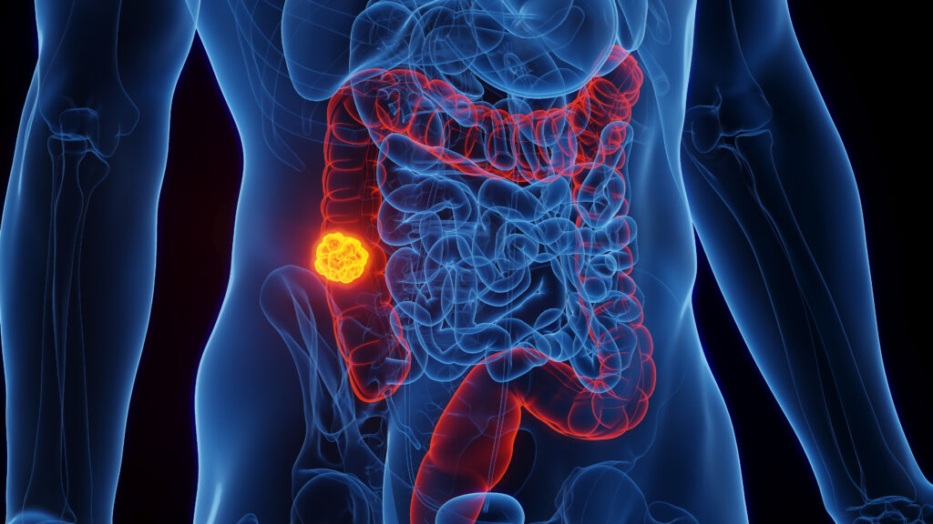
The liver is a common site of metastatic disease from some of the most prevalent malignancies, in particular gastrointestinal tumors for which metastatic deposits travel through the portal venous system. Approximately one-third of patients with solid tumors will develop liver metastases. In a large autopsy series the prevalence of liver involvement was 86 % for pancreatic cancer, 60 % for breast cancer, and 42 % in colorectal cancer.1
The liver is a common site of metastatic disease from some of the most prevalent malignancies, in particular gastrointestinal tumors for which metastatic deposits travel through the portal venous system. Approximately one-third of patients with solid tumors will develop liver metastases. In a large autopsy series the prevalence of liver involvement was 86 % for pancreatic cancer, 60 % for breast cancer, and 42 % in colorectal cancer.1
Hepatic involvement is often life-limiting and can result in severe morbidity. Approximately half of metastatic deaths from breast and prostate cancers are associated with liver metastases. Death due to colorectal cancer is frequently related to liver metastases, often as the only site of metastatic disease.
Background to the Role of Stereotactic Body Radiotherapy
Ablative Therapy of Liver Metastases
Systemic therapy is usually the primary therapy for metastatic liver disease,2 as it may allow for transitory responses and increased median survival. For patients with metastatic colorectal cancer treated with current palliative chemotherapy, median survival now approaches two years.3 Although whole liver radiotherapy has long been abandoned as an anti-cancer therapy, current (infrequent) use in symptomatic patients may still offer symptomatic relief.4
The goal of aggressive local treatment is long-term disease control for selected patients.5–8 Its benefits are now supported by large retrospective series. With a long history, surgical resection remains the gold standard local treatment of hepatic metastases—typically of colorectal origin.2,9 Surgery is associated with an acute risk of death (typically in less than 5 %)10 but can lead to five-year overall survival rates of up to 58 %.10–13 When Wilson and Adson retrospectively analyzed patients with limited liver metastases of colorectal cancer, approximately one-quarter of the resected patients were alive at five years. All long-term survivors in this series had solitary metastases, while none of the patients with multiple metastases or comparable non-resected patients were alive at five years.14 Adson et al. reported similar results, with 25 % five-year survival rate for resected patients versus 2.5 % in a non-resected group.15
Fong et al. studied 456 consecutive patients with liver metastases of colorectal cancer resections. They reported a 38 % five-year survival rate with a median survival of 46 months.10
Despite the apparent benefits of surgery, clinical and technical limitations narrow eligibility to metastasectomy. Limitations may relate to: location of the metastases; surgical plans unable to preserve sufficient liver parenchyma; medical comorbidities; or patient refusal. Thus only 10–25 % of patients with liver metastases from colorectal cancer can benefit from curative resection.2,16
New therapeutic options enable radical treatment instead of, or in combination with, surgery. Minimally invasive thermo-ablative procedures such as radiofrequency ablation (RFA), microwave ablation, cryotherapy, or laser-induced thermo-therapy have been developed. Complementing and competing with these is stereotactic body radiotherapy (SBRT)—a novel non-invasive approach with particular benefits: it can spare large vessels and, not being a thermal therapy, is insensitive to their cooling effects.9
Radiofrequency ablation—the most common alternative to surgery—is limited to lesions measuring less than 4–5 cm. It is generally avoided for lesions adjacent to large vessels and those in sub-capsular locations. Treatment of anatomically unfavorable lesions can be subject to a heat sink effect and/or carry a risk of thermal injury to vessels, biliary structures, or adjacent organs. Major complications are seen in 5 % of patients after RFA: portal thrombosis, hemoperitoneum, colic perforation, seeding of the catheter trajectory, and/or hepatic abscess. Mortality after RFA is rare, with a rate of 0.5–1.5 %.
Liver Irradiation
Radiotherapy for liver metastasis was first reported in 1954. In a series of 36 symptomatic liver metastases patients treated by Phillips et al. (19–36 Gy to whole liver), symptoms were reduced in over half the patients.17 A subsequent Radiation Therapy Oncology Group dose escalating trial (84–05) reported dose-dependant liver toxicity with a 10 % actuarial risk of severe liver toxicity at six months when whole-liver dose was increased from 27–33 Gy.18
In addition to total dose, liver tolerance to radiotherapy depends on underlying liver function, treatment volume, and dose fractionation. Patients with liver metastases are thus typically more tolerant than patients with primary liver tumors—where underlying liver cirrhosis is common. In standard fractionation (1.8–2 Gy per day) severe hepatic toxicity occurs with increasing frequency at whole-liver doses of ≥36 Gy.19,20 Radiation-induced liver disease (RILD) arises four to eight weeks after radiation therapy, manifesting with weight gain, increased abdominal girth, ascites, and a severe increase of alkaline phosphatase, with an associated mortality rate of 10–20 %. Pathophysiologically, RILD manifests as a veno-occlusive disease, liver architecture distortion, and fibrosis.9 Unfortunately, doses of 30–36 Gy are insufficient for tumor ablation.
Advances in imaging and radiotherapy have made partial liver irradiation feasible, allowing for differential dosing of tumor and normal liver. Stereotactic body radiotherapy can be defined as ‘a radiation therapy method used to very precisely deliver a high dose of radiation to an extracranial target within the body, using either a single dose or a small number of fractions’. It allows delivery of high doses per fraction to small volumes with geographic sparing of adjacent organs at risk, applying the principles of radiosurgery, long used for brain tumors. Although mean liver dose is a strong predictor of toxicity, the ‘threshold hypothesis’ of Jackson et al.21 would predict that the risk of RILD can be kept low, independently from the prescribed dose, as long as a certain volume of liver is spared. This is borne out by the work of Dawson et al. demonstrating clinically that much higher doses of radiation can be delivered to small liver volumes—beyond 100 Gy when the irradiated volume represents less than 25 % of the liver.22
Technical Challenges of Partial Liver Irradiation with Stereotactic Body Radiotherapy
Breathing Motion
Specific technical issues arise when irradiating focal liver targets, the most obvious and problematic is dealing with motion due to breathing. Cranio-caudal margins needed to accommodate for motion can range from 10 mm to 3–4 cm. A study by Kubas et al. showed a mean tumor position variation of 20 mm cranio-caudally, 5 mm laterally, and 9 mm antero-posteriorly.23 Without additional means to deal for liver motion, prohibitive irradiation of normal liver and surrounding bowel would often be required in order to achieve adequate tumor coverage.
Strategies have been developed to deal with breathing-dependent liver motion. Instructed breath-holding or shallow-breathing may help reduce diaphragm motion.24–26 Breath-hold valve devices (such as the active breathing coordinator®) are another option: the patient is helped to inhale to a predetermined volume and holds his breath for short intervals, during which radiation is delivered.27,28 The successful use of such devices, as with other breath-holding and shallow breathing techniques (with or without visual feedback), is dependent on patient compliance and demands attentive patient monitoring during treatment.
Abdominal pressure devices attached to a body cast are a simple mechanical means of constraining breathing motion. These devices, although potentially uncomfortable, can be efficient in reducing liver motion, typically allowing margins of 10 mm or less.29
Respiratory gating is a more recent strategy. Indicators are positioned on the anterior chest or abdominal wall where they are tracked by camera-based systems as surrogates to breathing phase and/or amplitude. More recent camera systems can also track surface anatomy directly without the use of surface reflectors or lights. At the time of treatment, a so-called gating window is defined during which radiation treatment is delivered.30–33 This strategy translates into treatment inefficiency and can be thwarted by breathing motion hysteresis and irregularities.
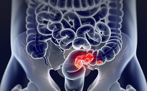
A more sophisticated means of dealing with motion is tumor tracking. Because liver tumors are invisible on planar imaging, tracking involves placement of one or multiple metal markers within the liver parenchyma (see Figure 1). The placement of fiducials is typically done percutaneously under CT-guidance prior to treatment planning. This additional procedure comes with its own unique set of risks and inconveniences but allows precise tracking of the fiducials as surrogates to the tumor.34
Radiotherapy Imaging
Treatment planning is typically based on a combination of 3D and 4D CT images: 4D CT scans oversample the anatomy and sort the images based on phase or amplitude in the breathing cycle. Such scans provide insight into tumor motion, relationship to external/internal fiducials, and potential deformation. Tumor (‘gross tumor volume’) is outlined on contrast imaging by the radiation oncologist, often with the help of additional studies (diagnostic CT and/or MRI) co-registered to the radiotherapy planning images. The role of PET in SBRT planning is still under investigation with new PET tracers (based on amino-acid metabolism instead of glucose) holding the promise of better biological tumor targeting.
During each treatment, precise patient positioning is attained using image-guidance (so-called image-guided radiotherapy) with 3D, 4D, and/or planar imaging.
Case Selection
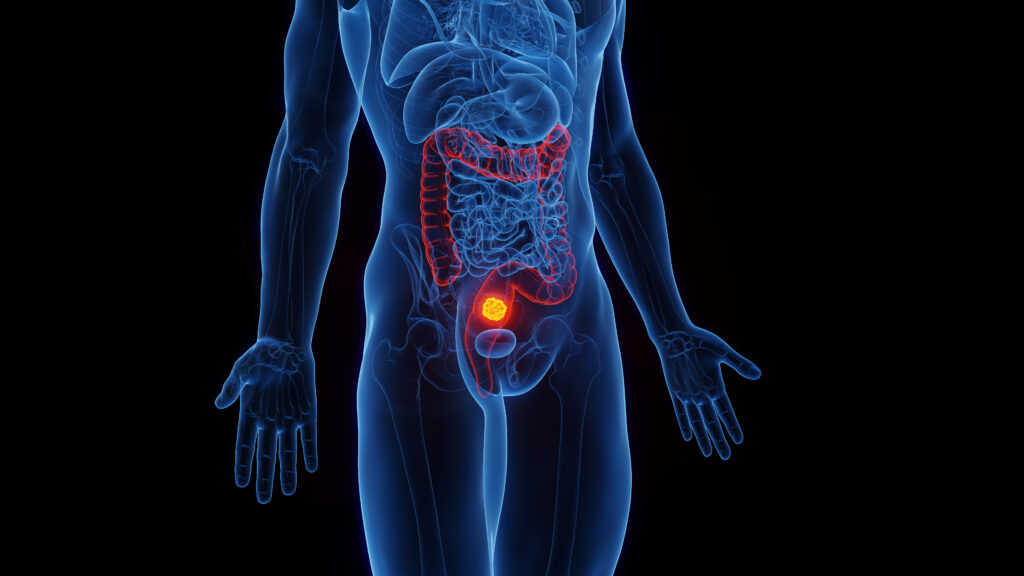
Patient selection is important. The best candidates for SBRT are patients with more than 1,000 cm3 of healthy liver and few (<3–4) reasonably-sized (<6 cm) lesions which do not touch the colon or duodenum (see Figure 2).
It is not rare for SBRT to complement a treatment plan where a caudate metastasis is irradiated and other tumors are dealt with using surgery or RFA. Poor candidates for SBRT have small or cirrhotic livers, numerous (>4) lesions, or lesions in broad contact with the bowel. If SBRT is used in these cases the risk of toxicity will be greater and the probability of ablation will be lower. There is an art to case selection but general guidelines are outlined in Table 1.
Clinical Outcomes with Liver Stereotactic Body Radiotherapy
Clinical Reports
The first liver SBRT report came from the Karolinska Institute in 199529 (subsequently updated in 199835). In this Swedish experience, fractionation and interval between fractions varied considerably: 1 x 10 Gy to 4 x 10 Gy delivered in 1–72 days for a mix of primary intrahepatic tumors and liver metastases.29 Low-grade toxicity was common—nausea and fever a few hours after treatment. There was one case of hemorrhagic gastritis and a post-treatment duodenal ulcer. Of concern, three of the eight hepatocellular carcinoma (HCC) patients—all with underlying cirrhosis—died days or weeks post-treatment. Eight of 22 patients had a significant decrease or complete response of their lesions after six to eight weeks (unlike thermal therapies, radiation results in gradual tumor regression over weeks or months).
A Phase I/II trial was initiated in Heidelberg in 1997.36 Sixty non-resectable liver tumors with adequate liver function were included. Dose (single fraction) was escalated from 14–26 Gy, taking into account normal tissue constraints. Reported actuarial one-year survival was 72 %. Local control at 18 months was 81 % when the first six patients (treated with 14–20 Gy) were excluded. A 2004 update37 reported an actuarial local control rate of 72 % at 12 months in 78 patients. This possibly lower control rate may be explained by a number of colorectal liver metastases, where local control appeared worse that for other histologies—45 compared to 91 %. This led the authors to postulate that higher doses and/or wider margins should be used for these patients. Side effects were mild, with transient (one to two weeks) nausea and anorexia in approximately one-third. All patients followed by CT showed a sharply demarcated focal radiation reaction after a median 1.8 months. This tended to decrease over time. It is speculated that these imaging changes correspond to focal veno-occlusive disease with a mean single-fraction threshold dose of 13.7 Gy (8.9–19.2 Gy).
Hoyer et al. published the results of a Danish Phase II study of SBRT for colorectal carcinoma metastases (located in the liver for 44 of 64 patients) treated with 45 Gy in three fractions, prescribed to the isocenter.38 One patient developed fatal—possibly iatrogenic—liver failure, two patients had duodenal ulcerations, and one, colonic ulceration. In addition, other mild to moderate toxicities were observed. Local control rates at one and two years were 89 and 79 %, with three-and five-year overall survival rates of 22 and 13 %.
Wulf et al. reported a one- and two-year actuarial local control rate of 92 % and 66 % in a study with five patients with primary liver cancer and 39 patients harboring 51 liver metastases.39 Two groups were treated with either ‘low-dose’ (3 x 10 Gy or 4 x 7 Gy) or ‘high-dose’ (3 x 12.5 Gy or 26 x 1 Gy) regimens. Dose was the only significant predictor of local control in a multivariate analysis.
A pooled analysis by Chang et al. studied the outcomes of 65 patients with 102 liver metastases from colorectal cancer after liver SBRT. These patients were treated from 2003 to 2009.40 The median prescribed dose was 42 Gy (22–60 Gy) in six fractions (1–6). Total dose, dose per fraction, and biologically effective dose correlated with local control. Eighteen-month local control diminished from 84–43 % when total dose was under 42 Gy. Treatments were well tolerated.
Vautravers-Dewas et al. evaluated the outcome, tolerance, and toxicity of SBRT for liver metastases in 42 patients with 62 lesions.41 Patients were treated with either 40 Gy in four fractions or 45 Gy in three fractions. There was a trend toward better local control with the higher dose (86 % with 40 Gy versus 100 % with 45 Gy at one-year), with similar toxicity, usually grade 1 or 2 nausea.
We reported our experience with 21 patients treated with SBRT for liver metastases with median dose 40 Gy (20–50 Gy) in one to 10 fractions.42 Treatment was well tolerated and toxicity was mild with the exception of one case of transient colitis. Actuarial overall one- and two-year survival rates were 94 and 60% and actuarial one- and two-year local control rates were 62 %.
Toxicity
After initial reports of severe bowel or liver toxicity, most contemporary trials incorporating specific normal tissue dose constraints typically report mild toxicity. Organ tolerance to focal radiation with short treatment schemes is however still incompletely understood. A Phase I multicenter prospective study was performed in the US to test the maximum tolerated dose (MTD) of SBRT for liver metastases.43 Eighteen patients were enrolled, four of which had multiple tumors. The initial prescribed dose was 36 Gy in three fractions, with subsequent cohorts receiving a dose escalation of 2 Gy per fraction up to 60 Gy in three fractions or MTD. The MTD was not reached and a Phase II study was initiated. Kavanagh et al. reported an interim analysis of the combined Phase I/II study, including the 18 patients from Phase I (doses of 36–60 Gy in three fractions) and another 18 patients treated to 60 Gy in the Phase II component.44 A high rate of durable in-field tumor control was achieved with only one case of grade 3 toxicity (no grade 4). In an expanded cohort, actuarial in-field local control rate at one and two tears were 95 and 92%, respectively, and two-year local control for lesions with 3 cm or less of 100 %.45
Lee et al. designed a Phase I study to determine the safety and efficacy of individualized six-fraction SBRT for liver metastases.46 Sixty-eight patients were treated with individualized regimens of 27.7–60 Gy in six fractions, prescribed based on risk of toxicity (5, 10, and 20 % risk), according to effective liver volume irradiated. The mean dose received by 700 ml of liver was 14.1 Gy in six fractions, and no serious toxicity was observed. A 12-month tumor control of 71 % was observed in this study which included larger liver lesions. Lower radiation doses were associated with worse local control and survival.
RTOG designed a Phase I trial to identify the MTD, with up to 50 Gy in 10 fractions of highly conformal radiation therapy prescribed in patients with metastatic liver cancer. It is now closed to accrual. Early results are that, when normal tissue constraints could be met, treatment of liver metastases with 50 Gy in 10 fractions is feasible and safe.47 Local control has yet to be reported.
An article by Sawrie et al. tried to compile information on normal tissue tolerance and toxicity with SBRT for liver metastases and HCC.48 Besides the liver, organs at risk of toxicity are essentially gastro-intestinal. Bowel and duodenum are close to the liver, and can be of concern depending on tumor location. Mild to severe diarrhea is the most common toxicity, nonetheless one colonic perforation and two duodenal ulcerations in 44 patients were reported by Hoyer et al.38 in patients with total bowel/duodenum dose of 67.5 Gy (BED) or higher. Gastric toxicity consists of nausea, treated symptomatically with anti-emetics or corticosteroids, or ulceration/perforation if the dose is not constrained. Acute dermatitis has been reported but is typically self-limiting. It can arise near the lesion, or in some unexpected locations, such as described in a case report in which a patient treated with SBRT for a HCC developed grade 3 dermatits in the contralateral axilla.49 There were no reported kidney, heart, or spinal cord toxicity.48 The mostly low to mild reported toxicity in this review is in keeping with the highly conformal dose distributions delivered.
Conclusion
Overall, the studies report local control rates ranging from 55–100 % at one to two years.36–42,44,46,50 This wide range can be due to patient, treatment, and follow-up heterogeneity across studies. Table 2 summarizes results of selected series. As with other local therapies, prospective evidence for an impact on overall survival is lacking for SBRT.40 Survival will be heterogeneous in these patients’ populations and depend on patient, disease, and treatment factors. Actuarial survival at one to two years with SBRT has been reported to range from 71–94 % and 30–62 %.41 Fractionation for these treatments varies among studies, from single dose to 10 fractions, and associated total doses of 14–60 Gy. Forty-five Gy in three fractions or 50 Gy in five fractions (over one to two weeks on non-consecutive days) are commonly used regimens.
Future Directions
Based on the results from published series, SBRT has evolved to a feasible, safe, and effective non-invasive treatment for selected liver metastases, complementing present treatment options. As these treatments become more widely available, published cohorts will become more substantive and the now heterogeneous treatment regimens will likely become better standardized. In an effort to help sort out the effect of number of sessions, the Stereotactic radiation therapy of liver metastases (StRaL) trial proposes to randomize patients with one to three lesions to a single dose of 28 Gy versus a hypofractionated approach with 3 x 12.5 Gy. Associations of chemotherapy or targeted therapy, will also likely be investigated in future trials. For now, clinical judgment is used in the timing of systemic therapy and SBRT. The most prudent physicians will prefer an interval of several weeks before and after SBRT, in other cases SBRT may be delivered on a normal ‘off week’ seen in many regimens or even concurrently with agents which are not strong radiation sensitizers.
In upcoming years, we expect to see evaluation of this technique in the treatment of oligometastases in cancers where ablative therapy of metastases is not yet a widespread standard (small cell lung, for example). Another direction is prospective comparison to other local modalities. In this sense, the most mature project is the ongoing Radiofrequency ablation versus stereotactic body radiation (RAS) trial, a multicenter randomized prospective trial for colorectal liver metastases. Patients are eligible for this trial if they are not suitable for surgical resection, and have one to four metastases with maximum 4 cm diameter, amenable to both RFA and SBRT. ■