touchONCOLOGY touchONCOLOGY
Haematological Malignancies, Lymphoma
Read Time: 12 mins

Novel Therapy of Relapsed and Refractory Classical Hodgkin’s Lymphoma

Copy Link
Published Online: Oct 11th 2011 European Oncology & Haematology, 2011;7(3):203-10 DOI: https://doi.org/10.17925/EOH.2011.07.03.203
Authors: Enrico Derenzini, Daniela Buglio, Anas Younes
Quick Links:
Abstract
Article
Article Information
Abstract:
Overview

Classical Hodgkin’s lymphoma (HL) is generally considered a highly curable disease, with approximately 80 % of patients cured with standard first-line chemotherapy. The standard treatment approach for relapsed/refractory patients is second-line salvage chemotherapy followed by autologous stem cell transplantation (ASCT). About half of all patients undergoing ASCT are rescued and definitely cured by such an approach, but the outcome in patients relapsing or refractory to second-line chemotherapy and ASCT is dismal, with a median survival of less than three years. Therapeutic options for this subset of patients comprise tandem ASCT, reduced-intensity allogeneic stem cell transplantation (allo-SCT) and novel agents. Median overall and progression-free survival rates following allo-SCT have ranged from 27 to 56 % and from 18 to 39 %, respectively, with a treatment-related mortality ranging from 15 to 25 %. Several new compounds have been identified as promising agents for the treatment of patients with relapsed classical HL. These drugs have shown promising activity in a subset of heavily pre-treated relapsed/refractory patients, with response rates and disease control rates exceeding 40 and 70 %. If approved, these compounds will probably change the standard of care, making it possible to develop combination regimens with chemotherapy or other new agents, thus improving efficacy with a decreased toxicity profile.

Keywords

Hodgkin’s lymphoma, panobinostat, SGN35, rituximab, everolimus, daclizumab, autologous stem cell transplantation, allogeneic stem cell transplantation

Article:

Although Hodgkin’s lymphoma (HL) is considered one of the most curable human cancers, the treatment of patients with relapsed and refractory disease, especially those who relapse after autologous stem cell transplantation (ASCT), remains challenging.1,2
Furthermore, because the median age of patients is in the mid-30s, the impact of early mortality on the number of years lost from productive life is remarkable. At the present time, patients with HL whose disease relapses after stem cell transplantation have no curative options. New drugs and novel treatment strategies that are based on our understanding of the disease biology and signalling pathways are needed to improve treatment outcomes in these patients. Ironically, because HL is a rare disease and has long been considered highly curable, the search for new therapies and treatment approaches has grown slowly in the past, with no new drugs approved in the last 30 years.

With recent advances in our understanding of HL pathology, biology and immunology, several therapeutic targets have been identified and are currently under preclinical and clinical investigation. Some of these new compounds have already been evaluated in large Phase II clinical trials seeking potential approval by regulatory agencies. This article focuses on current treatment strategies for patients with relapsed and refractory HL, including the role of stem cell transplantation and promising new monoclonal antibodies and small molecules.

Role of Stem Cell Transplantation Autologous Stem Cell Transplantation
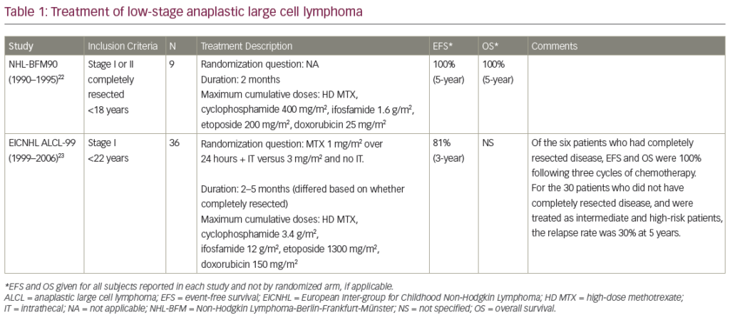
The superiority of high-dose chemotherapy with stem cell rescue over standard second-line chemotherapy alone was demonstrated in two randomised trials.3,4 About half of all patients undergoing ASCT are rescued and definitely cured by such an approach, but the outcome in patients relapsing or refractory to second-line chemotherapy and ASCT is dismal, with a median survival of less than three years.5 The need to identify patients who will not benefit from ASCT has resulted in a large number of studies describing various prognostic factors, including stage and time to relapse, B symptoms, extranodal disease, bone marrow involvement and chemosensitivity at relapse. Depending on the presence or absence of these factors, progression-free survival (PFS) and overall survival (OS) rates range from 10 to 83 % and from 13 to 90 %, respectively (see Table 1).6–12 So, the ideal patient who will benefit from ASCT is the one with limited stage, long time to relapse and chemosensitive disease, while the patient with advanced stage, short time to relapse and refractory disease has the worst prognosis. A recent report13 on the role of tandem ASCT suggests a potential OS and PFS benefit for poor-risk patients – with reported five-year PFS and OS of 36 and 45 %, respectively, in patients resistant to second-line chemotherapy – and these results compare favourably with other published series of patients refractory to second-line chemotherapy.12One of the most important and widely accepted prognostic factors for patients undergoing ASCT appears to be chemosensitivity at relapse,with patients responding to second-line chemotherapy having a much better outcome than patients with refractory disease, whose relapse rate approaches 80 % in some published series.9–12 The concept of chemosensitivity is strictly related to the imaging techniques and criteria used to assess the response to therapy. The advent of functional imaging techniques such as fluorodeoxyglucose-positron emission tomography (FDG-PET) has further improved the reliability of the response evaluation, allowing the early detection of chemoresistance. In fact, the FDG-PET scan after two cycles of chemotherapy (PET-2) is now considered the most powerful prognostic predictor in the first-line setting.14 Retrospective studies have suggested that this concept also applies to relapsed or refractory patients undergoing second-line chemotherapy and ASCT, but this finding needs to be confirmed in prospective studies (see Table 2).14,16–22

Allogeneic Stem Cell Transplantation In clinical practice, allogeneic stem cell transplantation (allo-SCT) has so far been offered to patients relapsing after ASCT. The evidence for a graft-versus-lymphoma (GVL) effect is indirect and comes from:

  • retrospective comparisons between relapse rates of allo-SCT recipients and ASCT recipients;
  • the fact that several groups reported disease regressions following donor lymphocyte infusions; and
  • reports on the protective effect of chronic graft-versus-host disease (GVHD).23

The treatment-related mortality observed with myeloablative conditioning regimens remains high and outweighs any survival advantage compared with ASCT.24 Reduced-intensity conditioning (RIC) regimens are currently being explored with promising results (see Table 3).24–30

In a recent paper from the European Group for Blood and Marrow Transplantation, the outcome in 89 HL patients who received RIC was compared with that in 79 patients who received myeloablative conditioning. The relapse rate was higher in the group receiving RIC (57 compared with 30 %), but the OS rate significantly favoured the RIC group (28 compared with 22 %), due to a much lower non-relapse mortality.24

Although the type of donor – matched related (MRD) or unrelated donor (URD) – does not seem to affect the prognosis, in a recent study, a consistent relapse-free survival advantage has been reported for haploidentical stem cell transplantation over MRD and URD.31

Monoclonal Antibodies XmAb2513 and SGN35 – Targeting CD30
CD30, a member of the tumour necrosis factor (TNF)-receptor superfamily, is an attractive target, because it is expressed nearly universally in Hodgkin’s and Reed–Sternberg (HRS) cells, with an extremely limited pattern of expression in normal tissues.32,33

Results from two clinical studies using first-generation naked anti-CD30 monoclonal antibodies in patients with relapsed HL have been disappointing, perhaps reflecting their poor antigen-binding and/or effector cell-activation properties.34 A second-generation humanised anti-CD30 antibody, XmAb2513, was recently developed to improve antigen binding and Fc-gamma receptor IIIA affinity and specificity.35,36 A Phase I study of XmAb2513 is currently enrolling patients in the US and encouraging preliminary results, with some responses, were recently reported.37

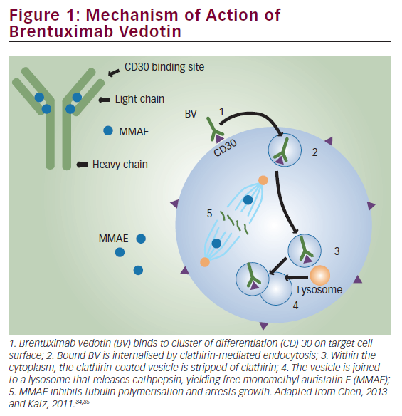
In an alternate strategy, the anti-CD30 antibody cAC10 was conjugated to a synthetic anti-microtubule agent, monomethylauristatin E (MMAE), resulting in a novel immunotoxin conjugate called SGN35.38 SGN35 was recently evaluated in two Phase I clinical trials in patients with relapsed HL and anaplastic large-cell lymphoma (ALCL). In the first study, SGN35 was administered by intravenous infusion every three weeks. Of 42 patients with relapsed/refractory disease, 17 (40 %) achieved partial or complete remission and 86 % of the patients had documented tumour reductions.39 A second Phase I study evaluated the safety and efficacy of SGN35 given on a weekly schedule. Of 17 evaluable patients, seven achieved complete response (CR) and one achieved partial response (PR), an overall response rate of 47 %.40 On the basis of this encouraging clinical activity, SGN35 is currently being evaluated in two Phase II pivotal trials seeking Food and Drug Administration (FDA) approval in patients with relapsed HL and ALCL.

Rituximab – Targeting CD20
The anti-CD20 monoclonal antibody rituximab was evaluated in the treatment of patients with classical HL. Although CD20 antigen is infrequently expressed by HRS cells, it is highly expressed by the reactive B cells in the microenvironment. Thus it was hypothesised that rituximab may induce clinical remission in classical HL by depleting B cells from the microenvironment, by directly killing the few cases of CD20-expressing HRS cells and perhaps by killing the putative HRS CD20-positive stem cells.41 In a pilot study, investigators from the University of Texas MD Anderson Cancer Center treated 22 patients with relapsed classical HL with six weekly doses of rituximab; of these 22 patients, six demonstrated CD20 expression by HRS cells.42 Five patients (23 %) achieved partial or complete remission and eight additional patients had stable disease (SD). Clinical remission was observed in patients regardless of CD20 expression by HRS cells and was limited in patients whose disease was confined to the lymph nodes.In a subsequent study, the same investigators combined rituximab with adriamycin–bleomycin–vinblastine–dacarbazine (ABVD) chemotherapy to treat patients with newly diagnosed classical HL.43 Fifty-two patients with newly diagnosed classical HL were treated in a Phase II study. With a median follow-up of 32 months, the estimated event-free survival (EFS) was 82 % and OS was 100 %. Importantly, the EFS improved for all risk categories: for patients with an internationally prognostic score of 0 to 1, the EFS was 92 %; for scores 0 to 2, 86 %; and for scores 3 to 5, 73 %. These findings are currently being confirmed in a multicentre, randomised study comparing ABVD with rituximab plus ABVD.

Daclizumab – Targeting CD25
CD25 is the interleukin-2 receptor (IL-2R) alpha subunit. IL-2R is a growth factor receptor linked to a variety of signalling pathways – Janus kinase/signal transducer and activator of transcription (Jak-STAT), AKT/mammalian target of rapamycin (mTOR) and mitogen-activated protein (MAP) kinase signalling – involved in inflammation, cell growth and proliferation. It is a transmembrane receptor protein present on only a few normal cells, such as activated T cells, activated B cells, some thymocytes and myeloid precursors, but also expressed in T-cell and B-cell malignancies, such as adult T-cell leukaemia, cutaneous T-cell lymphoma, ALCL and hairy cell leukaemia and on Reed–Sternberg and associated polyclonal T cells in HL. Abnormalities in CD25 expression have also been found in autoimmune diseases, allograft rejection and GVHD.45

The humanised anti-CD25 monoclonal antibody daclizumab was first approved by the FDA in 1997 for use in the prevention of renal allograft rejection. Putative mechanisms of action of daclizumab are blockade of IL-2R alpha signalling, a mechanism involving the FcR receptor and antibody-dependent, cell-mediated cytotoxicity (ADCC), and the recently documented rise of natural killer cells in the peripheral blood in humans.46–49 Daclizumab was proven to be effective and was mainly investigated in T-cell malignancies such as adult T-cell leukaemia/lymphoma and ALCL.45 Given the expression of CD25 in Reed– Sternberg cells, daclizumab was also tested in HL. A Phase I/II trial used the immunoconjugate composed of daclizumab and the Pseudomonas aeruginosa toxin PE38 in 59 patients with leukaemia/lymphoma expressing IL-2R alpha. Eight patients showed objective responses, one of whom was affected by HL.50 In a recent Phase II trial, daclizumab conjugated with the radionuclide yttrium-90 (90Y-daclizumab) was investigated in 30 relapsed/refractory HL patients.51 Radioimmunotherapy with 90Y-daclizumab was given once every six weeks at a dose of 15 mCi for a maximum of seven cycles. Twelve patients achieved CR, seven achieved PR and five had SD. The main side effects were haematological, with prolonged thrombocytopenia, as well as three patients who developed a myelodysplastic syndrome following treatment. These encouraging results, if confirmed in larger numbers of patients, make 90Y-daclizumab one of the most active new investigational drugs in relapsed/refractory HL.

Novel Small Molecules
HRS cells aberrantly express a variety of pro-survival proteins, such as nuclear factor-kappaB (NF-kappaB), Jak/STATs, Akt/mTOR, Notch-1 and extracellular signal-regulated kinase (ERK), that can be targeted by small molecules.52–54 These proteins can be targeted either by selective small-molecule inhibitors – Jak-2, mTOR and B-cell lymphoma (Bcl)-2 family inhibitors – or by broad inhibitors that modulate several unrelated molecules, such as histone deacetylase (HDAC), proteasome and heat shock protein 90 (HSP90) inhibitors (see Figure 1).

Histone Deacetylase Inhibitors
It is well established that post-transcriptional histone modification plays an important role in regulating gene transcription and that such modification is mediated by several enzymes, including histone acetyltransferases (HATs) and HDACs.55 The balance between HATs and HDACs is critical for regulating the expression and functional status of a variety of proteins involved in cell proliferation, survival, angiogenesis and immunity.56–60

Eighteen HDACs have been identified in humans and grouped into two major categories: zinc-dependent HDACs and nicotinamide adenine dinucleotide (NAD)-dependent HDACs.55,56,71 Furthermore, HDACs are classified into four major classes: class I (HDAC 1, 2, 3), class II (HDAC 4, 5, 6, 7, 9 and 10), class III (sirtuin 1–7) and class IV (HDAC 11). Class III is NAD-dependent, whereas classes I, II and IV are zinc-dependent.

MGCD0103 and etinostat (SNDX-275, formerly MS-275) preferentially inhibit class I HDACs (isotype-selective HDAC inhibitors). Vorinostat, ITF2357 and panobinostat (LBH589) inhibit HDAC classes I and II (pan-HDAC inhibitors) (see Table 4).

MGCD0103 is a novel oral non-hydroxamate benzamide-based HDAC inhibitor that selectively inhibits HDAC 1 and 2 (and, to a lesser extent, 3 and 11) isoforms. The safety and efficacy of MGCD0103 given orally three times per week (85–110mg starting doses) were recently evaluated in a study in patients with relapsed and refractory HL.61 Of 20 patients treated at the 110 mg dose level, seven (35 %) achieved partial or complete remission. However, this dose level was poorly tolerated, resulting in dose interruptions and reductions and discontinuation of therapy. Subsequently, three (30 %) of 10 patients enrolled on a reduced dose (85 mg) achieved partial remission, and the treatment was well tolerated. Overall, 80 % of the 30 evaluable patients had some decrease in their tumour measurements. Three pan-HDAC inhibitors were recently evaluated in patients with relapsed HL. In the first study, panobinostat was evaluated in a Phase I trial in patients with haematological malignancies that included relapsed HL.62 Five of 13 (38 %) HL patients achieved partial remission. On the basis of this promising clinical activity, two large international Phase II studies of panobinostat in relapsed HL are now enrolling patients to confirm these results.63,64 In a second study, the Southwest Oncology Group (SWOG) conducted a Phase II trial of vorinostat in patients with relapsed HL.65 Twenty-five patients were treated with 200 mg vorinostat given orally twice a day for 14 days every 21-day cycle. Unlike MGCD0103 and panobinostat, vorinostat produced only modest clinical activity, as only one patient (4 %) achieved partial remission.

Vorinostat was shown to induce cell cycle arrest and apoptosis in HL cell lines and to synergise with chemotherapy.66 Furthermore, vorinostat inhibited STAT6 phosphorylation and transcription in HL cell lines, an effect that was associated with a decrease in the expression and secretion of T-helper 2 (Th2)-type cytokines and chemokines, including thymus and activation-regulated chemokine (TARC/CCL17) as well as interleukin-5 and an increase in T-helper 1 (Th1)-type cytokines and chemokines, including a profound increase in IP-10 levels.66 This study confirms that HDAC inhibitors can also determine changes in the tumour microenvironment.

ITF2357 is a selective class I and class II HDAC inhibitor. The efficacy, safety and tolerability of daily 100 mg doses of ITF2357 in relapsed/refractory HL were investigated in a Phase II clinical trial at the National Tumour Institute of Milan. The preliminary results were presented at the 2008 American Society of Clinical Oncology (ASCO) meeting. Fifteen patients were enrolled and 13 were evaluable for response. Seven patients (54 %) had a stable disease, with a reduction in FDG-PET uptake in six patients (46 %) lasting at least three months; six patients had disease progression. Interestingly, a correlation was found between a decrease in serum TARC levels and the response to treatment in this study.67 On the basis of the single-agent activity of ITF2357 and the documented synergistic activity of ITF2357 and mechlorethamine in HL cell lines, a Phase II trial of ITF2357 combined with mechlorethamine was conducted by the same group. Nineteen patients who unsuccessfully underwent prior ASCT/allo-SCT were enrolled and preliminary data were presented at the 2008 American Society of Hematology (ASH) meeting. Seventeen patients were evaluable for response, with two CR (12 %), three PR (18 %) and five SD (29 %). The main toxicity was haematological, with seven patients experiencing grade 3/4 neutropenia and eight having thrombocytopenia; four patients experienced infections during treatment.68 Taken together, these data suggest that ITF2357 has encouraging clinical activity in relapsed/refractory HL.

Mammalian Target of Rapamycin Inhibitors
The phosphatidylinositol 3-kinase (PI3K)/Akt/mTOR signalling pathway (see Figure 1) is one of the most aberrantly activated survival pathways in cancer, making it an important target for drug development.69,70 This pathway is negatively regulated by the tumour-suppressor protein phosphatase and tensin homologue (PTEN). In vitro experiments demonstrated that inhibition of PI3K, Akt or mTOR by various small molecules can induce cell cycle arrest, autophagy and apoptosis in HRS-derived cell lines.71–73 In addition to a direct antitumour effect, mTOR inhibitors may induce clinical responses by enhancing the immune response and inhibiting angiogenesis.74,75

The therapeutic value of inhibiting the PI3K/Akt/mTOR axis has recently been studied using the oral mTOR inhibitor everolimus in 17 relapsed-refractory patients (intention to treat analysis) (see Figure 2).76 Of 15 evaluable patients with relapsed HL treated daily with 10 mg everolimus, seven (47 %) achieved partial responses (see Figure 2 and Table 5). If this finding is confirmed in a larger number of patients, everolimus may become one of the most active agents in relapsed HL. In vitro experiments suggested that mTOR inhibitors may synergise with chemotherapy, PI3K inhibitors and HDAC inhibitors in a variety of tumour models, including HL.71,77 A Phase I clinical trial combining the HDAC inhibitor panobinostat with the mTOR inhibitor everolimus is currently enrolling patients with non-Hodgkin’s lymphoma (NHL) and HL.

Proteasome Inhibitors
NF-kappaB plays a central role in regulating the expression of various genes involved in cell survival, apoptosis, carcinogenesis and inflammation, making it a potential therapeutic target. The first attempt to therapeutically inhibit NF-kappaB activation in HL used the proteasome inhibitor bortezomib. By inhibiting the degradation of cytoplasmic I-kappaB-alpha, bortezomib inhibits the activation of NF-kappaB.78 In preclinical studies, bortezomib inhibited HL cell line proliferation and induced cell cycle arrest and apoptosis in a time-dependent and dose-dependent manner and was effective even in HL cell lines that harboured mutations in the I-kappaB-alpha gene.79 Furthermore, bortezomib enhanced the effect of gemcitabine chemotherapy and potentiated the effect of anti-CD30 antibody and tnf-related, apoptosis-inducing ligand (TRAIL)/Apo2L.79,80 Despite these favourable preclinical results, bortezomib demonstrated no significant clinical activity in patients with relapsed HL.81.82 On the basis of these preclinical experiments, bortezomib-based combinations were evaluated in patients with relapsed classical HL. In the first study, a Phase I trial evaluated the combination of bortezomib with the ifosfamide–carboplatin–etoposide (ICE) regimen in relapsed/refractory HL.83 Bortezomib was given at doses of 1, 1.3 or 1.5 mg/m2 on days one and four of each ICE cycle. Twelve patients were enrolled, of whom six achieved PR and three achieved CR, for an overall response rate of 75 %. On the basis of these encouraging data, a randomised Phase II study comparing ICE with bortezomib plus ICE is currently enrolling patients to determine the contribution of bortezomib to the ICE regimen.

In a second study, 18 patients with relapsed HL were treated with bortezomib in combination with gemcitabine.84 Because of the relatively low response rate (22 %), coupled with treatment-related liver toxicity, the authors concluded that this regimen should not be further developed for the treatment of HL.

Heat Shock Protein 90 Inhibitor 17-AAG
Heat shock proteins are cellular chaperone proteins that are required for essential housekeeping functions such as protein folding, assembly and transportation across different cell compartments. HSP90 is frequently overexpressed in cancer cells and hence is an attractive target for cancer therapy. Similarly to findings in other cancers, HSP90 is overexpressed in primary and cultured HL cells.85,86 HSP90 chaperones several client proteins that promote HRS cell survival, including ERK, Akt and NF-kappaB.52,80,87

In a preclinical experiment, the HSP90 small-molecule inhibitor 17-allylamino-17-demethoxygeldanamycin (17-AAG) down-regulated Akt and cellular FLICE-inhibitory protein (cFLIP) and induced apoptosis in HL-derived cell lines.87 Furthermore, 17-AAG synergised with doxorubicin and agonistic anti-TRAIL death receptor antibodies. On the basis of these findings, a Phase II study of 17-AAG is currently enrolling patients with relapsed HL, and the results are expected to be available in the near future.

Lenalidomide
Two independent groups have evaluated the safety and efficacy of lenalidomide in patients with relapsed HL. In the first study, Fehniger et al. reported their experiences with 25 mg/day lenalidomide on days 1–21 of a 28-day cycle.88 Four of the 12 evaluable patients responded (one CR and three PR). Grade 3 and 4 neutropenia was observed in 47 % and thrombocytopenia was seen in 27 % of the patients. In a second study, Kuruvilla et al. treated 15 patients with relapsed HL using the same dose and schedule of lenalidomide as in the previous study.89 Two patients achieved PR and seven achieved SD, with a median time to progression of 3.2 months. Collectively, these data suggest that lenalidomide has promising single-agent activity in relapsed HL.

Conclusion
After more than three decades of quiet on the drug development front, several compounds have been identified as promising agents for the treatment of patients with relapsed classical HL. Future research should focus on identifying biomarkers, selecting patients who are likely to respond to these novel agents, incorporating these new agents with existing effective regimens and identifying predictive markers for treatment response. Ultimately, randomised clinical trials will be required to document the impact of these new agents on patients’ survival.

Article Information:
Disclosure

The authors have no conflicts of interest to declare.

Received

2010-09-20T00:00:00

References

  1. Diehl V, Franklin J, Pfreundschuh M, et al., Standard and increased-dose BEACOPP chemotherapy compared with COPP-ABVD for advanced Hodgkin’s disease, N Engl J Med, 2003;348:2386–95.
  2. Duggan DB, Petroni GR, Johnson JL, et al., Randomized comparison of ABVD and MOPP/ABV hybrid for the treatment of advanced Hodgkin’s disease: report of an intergroup trial, J Clin Oncol, 2003;21:607–14.
  3. Linch DC, Winfield D, Goldstone AH, et al., Dose intensification with autologous bone-marrow transplantation in relapsed and resistant Hodgkin’s disease: results of a BNLI randomised trial, Lancet, 1993;341:1051–4.
  4. Schmitz N, Pfistner B, Sextro M, et al., Aggressive conventional chemotherapy compared with high-dose chemotherapy with autologous haemopoietic stem-cell transplantation for relapsed chemosensitive Hodgkin’s disease: a randomised trial, Lancet, 2002;359(9323):2065–71.
  5. Horning S, Fanale M, deVos S, et al., Defining a population of Hodgkin lymphoma patients for novel therapeutics: an international effort, Ann Oncol, 2008;20:118.
  6. Moskowitz CH, Nimer SD, Zelenetz AD, et al., A 2-step comprehensive high-dose chemoradiotherapy second-line program for relapsed and refractory Hodgkin disease: analysis by intent to treat and development of a prognostic model, Blood, 2001;97(3):616–23.
  7. Moskowitz CH, Kewalramani T, Nimer SD, et al., Effectiveness of high dose chemoradiotherapy and autologous stem cell transplantation for patients with biopsy-proven primary refractory Hodgkin’s disease, Br J Haematol, 2004;124(5):645–52.
  8. Zinzani PL, Tani M, Gabriele A, et al., High-dose therapy with autologous transplantation for Hodgkin’s disease: the Bologna experience, Haematologica, 2003;88(5):522–8.
  9. Tarella C, Cuttica A, Vitolo U, et al., High-dose sequential chemotherapy and peripheral blood progenitor cell autografting in patients with refractory and/or recurrent Hodgkin lymphoma: a multicenter study of the Intergruppo Italiano Linfomi showing prolonged disease free survival in patients treated at first recurrence, Cancer, 2003;97(11):2748–59.
  10. Josting A, Rudolph C, Mapara M, et al., Cologne high-dose sequential chemotherapy in relapsed and refractory Hodgkin lymphoma: results of a large multicenter study of the German Hodgkin Lymphoma Study Group (GHSG), Ann Oncol, 2005;16(1):116–23.
  11. Popat U, Hosing C, Saliba RM, et al., Prognostic factors for disease progression after high-dose chemotherapy and autologous hematopoietic stem cell transplantation for recurrent or refractory Hodgkin’s lymphoma, Bone Marrow Transplant, 2004;33(10):1015–23.
  12. Sirohi B, Cunningham D, Powles R, et al., Long-term outcome of autologous stem-cell transplantation in relapsed or refractory Hodgkin’s lymphoma, Ann Oncol, 2008;19(7):1312–9.
  13. Morschhauser F, Brice P, Fermé C, et al., Risk-adapted salvage treatment with single or tandem autologous stemcell transplantation for first relapse/refractory Hodgkin’s lymphoma: results of the prospective multicenter H96 trial by the GELA/SFGM study group, J Clin Oncol, 2008;26(36):5980–7.
  14. Moskowitz AJ, Yahalom J, Kewarlramani T, et al., Pretransplantation functional imaging predicts outcome following autologous stem cell transplantation for relapsed and refractory Hodgkin lymphoma, Blood, 2010;116(23):4934–7.
  15. Gallamini A, Hutchings M, Rigacci L, et al., Early interim 2- [18F]fluoro-2-deoxy-D-glucose positron emission tomography is prognostically superior to international prognostic score in advanced-stage Hodgkin’s lymphoma: a report from a joint Italian-Danish study, J Clin Oncol, 2007;25(24):3746–52.
  16. Schot BW, Zijlstra JM, Sluiter WJ, et al., Early FDG-PET assessment in combination with clinical risk scores determines prognosis in recurring lymphoma, Blood, 2007;109(2):486–91.
  17. Filmont JE, Gisselbrecht C, Cuenca X, et al., The impact of pre- and post-transplantation positron emission tomography using 18-fluorodeoxyglucose on poor-prognosis lymphoma patients undergoing autologous stem cell transplantation, Cancer, 2007;110(6):1361–9.
  18. Svoboda J, Andreadis C, Elstrom R, et al., Prognostic value of FDG-PET scan imaging in lymphoma patients undergoing autologous stem cell transplantation, Bone Marrow Transplant, 2006;38(3):211–6.
  19. Spaepen K, Stroobants S, Dupont P, et al., Prognostic value of pretransplantation positron emission tomography using fluorine 18-fluorodeoxyglucose in patients with aggressive lymphoma treated with high-dose chemotherapy and stem cell transplantation, Blood, 2003;102(1):53–9.
  20. Cremerius U, Fabry U, Wildberger JE, et al., Pre-transplant positron emission tomography (PET) using fluorine-18-fluorodeoxyglucose (FDG) predicts outcome in patients treated with high-dose chemotherapy and autologous stem cell transplantation for non-Hodgkin’s lymphoma, Bone Marrow Transplant, 2002;30(2):103–11.
  21. Jabbour E, Hosing C, Ayers G, et al., Pretransplant positive positron emission tomography/gallium scans predict poor outcome in patients with recurrent/refractory Hodgkin lymphoma, Cancer, 2007;109(12):2481–9.
  22. Derenzini E, Musuraca G, Fanti S, et al., Pretransplantation positron emission tomography scan is the main predictor of autologous stem cell transplantation outcome in aggressive B-cell non-Hodgkin lymphoma, Cancer, 2008;113(9):2496–2503.
  23. Laport GG, Allogeneic hematopoietic cell transplantation for Hodgkin lymphoma: a concise review, Leuk Lymphoma, 2008;49(10):1854–9.
  24. Sureda A, Robinson S, Canals C, et al., Reduced-intensity conditioning compared with conventional allogeneic stemcell transplantation in relapsed or refractory Hodgkin’s lymphoma: an analysis from the Lymphoma Working Party of the European Group for Blood and Marrow Transplantation, J Clin Oncol, 2008;26(3):455–62.
  25. Thomson KJ, Peggs KS, Smith P, et al., Superiority of reduced-intensity allogeneic transplantation over conventional treatment for relapse of Hodgkin’s lymphoma following autologous stem cell transplantation, Bone Marrow Transplant, 2008;41:765–70.
  26. Armand P, Kim HT, Ho VT, et al., Allogeneic transplantation with reduced-intensity conditioning for Hodgkin and non-Hodgkin lymphoma: importance of histology for outcome, Biol Blood Marrow Transplant, 2008;14:418–25.
  27. Alvarez I, Sureda A, Caballero MD, et al., Nonmyeloablative stem cell transplantation is an effective therapy for refractory or relapsed Hodgkin lymphoma: results of a Spanish prospective cooperative protocol, Biol Blood Marrow Transplant, 2006;12:172–83.
  28. Robinson SP, Sureda A, Canals C, et al., Reduced intensity conditioning allogeneic stem cell transplantation for Hodgkin`s lymphoma: identification of prognostic factors predicting outcome, Haematologica, 2009;94:230–8.
  29. Anderlini P, Saliba R, Acholonu S, et al., Fludarabinemelphalan as a preparative regimen for reduced-intensity conditioning allogeneic stem cell transplantation in relapsed and refractory Hodgkin’s lymphoma: the updated M.D. Anderson Cancer Center experience, Haematologica, 2008;93:257–64.
  30. Peggs KS, Hunter A, Chopra R, et al., Clinical evidence of a graft-versus-Hodgkin’s-lymphoma effect after reduced intensity allogeneic transplantation, Lancet, 2005;365:1934–41.
  31. Burroughs L, O’Donnell P, Sandmaier B, et al., Comparison of allogeneic hematopoietic cell transplantation after nonmyeloablative conditioning with HLA-matched related, unrelated and related haploidentical donors for relapsed or refractory Hodgkin lymphoma, Blood, 2007;110:58a.
  32. Younes A, Carbone A, CD30/CD30 ligand and CD40/CD40 ligand in malignant lymphoid disorders, Int J Biol Markers, 1999;14:135–43.
  33. Younes A, Aggarwall BB, Clinical implications of the tumor necrosis factor family in benign and malignant hematologic disorders, Cancer, 2003;98:458–67.
  34. Bartlett NL, Younes A, Carabasi MH, et al., A phase 1 multidose study of SGN-30 immunotherapy in patients with refractory or recurrent CD30+ hematologic malignancies, Blood, 2008;111:1848–54.
  35. Hammond PW, Vafa O, Jacinto J, et al., A Humanized Anti- CD30 Monoclonal antibody, XmAbTM2513, with enhanced in vitro potency against CD30-positive lymphomas mediated by high affinity Fc-receptor binding, Blood (ASH Annu Meet Abstr), 2005;106:1470.
  36. Lawrence CE, Hammond PW, Zalevsky J, et al., XmAbTM2513, an Fc engineered humanized anti-CD30 monoclonal antibody, has potent in vitro and in vivo activities, and has the potential for treating hematologic malignancies, Blood (ASH Annu Meet Abstr), 2007;110:2340.
  37. Younes A, Zalevsky J, Blum KA, et al., Evaluation of the pharmacokinetics, immunogenicity, and safety of XmAb(R)2513 in the ongoing study XmAb2513-01: a phase 1 study of every other week XmAb2513 to evaluate the safety, tolerability, and pharmacokinetics in patients with Hodgkin lymphoma or anaplastic large cell lymphoma, Blood (ASH Annu Meet Abstr), 2008;112:5012.
  38. Oflazoglu E, Kissler KM, Sievers EL, et al., Combination of the anti-CD30-auristatin-E antibody-drug conjugate (SGN-35) with chemotherapy improves antitumour activity in Hodgkin lymphoma, Br J Haematol, 2008;142:69–73.
  39. Younes A, Forero-Torres A, Bartlett NL, et al., Robust antitumor activity of the antibody-drug conjugate SGN-35 when administered every 3 weeks to patients with relapsed or refractory CD30-positive hematologic malignancies in a phase 1 study. Haematologica, 2009;94(Suppl.2):205(Abs. 0503).
  40. Bartlett N, Forero-Torres A, Rosenblatt J, et al., Complete remissions with weekly dosing of SGN-35, a novel antibody-drug conjugate (ADC) targeting CD30, in a phase I dose-escalation study in patients with relapsed or refractory Hodgkin lymphoma (HL) or systemic anaplastic large cell lymphoma (sALCL), J Clin Oncol (ASCO Meeting Abstr), 2009;27:8500.
  41. Jones RJ, Gocke CD, Kasamon YL, et al., Circulating clonotypic B cells in classic Hodgkin lymphoma, Blood, 2009;113:5920–6.
  42. Younes A, Romaguera J, Hagemeister F, et al., A pilot study of rituximab in patients with recurrent, classic Hodgkin disease, Cancer, 2003;98:310–4.
  43. Wedgwood AR, Fanale MA, Fayad LE, et al., Rituximab + ABVD Improves Event-Free Survival (EFS) in Patients with Classical Hodgkin Lymphoma in All International Prognostic Score (IPS) Groups and in Patients Who Have PET Positive Disease after 2-3 Cycles of Therapy, Blood (ASH Annu Meet Abstr), 2007;110:215.
  44. Hasenclever D, Diehl V, A Prognostic Score for Advanced Hodgkin’s Disease. International Prognostic Factors Project on Advanced Hodgkin’s Disease, N Eng J Med, 1998:339(21):1506–14.
  45. Waldmann TA, Daclizumab (anti-Tac, zenapax) in the treatment of leukemia/lymphoma, Oncogene, 2007;26:3699–3703.
  46. Depper JM, Leonard WJ, Krönke M, et al., Regulation of interleukin 2 receptor expression: effects of phorbol diester, phospholipase C, and reexposure to lectin or antigen, J Immunol, 1984;133(6):3054–61.
  47. Zhang M, Zhang Z, Garmestani K, et al., Activating Fc receptors are required for antitumor efficacy of the antibodies directed toward CD25 in a murine model of adult t-cell leukemia, Cancer Res, 2004;64(16):5825–9.
  48. . Li Z, Lim WK, Mahesh SP, et al., Cutting edge: in vivo blockade of human IL-2 receptor induces expansion of CD56(bright) regulatory NK cells in patients with active uveitis, J Immunol, 2005;174(9):5187–91.
  49. Bielekova B, Richert N, Howard T, et al., Humanized anti-CD25 (daclizumab) inhibits disease activity in multiple sclerosis patients failing to respond to interferon beta, Proc Natl Acad Sci U S A, 2004;101(23):8705–8.
  50. Kreitman RJ, Wilson WH, White JD, et al., Phase I trial of recombinant immunotoxin anti-Tac(Fv)-PE38 (LMB-2) in patients with hematologic malignancies, J Clin Oncol, 2000;18(8):1622–36.
  51. O’Mahony D, Janik JE, Carrasquillo JA, et al., Yttrium-90 Radiolabeled Humanized Monoclonal Antibody to CD25 in Refractory and Relapsed Hodgkin’s Lymphoma, Blood (ASH Annu Meet Abstr), 2008;112:231.
  52. Zheng B, Fiumara P, Li YV, et al., MEK/ERK pathway is aberrantly active in Hodgkin disease: a signaling pathway shared by CD30, CD40, and RANK that regulates cell proliferation and survival, Blood, 2003;102:1019–27.
  53. Younes A, Garg A, Aggarwal BB, Nuclear transcription factorkappa B in Hodgkin’s disease, Leuk Lymphoma, 2003;44:929–35.
  54. Skinnider BF, Elia AJ, Gascoyne RD, et al., Signal transducer and activator of transcription 6 is frequently activated in Hodgkin and Reed-Sternberg cells of Hodgkin lymphoma, Blood, 2002;99:618–26.
  55. Glozak MA, Seto E, Histone deacetylases and cancer, Oncogene, 2007;26:5420–32.
  56. Heider U, Kaiser M, Sterz J, et al., Histone deacetylase inhibitors reduce VEGF production and induce growth suppression and apoptosis in human mantle cell lymphoma, Eur J Haematol, 2006;76:42–50.
  57. Wang S, Yan-Neale Y, Cai R, et al., Activation of mitochondrial pathway is crucial for tumor selective induction of apoptosis by LAQ824, Cell Cycle, 2006;5:1662–8.
  58. Brogdon JL, Xu Y, Szabo SJ, et al., Histone deacetylase activities are required for innate immune cell control of Th1 but not Th2 effector cell function, Blood, 2007;109:1123–30.
  59. Bolden JE, Peart MJ, Johnstone RW, Anticancer activities of histone deacetylase inhibitors, Nat Rev Drug Discov, 2006;5:769–84.
  60. Minucci S, Pelicci PG, Histone deacetylase inhibitors and the promise of epigenetic (and more) treatments for cancer, Nat Rev Cancer, 2006;6:38–51.
  61. Younes A, Pro B, Fanale M, et al., Isotype-Selective HDAC Inhibitor MGCD0103 Decreases Serum TARC Concentrations and Produces Clinical Responses in Heavily Pretreated Patients with Relapsed Classical Hodgkin Lymphoma (HL), Blood (ASH Annu Meet Abstr), 2007;110:2566.
  62. Prince HM, George D, Patnaik A, et al., Phase I study of oral LBH589, a novel deacetylase (DAC) inhibitor in advanced solid tumors and non-hodgkin’s lymphoma, J Clin Oncol (ASCO Meeting Abstr), 2007;25:3500.
  63. DeAngelo DJ, Spencer A, Ottmann OG, et al., Panobinostat has activity in treatment-refractory Hodgkin lymphoma, Haematologica, 2009;94(Suppl. 2):205(Abs. 0505).
  64. Younes A, Sureda A, Ben-Yehuda D, et al., Phase II study of oral panobinostat in patients with relapsed/refractory Hodgkin lymphoma (HL) after high-dose chemotherapy with autologous stem cell transplant (ASCT), Haematologica, 2009;94:34(Abs. 88).
  65. Kirschbaum MH, Goldman BH, Zain JM, et al., Vorinostat (Suberoylanilide Hydroxamic Acid) in Relapsed or Refractory Hodgkin Lymphoma: SWOG 0517, Blood (ASH Annu Meet Abstr), 2007;110:2574.
  66. Buglio D, Georgakis GV, Hanabuchi S, et al., Vorinostat inhibits STAT6-mediated TH2 cytokine and TARC production and induces cell death in Hodgkin lymphoma cell lines, Blood, 2008;112(4):1424–33.
  67. Viviani S, Bonfante V, Fasola P, et al., Phase II study of the histone-deacetylase inhibitor ITF2357 in relapsed/refractory Hodgkin`s lymphoma patients, J Clin Oncol (ASCO Meeting Abstr), 2008;26:8532.
  68. Stella CC, Guidetti A, Viviani S, et al., Phase II Trial of Combination of the Histone Deacetylase Inhibitor ITF2357 and Meclorethamine Demonstrates Clinical Activity and Safety in Heavily Pretreated Patients with Relapsed/Refractory Hodgkin Lymphoma (HL), Blood (ASH Annu Meet Abstr), 2008;112:2586.
  69. Ihle NT, Powis G, Take your PIK: phosphatidylinositol 3-kinase inhibitors race through the clinic and toward cancer therapy, Mol Cancer Ther, 2009;8:1–9.
  70. Franke TF, PI3K/Akt: getting it right matters, Oncogene, 2008;27:6473–88.
  71. Georgakis GV, Yazbeck VY, Li Y, Younes A, The mTOR Inhibitor Temsirolimus (CCI-779) Induces Cell Cycle Arrest and Autophagy in Hodgkin Lymphoma (HL) Cell Lines and Enhances the Effect of the PI3-Kinase Inhibitor LY294002, Blood (ASH Annu Meet Abstr), 2006;108:2259.
  72. Georgakis GV, Li Y, Rassidakis GZ, et al., Inhibition of the phosphatidylinositol-3 kinase/Akt promotes G1 cell cycle arrest and apoptosis in Hodgkin lymphoma, Br J Haematol, 2006;132:503–11.
  73. Jundt F, Raetzel N, Muller C, et al., A rapamycin derivative (everolimus) controls proliferation through down-regulation of truncated CCAAT enhancer binding protein {beta} and NF-{kappa} activity in Hodgkin and anaplastic large cell lymphomas, Blood, 2005;106:1801–7.
  74. Zheng Y, Collins SL, Lutz MA, et al., A role for mammalian target of rapamycin in regulating T cell activation versus anergy, J Immunol, 2007;178:2163–70.
  75. Del Bufalo D, Ciuffreda L, Trisciuoglio D, et al., Antiangiogenic potential of the Mammalian target of rapamycin inhibitor temsirolimus, Cancer Res, 2006;66:5549–54.
  76. Witzig E, Habermann T, Reeder C, et al., A phase II trial of the oral mTOR inhibitor everolimus in relapsed non-Hodgkin lymphoma (NHL) and Hodgkin disease (HD), Haematologica, 2009;94(Suppl. 2):436(Abs. 1081).
  77. Yazbeck VY, Buglio D, Georgakis GV, et al., Temsirolimus downregulates p21 without altering cyclin D1 expression and induces autophagy and synergizes with vorinostat in mantle cell lymphoma, Exp Hematol, 2008;36:443–50.
  78. Baud V, Karin M, Is NF-kappaB a good target for cancer therapy? Hopes and pitfalls, Nat Rev Drug Discov, 2009;8:33–40.
  79. Zheng B, Georgakis GV, Li Y, et al., Induction of cell cycle arrest and apoptosis by the proteasome inhibitor PS-341 in Hodgkin disease cell lines is independent of inhibitor of nuclear factor-kappaB mutations or activation of the CD30, CD40, and RANK receptors, Clin Cancer Res, 2004;10:3207–15.
  80. Boll B, Hansen H, Heuck F, et al., The fully human anti-CD30 antibody 5F11 activates NF-{kappa}B and sensitizes lymphoma cells to bortezomib-induced apoptosis, Blood, 2005;106:1839–42.
  81. Younes A, Pro B, Fayad L, Experience with bortezomib for the treatment of patients with relapsed classical Hodgkin lymphoma, Blood, 2006;107:1731–2.
  82. Blum KA, Johnson JL, Niedzwiecki D, et al., Single agent bortezomib in the treatment of relapsed and refractory Hodgkin lymphoma: cancer and leukemia Group B protocol 50206, Leuk Lymphoma, 2007;48:1313–9.
  83. Fanale MA, Fayad LE, Pro B, et al., A Phase I Study of Bortezomib in Combination with ICE (BICE) in Patients with Relapsed/Refractory Classical Hodgkin Lymphoma, Blood (ASH Annu Meet Abstr), 2008;112:3048.
  84. Mendler JH, Kelly J, Voci S, et al., Bortezomib and gemcitabine in relapsed or refractory Hodgkin’s lymphoma, Ann Oncol, 2008;19:1759–64.
  85. Georgakis GV, Li Y, Rassidakis GZ, et al., Inhibition of heat shock protein 90 function by 17-allylamino-17-demethoxygeldanamycin in Hodgkin’s lymphoma cells down-regulates Akt kinase, dephosphorylates extracellular signal-regulated kinase, and induces cell cycle arrest and cell death, Clin Cancer Res, 2006;12:584–90.
  86. Hsu PL, Hsu SM, Abundance of heat shock proteins (hsp89, hsp60, and hsp27) in malignant cells of Hodgkin’s disease, Cancer Res, 1998;58:5507–13.
  87. Neckers L, Ivy SP, Heat shock protein 90, Curr Opin Oncol, 2003;15:419–24.
  88. Fehniger TA, Larson S, Trinkaus K, et al., A Phase II Multicenter Study of Lenalidomide in Patients with Relapsed or Refractory Classical Hodgkin Lymphoma (cHL): Preliminary Results, Blood (ASH Annu Meet Abstr), 2008;112:2595.
  89. Kuruvilla J, Taylor D, Wang L, et al., Phase II Trial of Lenalidomide in Patients with Relapsed or Refractory Hodgkin Lymphoma, Blood (ASH Annu Meet Abstr), 2008;112:3052.

Further Resources

Share this Article
Related Content In Lymphoma
  • Copied to clipboard!
    accredited arrow-down-editablearrow-downarrow_leftarrow-right-bluearrow-right-dark-bluearrow-right-greenarrow-right-greyarrow-right-orangearrow-right-whitearrow-right-bluearrow-up-orangeavatarcalendarchevron-down consultant-pathologist-nurseconsultant-pathologistcrosscrossdownloademailexclaimationfeedbackfiltergraph-arrowinterviewslinkmdt_iconmenumore_dots nurse-consultantpadlock patient-advocate-pathologistpatient-consultantpatientperson pharmacist-nurseplay_buttonplay-colour-tmcplay-colourAsset 1podcastprinter scenerysearch share single-doctor social_facebooksocial_googleplussocial_instagramsocial_linkedin_altsocial_linkedin_altsocial_pinterestlogo-twitter-glyph-32social_youtubeshape-star (1)tick-bluetick-orangetick-red tick-whiteticktimetranscriptup-arrowwebinar Sponsored Department Location NEW TMM Corporate Services Icons-07NEW TMM Corporate Services Icons-08NEW TMM Corporate Services Icons-09NEW TMM Corporate Services Icons-10NEW TMM Corporate Services Icons-11NEW TMM Corporate Services Icons-12Salary £ TMM-Corp-Site-Icons-01TMM-Corp-Site-Icons-02TMM-Corp-Site-Icons-03TMM-Corp-Site-Icons-04TMM-Corp-Site-Icons-05TMM-Corp-Site-Icons-06TMM-Corp-Site-Icons-07TMM-Corp-Site-Icons-08TMM-Corp-Site-Icons-09TMM-Corp-Site-Icons-10TMM-Corp-Site-Icons-11TMM-Corp-Site-Icons-12TMM-Corp-Site-Icons-13TMM-Corp-Site-Icons-14TMM-Corp-Site-Icons-15TMM-Corp-Site-Icons-16TMM-Corp-Site-Icons-17TMM-Corp-Site-Icons-18TMM-Corp-Site-Icons-19TMM-Corp-Site-Icons-20TMM-Corp-Site-Icons-21TMM-Corp-Site-Icons-22TMM-Corp-Site-Icons-23TMM-Corp-Site-Icons-24TMM-Corp-Site-Icons-25TMM-Corp-Site-Icons-26TMM-Corp-Site-Icons-27TMM-Corp-Site-Icons-28TMM-Corp-Site-Icons-29TMM-Corp-Site-Icons-30TMM-Corp-Site-Icons-31TMM-Corp-Site-Icons-32TMM-Corp-Site-Icons-33TMM-Corp-Site-Icons-34TMM-Corp-Site-Icons-35TMM-Corp-Site-Icons-36TMM-Corp-Site-Icons-37TMM-Corp-Site-Icons-38TMM-Corp-Site-Icons-39TMM-Corp-Site-Icons-40TMM-Corp-Site-Icons-41TMM-Corp-Site-Icons-42TMM-Corp-Site-Icons-43TMM-Corp-Site-Icons-44TMM-Corp-Site-Icons-45TMM-Corp-Site-Icons-46TMM-Corp-Site-Icons-47TMM-Corp-Site-Icons-48TMM-Corp-Site-Icons-49TMM-Corp-Site-Icons-50TMM-Corp-Site-Icons-51TMM-Corp-Site-Icons-52TMM-Corp-Site-Icons-53TMM-Corp-Site-Icons-54TMM-Corp-Site-Icons-55TMM-Corp-Site-Icons-56TMM-Corp-Site-Icons-57TMM-Corp-Site-Icons-58TMM-Corp-Site-Icons-59TMM-Corp-Site-Icons-60TMM-Corp-Site-Icons-61TMM-Corp-Site-Icons-62TMM-Corp-Site-Icons-63TMM-Corp-Site-Icons-64TMM-Corp-Site-Icons-65TMM-Corp-Site-Icons-66TMM-Corp-Site-Icons-67TMM-Corp-Site-Icons-68TMM-Corp-Site-Icons-69TMM-Corp-Site-Icons-70TMM-Corp-Site-Icons-71TMM-Corp-Site-Icons-72