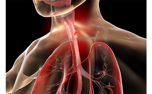
External beam radiotherapy (EBRT) is the most common form of radiation treatment offered to cancer patients. Currently, different types of EB therapy techniques are used. The goal of three-dimensional conformal RT (3-D CRT) is to deliver a full dose of irradiation to the target structure with as little radiation as possible affecting the surrounding normal tissue. Intensity-modulated RT (IMRT) is a further refinement of CRT that allows the dose within a target to be modified, so as to spare specific tissue and organs. In order to take respiratory motion of the target into account another approach is 4-D CRT, which can also be combined using intensity modulated fields. Many of the current advancements in EBRT involve techniques for tracking the motion of tumors—techniques that fall under the collective heading of image-guided radiation therapy (IGRT).
Historical Overview
Different approaches toward IGRT have been followed over the years. In the 1990s, the first electronic portal imaging devices (EPIDs) for linear accelerators (LINACs) were developed, initially with charge-coupled device (CCD) camera optics, later using liquid ion chamber technology, and now mostly based on amorphous silicon flat panels. The next step has been the introduction of room- or gantry-based kilovoltage (kV) radiograph and fluoroscopy devices, also allowing localization by means of bony structures or fiducial markers.
An initial way of obtaining 3-D information was achieved by placing a conventional computed tomography (CT) scanner in the treatment room in a known geometric relationship to the linear accelerator’s isocenter. Now, CT functionality has been integrated in the linear accelerator (LINAC) in order to eliminate the need for a separate scanner. These cone beam CT (CBCT) options are based on either an additional kV system or by using megavolt radiation from the therapy beam source. Most recent publications indicate that on-board imaging devices with a separate kV system offer the largest flexibility with regard to different modes such as radiography, fluoroscopy, and CT.
Gating/4-D Imaging
Most 3-D treatment planning systems utilize CT images based on a diagnostic CT scanner. These scanners limit how the patient can be positioned because of their relatively small opening; in order to overcome these issues, dedicated oncology scanners with a large bore have been developed. The new RT department at the Hirslanden Klinik Aarau was one of the first clinics in Europe to be equipped with a special RT scanner. Such multi-slice systems with up to 16 detector rows allow high-speed scans to be acquired. These scanners additionally offer 4-D functionality, which means the scans are obtained with co-registered respiratory signals.This technique entails the creation of multiple CT slices at each relevant table position for at least the duration of one full respiratory cycle, while simultaneously recording signals from a respiratory motion monitoring system.
Consequently, 4-D imaging is an integral part of IGRT because, as with gating, effective imaging is impossible without the 4-D imaging capability. The crucial element of the gating system is that, unlike some others, it is a non-invasive form of gating that operates via room-mounted cameras and a detector device that is placed on the patient’s torso. It records the patient’s breathing pattern during the scan resulting in a 4-D CT data set. An analysis of the data with regard to target motion during the different phases of the breathing cycle by the radiotherapist and medical physicist determines the minimum movement and leads to the therapeutic window defined by a lower and upper threshold.
On-board Imaging
Aarau was only the second European hospital to install an on-board imaging (OBI) system for IGRT. In July 2005, the RT department at the Hirslanden Klinik Aarau was equipped with one of the most advanced and sophisticated RT systems worldwide. In addition to the OBI, which is used to fine-tune patient positioning at the time of treatment, the system incorporates the realtime position management (RPM) respiratory gating system, which tracks tumor motion and turns the treatment beam on and off as the tumor moves in and out of range. By ‘gating’ the treatment beam, doctors are able to deliver a more precise dose to the tumor, and avoid more of the surrounding healthy tissues.
The OBI is a digital imaging device mounted on the treatment machine via robotically controlled arms that operate along three axes of motion. No other imaging device for RT has this range of motion.This allows the imager to be positioned optimally for the best possible view of the tumor and surrounding anatomy. The device produces high-resolution images of the tumor, and it can also track tumor motion to provide doctors with a clear indication of exactly how a tumor will move during treatment due to normal breathing and other physiological processes.
Prior to the advent of IGRT, and tools such as OBI and respiratory gating, radiation oncologists had to contend with variations in patient positioning, and with respiratory motion, by treating a larger margin of healthy tissue around the tumor. IGRT is expected to enable doctors to minimize the volume of healthy tissue exposed to the treatment beam.
In addition to such key IGRT solutions, the clinic’s radio-oncology department also implemented an entirely paperless and filmless working environment, utilizing a dedicated radio-oncology management system, offering verification and recording, as well as image management and functionality. It incorporates an additional electronic patient chart, which is totally customizable and allows a seamless data transfer, and it is linked to all the hospital and institute systems. Consequently, all patient data including images can be retrieved at every workstation.
The clinic’s goal was to combine the best possible technologies on the market for the benefit of cancer patients—it has achieved that.
Aarau has treated approximately 400 patients to date using the OBI, primarily for breast and prostate cancer, in the thorax region used in conjunction with gating. Aarau is the only clinic carrying out gating in Switzerland; it treats every breast, left or right side, and also treats every lung cancer with gating. For the latter it additionally uses the fluoroscopic pre-treatment set-up verification on the OBI. For the first time this modality allows a physician to look into the patient and to analyze the target volume movement prior to treatment. This means that the assumptions based on the 4-D CT scan information can be verified realtime with regard to possible changes in breathing pattern, i.e. due to treatment-induced tumor regression.To date, approximately 115 patients have undergone the gated therapy approach. All these patients experienced considerably fewer side effects and dosage was boosted, due to gated IGRT treatments.Without such techniques, it would have been very difficult, or even impossible, to treat left-sided breast tumors without exposing the heart to residual damage.
Currently, the clinic treats approximately 40 patients per day, 15 of them with gating. CBCT has been used for approximately 50 patients who have undergone a CBCT scan, but at the moment more experience with the image quality is needed and the possibilities of what can be obtained from it need to be explored before it is incorporated into routine clinical use. Initial problems with its stability have been solved, and the clinic has already used it for treatment planning. Comparisons between diagnostic CT scans and CBCT scans to the same patient have also been made and the relative dose distribution is approximately the same.
The fluoroscopic mode offers physicians the first chance they have ever had to really look into the patient and analyze whether the movement of the target for the gating threshold is actually correct. It allows them to decrease the margin, which obviously benefits patients, and it also reduces side effects.The clinic is treating every breast with a form of IMRT called e-compensation, which has enabled it to reduce the target dose maximum from values up to 125% to just 105%, with a minimum of 95%. Using this kind of modality, the skin reactions are decreased dramatically and the side effects are much better, bringing down the dose to the heart to zero in some cases. By using these imaging and treatment techniques, the patient experience is far better.
Future Perspectives
The next step for the clinic’s work with OBI will involve online 3-D/3-D matching.This would enable a genuine 3-D repositioning of the patient, as well as offering the best way to make use of the CBCT dataset—CBCT is the only way to gain genuine 3-D information about the anatomy.
Although limitations are currently set because the couch only allows 4º of movement, the clinic is exploring the possibility of gantry or collimator rotation, which could compensate for the problems of the 2º of motion missing in the basic therapy couch. Another approach considered is the integration of a six-axis couch, allowing a real 3-D repositioning. This solution—in a dynamic repositioning mode—would offer the possibility of realtime compensation for respiration-induced target motion. The integration of such a system is now evaluated in order to overcome the problem of treatment time prolongation due to the duty cycle (beam hold time), being dependent on the individual target volume movement.
Another goal is to achieve online re-planning, for which it is crucial to have automatic segmentation and follow the tumor shrinkage and changes in the structure.With this, an automatic re-contouring of the target structure is needed. It is currently possible to re-plan based on the CBCT slices, but is it not possible to do this within a three-minute-long process. Currently, online matching is only manual. Data can be exported into the Eclipse™ system, but this takes time. Offline planning takes 10 minutes, so it would double the treatment time, which is undesirable—if it takes this long, this type of re-planning cannot be introduced. At the moment, the clinic’s CBCT solution is integrated in a way that takes five to eight minutes, which is too long for regular use in routine clinical treatments. Anything that lengthens treatment times unduly must be avoided.
Online re-planning would involve placing the patient on the couch, carrying out a CT scan, and analyzing the scan to judge whether there is tumor shrinkage. The question of whether the field size is still correct or whether it should be increased or decreased now arises. With these new tools, all the assumptions that have been made to date could become things of the past. If a physician is treating lymphoma (which shrinks very quickly), for instance, it is questionable how the reduction is taken into account for the following sessions. The CT scan takes approximately one minute and a huge amount of data can be collected. As previously mentioned, the reconstruction can be accelerated so that it takes half the time it currently takes, although more is needed.Although this small amount of acceleration is better than no acceleration, physicians find themselves to be constantly one session behind, as they have to take the new situation into account for the following session, rather than the current session. However, this is still a major sign of progression.
Auto-segmentation is also a vision for the future— this involves an initial automatic contouring of the target volume depending on whether there is a clear border between the target and other anatomical structures. If not, there is the question of tumor response—it would make sense to integrate this for re-sizing the fields. Ideally, there would be a tool that supports this type of treatment to the target volume by allowing physicians to carry it out manually the first time, afterwards enabling them to analyze the electron density (Hounsfield units) of that target structure, look at the new CT scan, and determine whether the structure is smaller. They can then use this as the new contour structure for the target volume.
Dynamic adaptive RT (DART) is the next stage in personalized cancer care. The Hirslanden Klinik Aarau has the ability to dynamically adapt to the changing conditions in the patient to give them exactly the right kind of treatment—the right dose, in the right place, at the right time.
DART is being made possible by the technological convergence of image-guidance tools, integrated image and data management, sophisticated planning capabilities, such as auto-segmentation and image registration, and IMRT-enabled treatment delivery in a single system. ■