Recent advances have led to multifunctional nanoparticle probes for molecular and cellular imaging, nanoparticle drugs for targeted therapy and integrated nanodevices for early disease detection and screening.6-13 These developments have opened exciting opportunities for personalized oncology in which cancer detection, diagnosis, and therapy are tailored to each individual’s molecular profile, and also for predictive oncology in which genetic/molecular information is used to predict tumor development, progression, and clinical outcome.
For applications in oncology, nanotechnology is linked with biomolecular signatures or biomarkers that are correlated with tumor behavior and clinical outcome. These markers are commonly defined as mutant genes, RNA, proteins, lipids, carbohydrates, small metabolite molecules, and altered expressions of them.
For individualized therapy, biomarkers enable the characterization of patient populations and quantification of the extent to which new drugs reach their intended targets.14,15 One example is the drug trastuzumab (Herceptin®; Genentech/Roche), a monoclonal antibody designed to target amplified and over-expressed ERBB2 (also known as HER2) tyrosine kinase receptor found in only ~30% of breast cancers. In another example, the clinical response of lung cancer patients to the epidermal growth factor receptor (EGFR) tyrosine kinase inhibitor gefitinib (Iressa®; AstraZeneca) is associated with a small number of genetic mutations.16,17 Thus, a molecular diagnostic test could be used to identify patients that are most likely to respond to this drug.
In this article, the current status of cancer nanotechnology and its applications in molecular tumor imaging, diagnosis, and targeted therapy will be discussed. Together with cancer bioinformatics and biocomputing, nanotechnology is one of the most promising and enabling technologies and it should make a significant impact in clinical oncology. Quantum Dots for Cancer Molecular Profiling
A prototype nanotechnology is semiconductor quantum dots, which are tiny light-emitting particles on the nanometer scale and are under intense development as a new class of fluorescent probes for molecular imaging and medical diagnostics.6-13 In comparison with organic dyes and fluorescent proteins, quantum dots (QD) have unique optical and electronic properties such as size-tunable light emission, superior signal brightness, resistance to photobleaching, and simultaneous excitation of multiple fluorescence colors.
These ‘programmable’ properties are most promising for improving the sensitivity and the multiplexing capabilities of molecular histopathology and disease diagnosis. Recent research has led to highly bright and stable QD probes that are well-suited to profiling genetic and protein biomarkers in intact cells and clinical tissue specimens.6-13
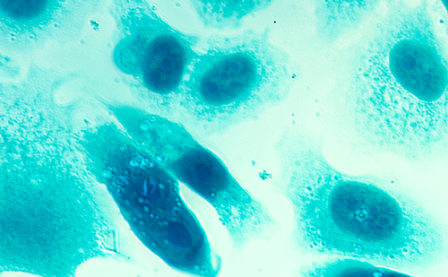
In contrast to in vivo imaging applications where the potential toxicity of quantum dots is a major concern, immunohistological studies are performed on in vitro or ex vivo clinical patient samples. The use of multicolor QD probes in immunohistochemistry (IHC) is one of the most important and clinically relevant applications in the short term.
In particular, nanoparticle probes can be used to quantify a panel of biomarkers on intact cancer cells and tissue specimens, allowing a correlation of traditional histopathology and molecular signatures for the same material.A single nanoparticle is large enough for conjugation to multiple ligands, leading to enhanced binding affinity and exquisite specificity through a ‘multivalency’ effect. These features are especially important for the analysis of cancer biomarkers that are present at low concentrations or in small numbers of cells. At present, clinical applications of QD-based IHC are still at an early stage of development. There is an urgent need for robust protocols and experimental procedures that define the key factors and steps such as QD-antibody bioconjugation, tissue specimen preparation, multicolor QD staining, image processing, and data quantification. In Vivo Tumor Imaging
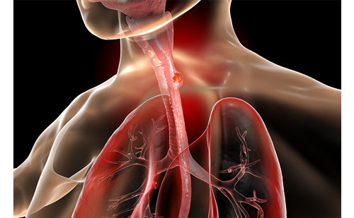
Traditional in vivo imaging probes or contrast agents include radioactive small molecules in positron emission tomography (PET) and single photo emission computed tomography (SPECT), gadolinium compounds in magnetic resonance imaging (MRI), and isotope-tagged antibodies. In comparison, bioconjugated QDs and targeted nanoparticles provide a number of unique features and capabilities that could significantly improve the sensitivity and specificity of disease-imaging and diagnosis.11-13
Firstly, the size-dependent optical and electronic properties of QDs can be tuned continuously by changing the particle size.This ‘size effect’ permits the use of a broad range of nanoparticles for simultaneous detection of multiple cancer biomarkers. Secondly, nanoparticles have more surface area to accommodate a large number or different types of functional groups that can be linked with both diagnostic and therapeutic agents. This opens the opportunity to design multifunctional ‘smart’ nanoparticles for multimodality imaging, as well as for integrated imaging and therapy.
Thirdly, extensive research has shown that nanoparticles in the size range of 10–100nm are accumulated preferentially at tumor sites through an effect called enhanced permeability and retention (EPR).18-21 This effect is believed to arise from two factors:
• growing tumors produce vascular endothelial growth factors (VEGF) that promotes angiogenesis; and
• many tumors lack an effective lymphatic drainage system, which leads to subsequent macromolecule or nanoparticle accumulation.
This latter causes tumor-associated neovasculatures to be highly permeable, allowing the leakage of circulating macromolecules and nanoparticles into the tumor tissue. These novel properties provide exciting opportunities for developing new and advanced nanoparticle probes for biomedical imaging, especially for tumor imaging and targeting. In recent work, Gao et al. developed multifunctional probes for simultaneous targeting and imaging of tumors in live animals.22 This class of QD conjugates contains an amphiphilic triblock copolymer that provides protection against aggregation and degradation in vivo and in functional groups for targeting ligands for tumor antigen recognition.
The addition of multiple polyethylene glycol (PEG) molecules provides improved biocompatibility and blood retention time. The use of an ABC triblock copolymer has solved the problems of particle aggregation and fluorescence loss previously encountered for QDs stored in physiological buffer or injected into live animals. Detailed studies were reported on the in vivo behaviors of QD probes, including biodistribution, non-specific uptake, cellular toxicity, and pharmacokinetics.
For active tumor targeting, antibody-conjugated QDs were used to recognize a prostate-specific membrane antigen (PSMA), a cell surface marker for both prostate epithelial cells and neovascular endothelial cells.23 PSMA has been selected as an attractive target for both imaging and therapeutic intervention of prostate cancer.Accumulation and retention of PSMA antibody at the site of tumor growth is the basis of radioimmunoscintigraphic scanning, e.g. ProstaScint scanning, and targeted therapy for human prostate cancer metastasis.24
Nanoparticle Therapeutics
The use of nanoparticles for drug delivery and targeting is one of the most exciting and clinically important areas in cancer nanotechnology. Nanotechnology is used to improve the efficacy and toxicity profiles of chemotherapeutic agents, because these agents can be encapsulated, covalently attached, or adsorbed onto nanoparticles.
It is also being used to overcome drug solubility problems, because more than 40% of active substances being identified through combinatorial screening programs are not fully soluble in water.
In particular, nanoparticle probes can be used to quantify a panel of biomarkers on intact cancer cells and tissue specimens, allowing a correlation of traditional histopathology and molecular signatures for the same material.A single nanoparticle is large enough for conjugation to multiple ligands, leading to enhanced binding affinity and exquisite specificity through a ‘multivalency’ effect. These features are especially important for the analysis of cancer biomarkers that are present at low concentrations or in small numbers of cells. At present, clinical applications of QD-based IHC are still at an early stage of development. There is an urgent need for robust protocols and experimental procedures that define the key factors and steps such as QD-antibody bioconjugation, tissue specimen preparation, multicolor QD staining, image processing, and data quantification.In Vivo Tumor Imaging
Traditional in vivo imaging probes or contrast agents include radioactive small molecules in positron emission tomography (PET) and single photo emission computed tomography (SPECT), gadolinium compounds in magnetic resonance imaging (MRI), and isotope-tagged antibodies. In comparison, bioconjugated QDs and targeted nanoparticles provide a number of unique features and capabilities that could significantly improve the sensitivity and specificity of disease-imaging and diagnosis.11-13
Firstly, the size-dependent optical and electronic properties of QDs can be tuned continuously by changing the particle size.This ‘size effect’ permits the use of a broad range of nanoparticles for simultaneous detection of multiple cancer biomarkers. Secondly, nanoparticles have more surface area to accommodate a large number or different types of functional groups that can be linked with both diagnostic and therapeutic agents. This opens the opportunity to design multifunctional ‘smart’ nanoparticles for multimodality imaging, as well as for integrated imaging and therapy.
Thirdly, extensive research has shown that nanoparticles in the size range of 10–100nm are accumulated preferentially at tumor sites through an effect called enhanced permeability and retention (EPR).18-21 This effect is believed to arise from two factors:
• growing tumors produce vascular endothelial growth factors (VEGF) that promotes angiogenesis; and
• many tumors lack an effective lymphatic drainage system, which leads to subsequent macromolecule or nanoparticle accumulation.
This latter causes tumor-associated neovasculatures to be highly permeable, allowing the leakage of circulating macromolecules and nanoparticles into the tumor tissue. These novel properties provide exciting opportunities for developing new and advanced nanoparticle probes for biomedical imaging, especially for tumor imaging and targeting.
In recent work, Gao et al. developed multifunctional probes for simultaneous targeting and imaging of tumors in live animals.22 This class of QD conjugates contains an amphiphilic triblock copolymer that provides protection against aggregation and degradation in vivo and in functional groups for targeting ligands for tumor antigen recognition.
The addition of multiple polyethylene glycol (PEG) molecules provides improved biocompatibility and blood retention time. The use of an ABC triblock copolymer has solved the problems of particle aggregation and fluorescence loss previously encountered for QDs stored in physiological buffer or injected into live animals. Detailed studies were reported on the in vivo behaviors of QD probes, including biodistribution, non-specific uptake, cellular toxicity, and pharmacokinetics.
For active tumor targeting, antibody-conjugated QDs were used to recognize a prostate-specific membrane antigen (PSMA), a cell surface marker for both prostate epithelial cells and neovascular endothelial cells.23 PSMA has been selected as an attractive target for both imaging and therapeutic intervention of prostate cancer.Accumulation and retention of PSMA antibody at the site of tumor growth is the basis of radioimmunoscintigraphic scanning, e.g. ProstaScint scanning, and targeted therapy for human prostate cancer metastasis.24Nanoparticle Therapeutics
The use of nanoparticles for drug delivery and targeting is one of the most exciting and clinically important areas in cancer nanotechnology. Nanotechnology is used to improve the efficacy and toxicity profiles of chemotherapeutic agents, because these agents can be encapsulated, covalently attached, or adsorbed onto nanoparticles.
It is also being used to overcome drug solubility problems, because more than 40% of active substances being identified through combinatorial screening programs are not fully soluble in water.
Conventional and most current formulations of such drugs are frequently plagued with problems such as poor and inconsistent bioavailability. For example, paclitaxel (Taxol™) is one of the most widely used anticancer drugs in the clinic. It is a microtubulestabilizing agent that promotes tubulin polymerization, thus disrupting cell division and leading to cell death.25,26 It displays neoplastic activity against primary epithelial ovarian carcinoma, breast, colon, and lung cancers. Owing to its inadequate water-soluability, the formulation available currently is Chremophor EL (polyethoxylated castor oil) and ethanol.27
One of the most significant advances has been the development and Food and Drug Administration (FDA) approval of albumin-conjugated paclitaxel (Taxol™), a two-component ‘binary’ nanoparticle (Abraxane™) for treatment of taxane-refractory metastatic breast cancer.28,29 This nanoparticle formulation is shown to be effective in circumventing side effects of the highly toxic Chremophor EL such as hypersensitivity reactions, nephrotoxicity and neurotoxicity.27,30
Recent work by Nie and colleagues (S M Nie,G Kim, and D Shin, unpublished data) has developed a more sophisticated ‘ternary’ nanoparticle structure by linking both a hydrophobic cancer drug (Taxol) and a tumor-targeting ligand to a hydrophilic and biodegradable polymer.
This chemical conjugation produces a graft amphiphilic polymer that is able to self-assemble into nanostructures. In vitro cellular toxicity studies show that this new class of nanoparticle drug exhibits only similar to or less toxic than free Taxol, but in vivo introduction to animal models bearing human tumors affected a 10-fold efficacy increase in shrinking and arresting tumors when compared to free Taxol.Future Directions
Nanotechnology has become an enabling technology for personalized oncology in which cancer detection, diagnosis, and therapy are tailored to each individual’s tumor molecular profile, and also for predictive oncology in which genetic/molecular markers are used to predict disease development, progression, and clinical outcomes.
In recognition of its potential impact in cancer research, the US National Cancer Institute (NCI) has recently funded eight national Centers of Cancer Nanotechnology Excellence (CCNE) (www.nano. cancer.gov). Looking to the future, there are a number of research themes or directions that are particularly promising but require concerted effort for success. These are outlined below.
Design and Development of Nanoparticles with Dual, Triple, and Multiple Functions
The first direction area is the design and development of nanoparticles with dual, triple, and multiple functions. For cancer and other medical applications, important functions include imaging—single or dualmodality; therapy—single drugs or combinations of two or more drugs; and targeting—one or more ligands. With each added function, nanoparticles could be designed to have novel properties and applications. For example, binary nanoparticles with two functions could be developed for molecular imaging, targeted therapy, or for simultaneous imaging and therapy without targeting. Bioconjugated QD with both targeting and imaging functions will be used for targeted tumor imaging and molecular profiling applications.
Conversely, ternary nanoparticles with three functions could be designed for simultaneous imaging and therapy with targeting, targeted dual-modality imaging, or for targeted dual-drug therapy. Quaternary nanoparticles with four functions can be conceptualized in the future to have the abilities of tumor targeting, dual-drug therapy, and imaging.
Nanoparticle Molecular Profiling for Clinical Oncology
The second direction is nanoparticle molecular profiling (nanotyping) for clinical oncology.That is, the use of bioconjugated nanoparticle probes to predict cancer behavior, clinical outcome, treatment response, and individualize therapy. This should start with retrospective studies of archived specimens because the patient outcome is already known for these specimens. The two key hypotheses to be tested are, firstly, that nanotyping a panel of tumor markers will allow more accurate correlations than single tumor markers; and secondly, that the combination of nanotyping tumor gene expression and host stroma are both important in defining the aggressive phenotypes of cancer, as well as determining the response of early stage disease to treatment—chemotherapy, radiation, or surgery.
Nanoparticle Distribution, Excretion, Metabolism, and Pharmacodynamics
The third important direction is to study nanoparticle distribution, excretion, metabolism, and pharmacodynamics in in vivo animal models. These investigations will be very important in the development of nanoparticles for clinical applications in cancer imaging or therapy.
Acknowledgements
We are grateful to Ms Gloria Kim, Dr Leland Chung,Dr Ruth O Regan and Dr Dong Shin for insightful discussions. This research was supported by NIH grants (P20 GM072069, R01 CA108468-01, and U54CA119338) and the Georgia Cancer Coalition Distinguished Cancer Scholars Program (to SN and to MDW).