Esophageal cancer (EC) is a major global health concern, ranking as the 11th most commonly diagnosed cancer and the seventh leading cause of cancer-related deaths worldwide, with an estimated 494,320 new cases and 441,994 deaths in 2024.1 Esophageal squamous cell carcinoma (ESCC) remains the predominant subtype globally (84% of new cases), while esophageal adenocarcinoma (EAC) now accounts for the majority of cases in high-income Western countries. ESCC predominates in Eastern Asia and parts of Africa, whereas EAC has risen sharply in the USA, Australia, Canada and several countries in Western and Northern Europe.2 These differences in geography and risk factor exposure suggest that ESCC and EAC are not simply histologic variants of one disease but biologically distinct malignancies shaped by different environmental exposures, host factors and local tissue microenvironments.
Despite improvements in multimodality therapy, outcomes for EC remain poor. According to U.S. Surveillance, Epidemiology and End Results program data, the overall 5-year relative survival rate for EC is 22.2%, while the 5-year survival rate is 5.3% for distant disease. The near convergence of global incidence and mortality underscores how frequently EC remains lethal. These figures underscore the need for earlier detection, better risk stratification and a more precise understanding of underlying pathophysiology.3
The two histologies also differ in anatomy and pathophysiology. ESCC arises in the upper or mid esophagus and, similar to other aerodigestive malignancies, is linked to tobacco use, alcohol consumption, dietary carcinogens and chronic mucosal injury.4 EAC develops in the distal esophagus or gastroesophageal junction (GEJ) due to chronic gastroesophageal reflux disease, associated with central obesity, Barrett’s esophagus (BE) and male sex. BE is the only established precursor lesion for EAC and represents the metaplastic consequence of long-standing reflux injury. EAC is therefore a particularly useful model for microbiome research because its pre-malignant progression sequence is endoscopically visible and accessible to biopsy. In certain patients, reflux esophagitis progresses to Barrett’s metaplasia, followed by dysplasia and ultimately invasive adenocarcinoma. This stepwise progression provides an opportunity to assess whether microbial changes merely reflect epithelial transformation or actively participate in it. These differences between the two histologies matter clinically and therapeutically because the mechanisms driving initiation, evolution and treatment resistance are unlikely to be identical across subtypes, and any microbiome-based model of esophageal carcinogenesis must be interpreted within a histology-specific framework.5 The EAC microbiome literature is less mature than that for ESCC but is most comprehensive around the BE–dysplasia–EAC sequence, where tissue, brushing, saliva and multi-omics studies now support an association between microbial community shifts and neoplastic progression.6 The microbiome has emerged as a promising line of investigation in this space because it is dynamic, measurable, mechanistically active and potentially modifiable. Microbial signatures could help refine cancer risk assessment, identify biologically relevant disease states and inform prevention or therapeutic strategies, as summarized in the graphical abstract (Figure 1).
Figure 1: Graphical abstract
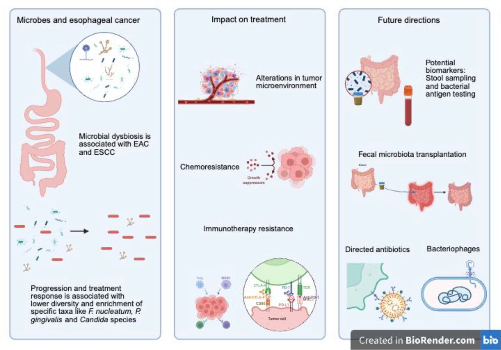
Figure created in BioRender.com (Toronto, ON, Canada)
EAC = esophageal adenocarcinoma; ESCC = esophageal squamous cell carcinoma; F. nucleatum = Fusobacterium nucleatum; = P. gingivalis = Porphyromonas gingivalis; CTLA-4 = cytotoxic T-lymphocyte-associated protein 4, PD-1 = programmed cell death protein 1; PD-L1 = programmed cell death-ligand 1, TCR = T-cell receptor.
Methods
For this narrative article, a literature search was performed using PubMed and ClinicalTrials.gov. The search strategy included the keywords ‘microbiome,’ ‘mycobiome,’ ‘esophageal squamous cell carcinoma’ or ‘esophageal adenocarcinoma,’ yielding 334 results. Studies published between January 1, 1997, and May 15, 2026, were evaluated. Studies were independently analyzed by two reviewers, and disagreements on inclusion were resolved through discussion. Peer-reviewed, English-language studies that included adult patients (≥18 years) with either EAC or ESCC were considered. Sixteen studies evaluating microbiome or mycobiome profiles using human tissue or biospecimens from adult patients were included in the primary narrative synthesis (Tables 1 and 2).7–22 Meta-analyses, systematic reviews, preclinical studies and relevant conference abstracts were additionally incorporated to provide context and further explore potential mechanisms of disease. Findings were synthesized and presented descriptively.
Table 1: Microbiome impact on response to therapy in esophageal squamous cell carcinoma and esophageal adenocarcinoma7–18
| Study | Participants | Therapy | Sampling | Microbial identification | Results |
| Chemotherapy response | |||||
| Ohsawa et al., 2026 Japan 8 | ESCC (n=74) | Cisplatin/5-FU, nedaplatin/5-FU or docetaxel/cisplatin/5-FU ± radiation followed by esophagectomy | Oral tongue coating | 16S rRNA (V1–V2) | Alpha diversity correlated with pathologic response (OR, 11.46; 95% CI, 2.49–52.64; p=0.002), RFS (HR, 3.89; 95% CI, 1.57–9.65; p=0.005) and OS (p=0.044) Poor responders had higher abundance of Streptococcaceae and Corynebacteriaceae |
| Wu et al., 2024 China 9 | ESCC (n=180); 48 paired tissue samples (pre- and post-neoadjuvant chemoradiotherapy) from 24 patients | Platinum combined with paclitaxel chemotherapy + radiotherapy | Esophagectomy tissue samples | 16S rRNA (V3–V4) | A microbiota-based risk prediction model on the genera of TM7x, Sphingobacterium and Prevotella was predictive of disease-free survival (p=0.00074 in internal validation set and p=0.0017 in independent validation set) and OS (p=0.00023 in internal validation set and p=0.11 in independent validation set) |
| Li et al., 2024 China 10 | ESCC (n =31) | Paclitaxel and cisplatin | Fecal samples pre- and post-treatment | 16S rRNA (V4) | Bacteroides plebeius was more abundant in baseline samples from responders than non-responders (p=0.045) |
| Yamamura et al., 2019 Japan 11 | ESCC (n=551); 101 of those patients had received neoadjuvant chemotherapy | Training cohort: surgical resection alone (n=394) Validation cohort: neoadjuvant chemoradiation (n=41), neoadjuvant chemotherapy (n=101) with DCF regimen, neoadjuvant chemotherapy unspecified regimen (n=15) | Esophagectomy surgical samples | qPCR | Higher abundance of Fusobacterium nucleatum was associated with poor RFS in both cohorts (training cohort HR=1.61; p=0.03, validation cohort HR=1.96; p=0.004) Higher abundance of F. nucleatum was associated with worse chemotherapeutic response for all evaluation methods: RECIST (p=0.04), SUVmax change in PET/CT (p=0.0004) and TRG (p=0.003) |
| Liu et al., 2021 Japan 12 | ESCC (n=120 post-treatment samples, n=30 pre-treatment samples) | Neoadjuvant cisplatin and 5-FU ± docetaxel followed by surgical resection | Esophagectomy surgical samples for post-treatment; endoscopic biopsy for pre-treatment | qPCR | Fewer responders to chemotherapy in patients with high F. nucleatum burden compared with low burden (37.0% (10/27) versus 88.2% (60/68) in the low F. nucleatum group; p<0.001) |
| Yoshikawa et al., 2025 Japan 13 | ESCC (n=305) | Neoadjuvant CF or DCF or CRT with CF or DCF | Serum IgG-Fn | IgG-Fn | Positive group had significantly lower rates of OS and CSS as compared with patients who tested negative (OS; p=0.01, CSS; p=0.02) IgG-Fn-positive group showed a higher rate of pathologic progression and inadequate response to neoadjuvant treatment compared with the IgG-Fn-negative group (OR, 0.48; 95% CI, 0.24–0.92; p=0.04) |
| Immunotherapy response | |||||
| Xu et al., 2022 China 14 | ESCC (n=46) | Camrelizumab combined + carboplatin and albumin paclitaxel prior to surgical resection | Fecal samples | 16S rRNA (V3–V4) | Alpha diversity declined after neoadjuvant chemoimmunotherapy Patients exhibiting pCR had different species and proportions of bacteria in pre-treatment samples compared with non-responders, although alpha diversity was not significantly different |
| Zhou et al., 2026 China 15 | ESCC (n=43); 122 fecal samples | Neoadjuvant cisplatin + paclitaxel; immunotherapy included pembrolizumab, sintilimab, toripalimab, camrelizumab or adrebelimab | Fecal samples | 16S rRNA | Alpha diversity was higher in the responder group (p<0.05) Ligilactobacillus salivarius was enriched in non-responders (p<0.05) |
| Li et al., 2023 China 7 | ESCC (n=98) | PD-1 inhibitor | Serum IgG-Fn, surgical resection samples | IgG-Fn, qPCR | Patients with progressive disease had higher serum anti-Fn IgG levels and Fn DNA compared with partial responders (p=0.003) and those with stable disease (p=0.0045) |
| Wu et al., 2023 China 16 | ESCC; received neoadjuvant chemoimmunotherapy (n=25); received surgery without neoadjuvant therapy (n=15) | Neoadjuvant chemoimmunotherapy (exact regimen unspecified), PD-1 inhibitor | Surgical resection samples, fecal samples | qRT-PCR | Abundance of Streptococcus positively correlated with disease-free survival (p=0.034) and GrzB+ and CD8+ T-cell infiltration in tumor tissues |
| Van den Ende et al., 2024 Netherlands 17 | n=172 with resectable esophageal carcinoma (EAC + ESCC); 132 received CROSS alone, 40 received CROSS + anti-PD-L1. Mixed histology; CRT-only arm included ESCC; ICI arm was EAC/GEJ-dominant | Neoadjuvant CROSS (carboplatin + paclitaxel + 41.4 Gy) ± anti-PD-L1 (atezolizumab), followed by esophagectomy | Fecal samples at baseline, week 3 and pre-surgery; duodenal biopsies in the ICI arm | 16S rRNA (V3–V4) | Distinct longitudinal fecal microbiome trajectories in responders versus non-responders; specific Lachnospiraceae taxa associated with pathologic response; intratumoral microbiota also shifted during treatment |
| Shaikh et al., 2024 USA 18 | Pilot cohort of patients with stage II–III resectable esophageal or GEJ carcinoma (EAC/GEJ) | Neoadjuvant ICI (anti-PD-1/PD-L1) combined with chemoradiation, prior to surgical resection | Paired fecal and tumor samples before and during treatment; concurrent fecal metabolomics | 16S rRNA + untargeted fecal metabolomics | Patients achieving pCR showed enrichment of sphingolipid and primary bile-acid pathways with elevated Roseburia inulinivorans, Ruminococcus callidus and Fusicatenibacter saccharivorans; non-pCR patients lacked these SCFA-producing taxa |
CF = cisplatin/5-fluorouracil; CI = confidence interval; CROSS = ChemoRadiotherapy for Oesophageal cancer followed by Surgery Study, refers to regimen of chemoradiotherapy with carboplatin and paclitaxel; CRT = chemoradiotherapy; CSS = cancer-specific survival; DCF = docetaxel, cisplatin and 5-fluorouracil; EAC = esophageal adenocarcinoma; ESCC = esophageal squamous cell carcinoma; F. nucleatum = Fusobacterium nucleatum; 5-FU = 5-fluorouracil; GEJ = gastroesophageal junction; GrzB+ = granzyme B serine protease; HR = hazard ratio; ICI = immune checkpoint inhibitor; IgG-Fn = IgG antibody titer level against Fusobacterium nucleatum; OR = odds ratio; OS = overall survival; pCR = pathologic complete response; PD-1 = programmed cell death protein 1; PD-L1 = programmed death-ligand 1; PET/CT = positron emission tomography–computed tomography; qPCR = quantitative polymerase chain reaction; qRT-PCR = quantitative reverse transcription polymerase chain reaction; RECIST = Response Evaluation Criteria in Solid Tumors; RFS = recurrence-free survival; SCFA = short-chain fatty acid;16S rRNA = bacterial ribosomal RNA; SUVmax = standardized uptake value; TRG = tumor regression grade.
Table 2: Mycobiome in esophageal cancer19–22
| Study, country | Participants | Sampling | Microbial identification | Results |
| Yuan et al., 2025 China 19 | HC (n=13), ESCC (n=12), ESIN (n=10) | Endoscopic biopsies (HC), biopsies of ESCC or ESIN lesions and adjacent tissue obtained in patients with ESCC and ESIN | ITS sequencing and 16S rRNA amplicon sequencing (V3–V4) | Alternaria, Neosetophoma and Monascus were more prevalent in the ESCC group, although not significantly different between samples with disease and adjacent tissue Aspergillus was dominant in the ESIN group and was enriched in samples with disease compared with adjacent tissue Coniochaeta and Sclerostagonospora were more prevalent in the HC group Tumor tissue had lower alpha diversity compared with adjacent tissue (p=0.001) |
| Rao et al., 2023 China 20 | ESCC (n=66), no HC | Paired tumor and tumor-adjacent surgical samples were obtained from patients with ESCC undergoing esophagectomy | ITS sequencing and 16S rRNA amplicon sequencing (V3–V4), qRT-PCR | Rhodotorula toruloides, Malassezia dermatis, Hanseniaspora lachancei and Spegazzinia tessarthra were more abundant in tumor samples Preussia persica, Fusarium solani, Nigrospora oryzae, Acremonium furcatum, Golovinomyces artemisiae and Tausonia pullulans were more abundant in tumor-adjacent samples Expression of MET proto-oncogene, receptor tyrosine kinase (MET) was positively correlated with S. tessarthra |
| Liang et al., 2025 China 21 | ESCC (n=68), HC (n=19) | Fecal samples | ITS sequencing | Baseline samples in ESCC showed reduced alpha diversity (p<0.001), with enrichment of pathogenic fungi Rhodotorula minuta, Actinomucor elegans and Candida zeylanoides Responders to neoadjuvant immunotherapy showed higher fungal diversity and enrichment of beneficial strains (Candida boidinii, Meyerozyma guilliermondii and Trichosporon dermatis), correlating with elevated cytotoxic markers Non-responders to immunotherapy showed enrichment of immunosuppressive fungi (Candida albicans, Candida parapsilosis, Candida glabrata and Saccharomyces), correlating with Th2 skewing and increased regulatory cytokines |
| Dohlman et al., USA 22 | Pan-cancer mycobiome reanalysis; EC n=142 primary tumors (mixed EAC + ESCC, not separated) | Primary tumor tissue (TCGA WGS reads) | PathSeq pipeline on TCGA WGS; prevalence-based decontamination; external ITS validation; culture-dependent confirmation | Esophageal tissue had lower fungal DNA abundance than colon/stomach/head-neck. GI tumors classifiable as Candida-dominant (Ca-type) or Saccharomyces-dominant (Sa-type). Multiple Candida species (C. albicans, C. tropicalis, C. glabrata) enriched in tumors versus adjacent normal; tumor-associated Candida DNA predictive of decreased survival across GI sites |
EAC = esophageal adenocarcinoma; EC = esophageal cancer; ESCC = esophageal squamous cell carcinoma; ESIN = esophageal squamous intraepithelial neoplasia; GI = gastrointestinal; HC = healthy controls; ITS = internal transcribed spacer; MET = mesenchymal–epithelial transition proto-oncogene, receptor tyrosine kinase; qRT-PCR = quantitative reverse transcription polymerase chain reaction; 16S rRNA = bacterial ribosomal RNA; TCGA = The Cancer Genome Atlas; Th2 = subset of T helper cell involved in antibody-mediated immunity; WGS = whole-genome sequencing.
Microbiome and esophageal adenocarcinoma
Dysbiosis in esophageal metaplasia and dysplasia
The traditional view of the esophagus as a nearly sterile conduit was replaced by a model in which the normal esophagus harbors a reproducible microbial community resembling the oral microbiome but with its own mucosa-associated features. Early bacterial ribosomal RNA (16S rRNA) gene-based studies identified a ‘type I’ esophageal microbiome dominated by gram-positive organisms, particularly Streptococcus, in histologically normal tissue. In contrast, reflux esophagitis and BE were more often associated with a ‘type II’ microbiome enriched for gram-negative anaerobes and microaerophiles. This conceptual shift is important because it links microbial composition to disease state rather than simply to luminal contamination. It also provided one of the earliest frameworks for understanding how microbial populations might track the reflux–metaplasia axis that precedes EAC.23
Subsequent studies examining the BE–EAC sequence have supported the presence of dysbiosis, although the specific taxa associated with progression vary. Cross-sectional studies sampling controls, BE, dysplasia and EAC have shown that the microbiome changes as BE/pre-neoplasia advances, with reduced diversity in EAC compared with the Firmicutes–enriched pattern in healthy tissues. Other cohorts identified significant compositional differences among control mucosa, BE, dysplastic BE and adenocarcinoma, reinforcing the idea that microbial composition changes during epithelial transformation rather than only at the endpoint of invasive cancer.6
Although no single taxonomic signature is replicated across every cohort, several recurrent themes exist related in the progression from BE to EAC. Relative loss of Streptococcus and other gram-positive commensals and gain of gram-negative or oropharyngeal-associated taxa occur. In one cohort, Streptococcus and Granulicatella were less abundant in EAC mucosa than in BE mucosa, whereas other studies reported enrichment of taxa such as Veillonella, members of the Enterobacteriaceae, Akkermansia and Lactobacillus fermentum in EAC samples. The overall process reflects the development of EAC in the setting of a community-level departure from the Streptococcus-dominant pattern seen in the healthy distal esophagus.24–26
A multi-omics study by Deshpande et al. showed that the progression of BE is associated not only with community-level shifts but also with strain-level microbial signatures that are linked to host transcriptional changes. Enrichment of Campylobacter species was a progression-relevant signal that demonstrated that the esophageal microbiome occupies an ecological niche that is independent of the oropharynx.27
Potential mechanisms for dysbiosis-mediated carcinogenesis
The current mechanistic hypothesis linking dysbiosis to EAC centers on the chronic activation of innate immunity. The BE-related shift from a gram-positive, Streptococcus-dominant community toward gram-negative organisms may be due to increased local exposure to bacterial lipopolysaccharide (LPS). Translational research focuses on the LPS–toll-like receptor 4 (TLR4)–nuclear factor κB (NF-κB axis). Activation of TLR4 in BE is linked to NF-κB signaling activation and interleukin-8 (IL-8) induction. This provides a plausible route by which dysbiosis could sustain low-grade inflammation, epithelial injury and pro-neoplastic signaling over time. In this model, the microbiome is not just a biomarker of reflux injury but a biologically active amplifier.28
Campylobacter concisus is a candidate organism for activating this process. It is present in BE and is proposed as a progression-relevant organism. Mechanistic studies demonstrate a functional role rather than incidental colonization. Exposure of BE cell lines to C. concisus alters the expression of IL-18, tumor necrosis factor alpha (TNF-α) and p53 and upregulates programmed death-ligand 1 messenger RNA (PD-L1 mRNA), following C. concisus exposure.29 This suggests that specific microbes influence inflammatory and immune-regulatory programs relevant to carcinogenesis.
A second mechanism involves the interaction between reflux chemistry, microbial metabolism and host epithelial signaling. Exposure to refluxed bile acids is a known cause of BE; however, newer studies suggest a causative interaction between bile acids and the microbiome. Increased refluxed bile acids correlate with BE-associated microbial configurations and host gene expression programs potentially relevant to EAC development. Complementary experimental work showed that microbiota-metabolized bile acids accelerate EAC through a diet–microbiome–metabolome axis.30 These findings are still early and require further validation, but they point toward functional microbial metabolites as potential mediators of risk.
These findings support a working model in which EAC-associated dysbiosis contributes to carcinogenesis through three overlapping processes: the maintenance of chronic mucosal inflammation, modulation of epithelial stress and immune signaling and alteration of metabolite exposure within the distal esophageal niche.28 Although most evidence remains associative, findings thus far suggest that microbial shifts may both influence and reflect carcinogenic change (Figure 2).
Figure 2: Impact of microbiome on esophageal dysplasia
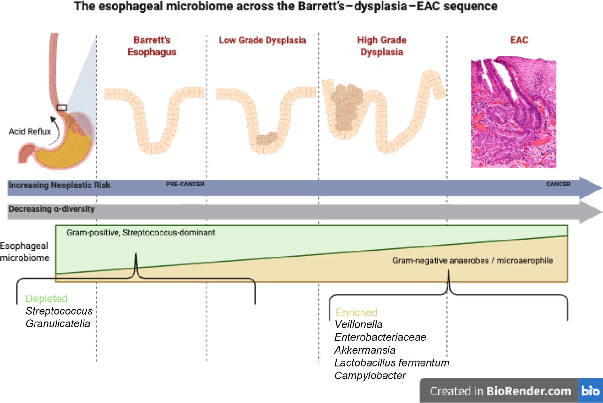
Figure created in BioRender.com (Toronto, ON, Canada)
EAC = esophageal adenocarcinoma.
There are several limitations to these proposed mechanisms. First, most studies are cross-sectional and based on small cohorts. Second, low-biomass sampling creates substantial susceptibility to contamination, batch effects and platform-dependent differences. Third, studies vary widely in source material, including saliva, oral swabs, esophageal brushings, mucosal biopsies, resection specimens and stool; one recent study showed that tissue source itself can influence the observed esophageal flora.26 Fourth, treatment and exposure confounders, such as proton pump inhibitors, antibiotics, reflux severity and diet, are difficult to standardize and may alter the very microbial signatures under study.31
These limitations help explain why the EAC literature often converges at the level of broad ecological change but diverges at the level of individual taxa. For the field to move forward, longitudinal studies that start in non-dysplastic BE, use standardized sampling and contamination controls, incorporate metabolomic or transcriptomic correlates and follow patients through clinically meaningful endpoints will be essential. Until then, the microbiome should be considered a biologically credible and clinically promising component of EAC pathogenesis, but not yet a validated stand-alone biomarker or therapeutic target.
Microbiome and esophageal squamous cell carcinoma
Intestinal dysbiosis in esophageal squamous cell carcinoma
There is a comparatively more robust body of literature on microbiome alterations and dysfunction in ESCC. Similar to BE and EAC, there is histological progression in ESCC starting with dysplasia. Fecal samples demonstrate dysbiosis of the intestinal microbiome in patients with ESCC compared with healthy controls, as well as common patterns of bacterial abundance across cohorts. Fusobacterium, Streptococcus and Bacteroides are typically more prevalent.32–40 In contrast, taxa that have been associated with gut homeostasis are often depleted, such as Prevotella, Lachnospira, Butyricicoccus and Faecalibacterium.33,35–37,40,41
There are notable trends where patients with ESCC tend to have lower levels of bacteria that produce short–chain fatty acids (SCFAs), including butyrate.36,37 There is a well-established link between butyrate depletion and gut barrier disruption via decreased expression of occludin and claudin-1, destabilization of hypoxia-inducible factor 1 and promotion of IL-22.42 This yields a plausible mechanism for increased mucosal permeability, inflammation and bacterial translocation, although it has not been directly validated in ESCC. There is also an enrichment of LPS-producing bacteria in ESCC.36,43 Notably, serum levels of LPS have been found to be elevated in patients with ESCC.44 As a natural agonist of TLR4, LPS may activate pro-inflammatory signaling cascades (TNF-α, transforming growth factor-beta [TGF-β] and inhibition of IL-10) and cell proliferation (NF-kB, extracellular signal-regulated kinase and p38 mitogen-activated protein kinase).45
Dysregulated biosynthetic pathways may also contribute. Reduced levels of L-aspartate and pantothenic acid, with subsequent downregulation of the pantothenate and coenzyme A biosynthesis pathway, correlate with an increased abundance of Enterobacteriaceae and Lactobacillus.34 One limitation of fecal sampling is its indirect representation of the esophageal tumor microenvironment, as it is more reflective of the intestinal microbiome than the microbiome within or adjacent to tumor tissue. However, there are clear practical advantages of fecal sampling, including its non-invasive nature and feasibility of longitudinal sampling, making it an important target for further research.
Esophageal dysbiosis in esophageal squamous cell carcinoma
In ESCC, the pathogenic bacteria of the genus Fusobacterium are extensively studied. Fusobacterium nucleatum (Fn), a gram-negative anaerobic bacterium commonly located in the oropharynx, colonizes the entire gastrointestinal tract, including the esophagus.46,47 Multiple studies consistently demonstrate higher levels of F. nucleatum in ESCC compared with healthy esophageal mucosa.48–53 F. nucleatum is implicated in ESCC progression, with its abundance correlating with the progression of clinical stage and poorer prognosis.50,52,54–56
Given the high association of F. nucleatum with the pathogenesis and progression of ESCC, multiple mechanisms are proposed to explain carcinogenesis. F. nucleatum is a facultative intracellular bacterium that can invade ESCC cells.7,56 Methyltransferase-like protein 3 transcription is upregulated during intracellular infection, resulting in increased methylation and stabilization of the mRNA for oncogene c-Myc, a transcription factor, which may contribute to ESCC metastasis.57 Intracellular F. nucleatum also activates the immune receptor nucleotide-binding oligomerization domain-containing protein 1 (NOD1), promoting the pro-inflammatory NOD1/receptor-interacting protein kinase 2 (RIPK2)/NF-κB pathway, thus providing a plausible link to uncontrolled cell proliferation, resistance to apoptosis and induction of the epithelial–mesenchymal transition (EMT), thereby enabling metastasis.58,59 It also upregulates IL-32/proteinase 3, subsequently inducing the highly oncogenic phosphoinositide 3-kinase (PI3K)/protein kinase B (Akt) and aryl hydrocarbon receptor (AHR)/cytochrome P450 monooxygenase (CYP1A1)/Akt signaling pathways.55,60
A similar pattern emerges with other oral bacteria. P. gingivalis, which is implicated in periodontal disease, is also associated with ESCC.61 Similar to F. nucleatum, it is enriched in tumor tissue and correlates with disease progression and poor prognosis.62–64 Numerous potential mechanisms for this interaction are known, including activation of TGF-β/Smad signaling, enrichment of the PI3K/Akt pathway, inhibition of apoptosis via decreased purinergic receptor 7 (P2X7), promotion of the EMT through IL-6 activation, resistance to apoptosis dependent on fimbriae and signal transducer and activator of transcription 3 and TLR4/myeloid differentiation primary response 88 (MYD88)/c-Jun N-terminal kinase signaling via LPS, in addition to others.64–74 Although F. nucleatum and P. gingivalis are the most extensively characterized, other taxa may also be associated with ESCC, including Fusobacterium periodonticum, streptococcal species, Prevotella and Campylobacter.75–80 Although their role mechanistically is less defined, recurrent identification of these taxa suggests a broader microbial network that contributes to tumorigenesis and progression.
When considering the influence of the local esophageal microbiome on host metabolic pathways, specifically aldehyde metabolism, a mixed picture appears. Acetaldehyde, a byproduct of ethanol breakdown, is an established carcinogen and driver of ESCC development. Deficiency of the enzyme responsible for elimination of acetaldehyde, aldehyde dehydrogenase 2, increases the risk of ESCC in individuals who consume alcohol.81 The local effects of acetaldehyde, including DNA damage and increased oxidative stress in tissues exposed to alcohol, have been proposed to mediate carcinogenesis of upper gastrointestinal tract cancers.82 Several common oropharyngeal bacteria can produce significant quantities of acetaldehyde when exposed to ethanol, including Neisseria, Streptococcus, Rothia and Prevotella. Notably, these species are enriched in healthy mucosa, with only certain Streptococcus species inconsistently enriched in ESCC tumor tissue.19,83 F. nucleatum and Porphyromonas do not produce acetaldehyde in significant quantities.84 These expression patterns suggest that acetaldehyde-producing microbial populations may play a greater role in initial carcinogenesis than in tumor progression. Further studies are needed to elucidate the exact nature of this relationship, as numerous factors, such as environmental exposures, genetics and behavioral patterns of alcohol consumption, all influence this metabolic pathway.85
Influence of the microbiome on response to therapeutics in esophageal cancer
Chemotherapy resistance patterns
Beyond the role of the microbiome in ESCC development, there is increasing evidence that microbial composition may influence pathologic response. Multiple studies have identified alterations in response to neoadjuvant chemotherapy based on patterns of microbial diversity and the abundance of certain bacterial taxa, particularly F. nucleatum (Table 1). Higher bacterial alpha diversity in oral samples correlates with pathologic response, recurrence-free survival and overall survival in patients treated with platinum/5-fluorouracil (FU)-based regimens. Responders had enrichment of Lactobacillales, Peptostreptococcales–Tissierellales, Bifidobacteriaceae, Erysipelotrichaceae, Lactobacillaceae, Anaerovoracaceae, Staphylococcaceae and Aerococcaceae, whereas Streptococcaceae and Corynebacteriaceae were enriched in non-responders.8 These flora are derived from the oral microbiome rather than from direct sampling of the esophageal mucosa but may be anatomically relevant, as oral microbial species can move to the esophageal and gastrointestinal tracts via microaspiration, reflux or bacteremia.86 Although further investigation is needed to clarify the relationship between the oral microbiome and esophageal pathology, oral microbial sampling could represent a minimally invasive and highly accessible method for predicting treatment response. It also raises intriguing questions regarding the potential role of low-cost interventions aimed at improving oral health in enhancing treatment response.87,88
Multiple pre-treatment bacterial taxa are associated with either enhanced or impaired therapeutic response. A microbiota-based risk prediction model incorporating the genera TM7x, Sphingobacterium and Prevotella in esophageal tissue samples predicted disease-free survival (DFS) in patients treated with cisplatin-based chemotherapy combined with radiotherapy.9 Similarly, fecal microbiome analyses have shown that the abundance of Bacteroides plebeius correlates with chemotherapy response to cisplatin/paclitaxel, suggesting a protective or sensitizing effect of certain microbial populations.10 Conversely, P. gingivalis is associated with high expression of the inflammatory protein glycogen synthase kinase-3 beta (GSK3β) in ESCC cells, which could induce chemoresistance and disease progression via GSK3β-mediated mitochondrial oxidative phosphorylation.89,90
As previously discussed, F. nucleatum is implicated in ESCC development through multiple mechanisms.55,56,91 It may also influence the response to chemotherapy. Treatment with docetaxel, cisplatin and 5-FU (DCF) in patients with a higher abundance of F. nucleatum in esophagectomy specimens is associated with advanced clinical stage, poor recurrence-free survival and reduced therapeutic response, as measured by multiple modalities (standard uptake value, positron emission tomography/computed tomography and pathologic tumor regression grade).11 Similar findings were observed in patients treated with cisplatin/5-FU (CF) ± docetaxel who had lower response rates with a high F. nucleatum burden.12 As an alternative sampling modality, serum immunoglobulin G (IgG) antibody titer levels against F. nucleatum (IgG-Fn) predict the response to neoadjuvant chemotherapy (CF and DCF regimens with or without radiotherapy). Positive IgG-Fn correlates with poor cancer-specific and overall survival.13 Although further validation is necessary, a serum F. nucleatum antigen test may be a non-invasive biomarker for risk stratification and treatment planning. Notably, an IgG test would not differentiate current colonization from past exposure to F. nucleatum in a given individual.92
The mechanisms by which F. nucleatum impacts therapeutic response are not fully understood. Bacterium-induced drug resistance occurs in colorectal cancer models via TLR4 and MYD88 signaling and microRNA (miRNA), a class of non-coding RNA production, as well as in pancreatic cancer via the conversion of gemcitabine into an inactive metabolite by Gammaproteobacteria.93,94 In vitro and in vivo models demonstrate that high NOD-like receptor protein 3 expression and the presence of myeloid-derived suppressor cells in ESCC are associated with a decline in cisplatin sensitivity, suggesting an immunomodulatory component of chemoresistance.95 Autophagy may also contribute to resistance, given that F. nucleatum induces autophagosome formation and increases Autophagy related 7 gene (ATG7) and Beclin 1 levels in ESCC cells. Inhibition of autophagy via ATG7 knockdown reversed chemoresistance.12 F. nucleatum invades chemotherapy-induced senescent ESCC cells, stimulating paracrine signaling via the senescence-associated secretory phenotype promoting progression, recurrence and chemoresistance.96
Immunotherapy resistance patterns
Since the publication of landmark studies that established the efficacy of chemoimmunotherapy (CIT) and its increasing incorporation into clinical practice, there are on-going attempts to characterize the impact of the microbiome on immunotherapy response.97–102 Poor responders to CIT demonstrate enrichment of distinct bacterial taxa in pre-treatment fecal samples compared with patients who achieve a pathologic complete response. Exposure to neoadjuvant CIT reduces alpha diversity in tumor samples.14 This contrasts with findings of improved response to chemotherapy in individuals with higher alpha diversity in pre-treatment specimens and with findings in immunotherapy responders.8,91 This suggests a complex and potentially bidirectional relationship in which pre-treatment microbial diversity influences response, while subsequent treatment with CIT may produce treatment–favorable microbial populations. Supporting this concept, chemotherapy disrupts the microbiome independently of antibiotic exposure, indicating that treatment-related microbial alterations may be due to a direct biological effect rather than a consequence of supportive care measures.103
Specific bacterial strains are involved in the immunotherapy response, particularly F. nucleatum. In patients treated with a programmed cell death protein 1 (PD-1) inhibitor, both higher levels of F. nucleatum DNA in tumor tissue samples and serum Fn antibodies were identified in non-responders. Proposed mechanisms for F. nucleatum interference in immunotherapy include inhibition of T-cell cytokine secretion and binding of the virulence factor Fn-DNA hunger/stationary phase protective protein to the PD-L1 gene promoter. This upregulates PD-L1 expression via activation of transcription factor 3, allowing tumor cells to evade the T-cell-mediated immune responses.7 Alternatively, F. nucleatum may promote immune evasion and tumor growth through stromal remodeling. F. nucleatum activates the NF‐κB pathway in tumor cells via NOD1/RIPK2.58 CD8+ T-cell infiltration is subsequently inhibited, and there is an increase in cancer‐associated fibroblasts within the tumor microenvironment, providing a plausible mechanism for the suppression of PD-1 inhibitor activity.91
Additional bacterial taxa modulate PD-1 inhibition. Ligilactobacillus salivarius is amplified in non-responders. In vitro and in vivo models have demonstrated that indole-3-lactic acid, produced by L. salivarius, suppresses NKG7-expressing CD8+ Tpex (precursor exhausted T) cells within tumor tissue, thereby exacerbating PD-1 resistance.15 Conversely, certain bacterial populations may enhance immunotherapeutic efficacy. Higher levels of Streptococcus correlate with longer DFS , granzyme B serine protease and CD8+ T-cell infiltration of tumor tissue. In murine models, fecal microbiota transplantation led to higher Streptococcus levels in tumor tissue associated with increased CD8+ T-cell infiltration and an improved response to PD-1 inhibition.16 C chemokine receptor 1 (CCR1) is implicated in T-cell exhaustion and poor response to PD-1 inhibition.104 Bacterial LPS, a metabolite of gram-negative bacilli, upregulates the expression of both CCR1 and PD-L1 in mouse models. There is a predominance of gram-negative bacteria in the esophageal flora, supporting another explanation for poor response to immunotherapy.43 Together, these data support the hypothesis that the microbiome modulates PD-1 inhibitor efficacy through multiple interconnected pathways, including mediation of T-cell function, production of metabolites and regulation of checkpoint-associated signaling. Despite advances in EAC treatment with CIT, few studies have examined the modulation of treatment response by the microbiome.105 Similar to the patterns demonstrated in ESCC, fecal alpha diversity is higher in responders. Low taxonomic diversity and a high burden of Fusobacterium are associated with poor progression–free survival (PFS).17 There are also alterations in metabolic profiles, with responders showing enrichment of sphingolipid and primary bile-acid pathways, along with elevated levels of Roseburia inulinivorans, Ruminococcus callidus and Fusicatenibacter saccharivorans, whereas non-responders lacked these SCFA-producing taxa.18 Additional studies are needed to further characterize these complex relationships in EAC.
Emerging role of the fungal mycobiome
The role of the fungal mycobiome in ESCC is an emerging field of study (Table 2). Technological advancements have improved fungal profiling, which has led to a greater understanding of the crucial role the mycobiome plays in immune regulation in healthy individuals.106–108 This occurs through the modulation of cytokine production, which influences T-cell responses, and interactions with pattern recognition receptors such as Dectin-1 and TLRs.109–111 Importantly, fungal dysbiosis is implicated in other gastrointestinal malignancies, including pancreatic and colorectal cancers.112,113 Although current ESCC literature is limited, early evidence suggests that similar patterns of fungal dysbiosis, lack of species diversity, mycotoxin production and modulation of immune response and metabolic pathways may influence development of ESCC.
In a study comparing esophageal biopsy samples from ESCC, esophageal squamous intraepithelial neoplasia (ESIN) and healthy controls, higher abundances of Alternaria, Neosetophoma and Monascus were demonstrated in the ESCC group.19 Neosetophoma and Monascus were not previously implicated in carcinogenesis. Alternaria alternata is an emerging mycotoxin-producing fungus, which is linked to EC as the main fungal contaminant of grain in areas of China with high ESCC burden.114,115 Alternaria produces alternariol monomethyl ether and alternariol, which induce transformation of mouse embryonic fibroblast cells, promote proliferation of human esophageal epithelium, interact with DNA topoisomerase and increase the production of reactive oxygen species in vitro.116–118 Notably, Aspergillus was the key fungal genus identified in ESIN samples. Aspergillus has not been studied in EC, although it is associated with the suppression of cytotoxic T cells in lung adenocarcinoma through the activation of myeloid–derived suppressor cells, mediated by IL-1β secretion via the β-glucan/Dectin-1/CARD9 pathway, which is involved in the recognition of fungal antigens by the innate immune system.119 While direct evidence in esophageal malignancy is currently lacking, the identification of Aspergillus in pre-malignant lesions and other tumor types suggests that fungal-mediated immune modulation may drive malignant transformation and subsequent immune evasion.
Another study demonstrated higher levels of Rhodotorula toruloides, Malassezia dermatis, Hanseniaspora lachancei and Spegazzinia tessarthra in ESCC tumor samples compared with adjacent tissue in esophagectomy specimens.20 The composition of fungal populations was associated with the geographic origin of patients, which is consistent with the known relationship between the environment and an individual’s mycobiome.120 One enriched species, Malassezia, is implicated in tumorigenesis in pancreatic ductal adenocarcinoma. One mechanistic driver in pancreatic cancer is activation of the complement cascade via mannose-binding lectin.113 R. toruloides is a recognized opportunistic pathogen that is thought to be involved in multiple metabolic processes, such as fatty acid biosynthesis and glucose metabolism, although it is not yet implicated in oncogenesis.121–124 Species richness and diversity were lower in tumor samples compared with adjacent tissue. Using co-expression analysis of biomarkers, the abundance of certain species correlated with the expression of genes MTOR and MET. Higher levels of expression were observed in tumor–adjacent samples with abundant Nigrospora oryzae and Tausonia pullulans, as well as in tumor samples with abundant S. tessarthra.20 This raises the question of whether certain fungal strains may impact gene expression in both healthy tissue and the tumor microenvironment, influencing the development and progression of ESCC.
Despite the mycobiome representing a relatively new focus of study, efforts are underway to analyze whether it meaningfully impacts host response to immunotherapy. Liang et al. compared fecal samples from patients with ESCC with those from healthy controls. Baseline samples revealed a lack of alpha diversity and enrichment of pathogenic fungi in ESCC samples, specifically Rhodotorula minuta, Actinomucor elegans and Candida zeylanoides. These baseline profiles predicted response to immune checkpoint inhibitor (ICI) therapy, with responders exhibiting higher diversity and an abundance of beneficial taxa such as Candida boidinii, Meyerozyma guilliermondii and Trichosporon dermatis. Non-responders exhibited a higher abundance of immunosuppressive fungi (Candida albicans, Candida parapsilosis, Candida glabrata, Saccharomyces). Furthermore, responders had increased expression of cytotoxic markers (interferon-γ, IL-2) and greater Th1-polarized immunity compared with higher levels of regulatory cytokines (IL-4, IL-10, IL-13) and Th2 skew in non-responders. Th1 refers to type 1 helper T cells promoting cell-mediated immunity, while Th2 refers to type 2 helper T cells involved in antibody-mediated immunity. In this trial, participants were excluded if they had recent exposure to antibiotics or immunosuppressants within 4 weeks of sample collection. Participants selected as healthy controls were cohabitants of the patients with ESCC to control for variation in lifestyle or dietary habits. There are clear limitations in drawing broad conclusions on the association between fungal dysbiosis and immunotherapy response given the limited data available in this singular retrospective cohort study.21 These initial findings merit further investigation to characterize how fungal community composition may correlate with both treatment response and the broader immune milieu associated with effective antitumor immunity in different patient populations.
The overexpression of Candida species in immunotherapy non-responder samples is particularly notable, as these yeasts have also been associated with increased metastatic potential in other tumor types and may suppress cytotoxic T-cell activity, thereby promoting immune evasion.125–128 It has also been linked with ESCC development, with one retrospective cohort study of 207 patients with achalasia identifying an association between prior esophageal Candida infection and development of EC (adjusted hazard ratio, 8.24; 95% confidence interval, 2.97–22.89; p<0.001).129 A higher abundance of Candida species enriched at tumor sites is associated with decreased survival across multiple gastrointestinal cancer types, including EAC and ESCC.22 Candida species have been shown to produce carcinogenic concentrations of acetaldehyde following ethanol exposure.82,130 Notably, Candida species have also been shown to produce acetaldehyde when exposed to glucose, revealing an important functional distinction from oropharyngeal bacteria, in which production appears to be dependent on ethanol exposure.84 This represents another potential mechanism for carcinogenesis via disruption of host metabolic pathways and local tissue injury, although studies have primarily focused on oral cancers, and data in EC are limited.131,132 Smoking and alcohol consumption may upregulate yeast acetaldehyde production, underscoring the complex interplay between the mycobiome, environmental exposures and host behavior.131 Although these findings are preliminary, they highlight a potentially important role for fungal-mediated regulation of host immunity and metabolism and support further investigation into the mycobiome as a modulator of immunotherapeutic response in EC.
Future directions
Fecal microbiota transplant (FMT) is under study as a potential intervention to address cancer-related dysbiosis in multiple tumor types.133 FMT is approved for recurrent Clostridioides difficile infection.134,135 Investigation of FMT in an oncologic context is in its early stages. Two phase I studies conducted by our collaborators, Drs Boursi and Davar, showed that FMT can overcome primary resistance in advanced melanoma. In the first phase I study of 10 patients, FMT restored anti-PD-1 response (partial response n=2, complete response n=1) and increased CD8+ T-cell activation.136 The second trial demonstrated benefit in 6 of 15 patients with melanoma who received FMT. Responders had enrichment of taxa associated with anti-PD-1 response and changes in proteomic and metabolomic signatures, suggesting beneficial alterations in the tumor microenvironment that promoted anti-PD-1 sensitivity.137 Subsequent trials in melanoma found positive response rates in treatment–naive patients receiving concurrent donor FMT and anti-PD-1 therapy (objective response rate [ORR], 65%) and dual anti-PD-1 and cytotoxic T-lymphocyte-associated protein 4 (CTLA-4) therapy (ORR, 75% with elimination of deleterious bacterial strains).138,139 Promising results, albeit in relatively small sample sizes, were seen in other solid tumors, including non-small cell lung cancer and renal cell carcinoma.139–142 Within the scope of advanced gastrointestinal cancers, two phase I trials have examined FMT in patients resistant to anti-PD-1 therapy, with disease control rates of 46.2% and 40%.143,144 To date, there are no large–scale randomized controlled trials (RCTs) that investigate FMT in any tumor type, and the trials that have been published largely lack statistical power for assessment of efficacy, creating a barrier to adoption in clinical practice. These phase I and phase II trials have demonstrated the safety and feasibility of FMT, supporting the need for larger RCTs to fully assess the therapeutic potential of FMT.145 Exactly which microbial populations constitute the optimal FMT donor profile remains to be determined. An additional consideration is the influence of geography, ethnicity, diet and environmental exposures on host microbial populations. These crucial variations may influence both the composition and efficacy of microbiome-based interventions, including FMT, and should be individualized to the patient.
Given the strong evidence for the pro-oncogenic role played by certain bacterial taxa, such as F. nucleatum, there may also be therapeutic benefit in the eradication of deleterious bacteria. A meta-analysis of 41,663 patients from 123 cohorts found that administration of antibiotics around the time of ICI treatment was associated with shorter overall survival (hazard ratio [HR], 1.61; 95% CI, 1.48–1.76) and PFS (HR, 1.45; 95% CI, 1.32–1.60), suggesting that a broad-spectrum antimicrobial approach would not be optimal.146 Using antibiotics that specifically target deleterious bacteria may represent an alternative approach. In a retrospective analysis of 36,105 patients with colorectal cancer, those who received antibiotic therapy targeting anaerobic bacteria had a 25.5% lower rate of cancer recurrence or death (HR, 0.745; 95% CI, 0.57–0.98).147 Targeted drug delivery may be a solution to the off-target effects of broad-spectrum antibiotics, sparing host microbial populations and preserving immune function. PEGylated metronidazole, a nano-antibiotic designed to target intratumoral F. nucleatum while leaving the gastrointestinal microbiome undisturbed, augmented the effects of oxaliplatin and immunotherapy on colorectal cancer cells in vitro, suggesting a reversal of bacteria-induced drug resistance.148 Similarly, the use of a silver–tinidazole liposomal complex targeted to intratumoral bacteria resulted in greater duration of survival in colorectal cancer murine models colonized with F. nucleatum. Antibiotic treatment led to the generation of microbial neoantigens, priming CD8+ T cells to recognize and target both infected and uninfected tumor tissue.147 An alternative strategy for targeted antimicrobial activity may be found in bacteriophages, which are viruses that infect and eliminate specific bacteria. In colorectal cancer models, a bacteriophage successfully targeted F. nucleatum, resulting in activation of the host immune response against tumor cells in vitro and prolonged survival in murine models.149 Although the role of targeted antibacterial therapy has only been explored in the preclinical settings, further investigation is warranted, particularly given the beneficial activation of host antitumor immune response observed in multiple studies after F. nucleatum elimination.
One potential application of this literature is biomarker development. EAC and ESCC are frequently diagnosed at advanced clinical stages after years of silent progression, generating strong interest in non-invasive or minimally invasive sampling that could improve detection or risk stratification. Several studies have shown distinct microbial signatures in oral samples in ESCC and EAC alike, with advanced neoplasia characterized by greater dysbiosis and microbial metabolic shifts.6,8,150 Similarly, fecal sampling in ESCC has identified certain patterns of microbial enrichment that appear consistently across multiple studies, as well as serum antigen testing in the case of F. nucleatum.32–34 Although further investigation is necessary before clinical implementation, oral and fecal sampling offer important practical advantages over invasive endoscopic biopsy, particularly by enabling less invasive longitudinal monitoring and potentially more feasible large-scale screening strategies. Direct esophageal cell sampling via non-endoscopic ‘swallowed capsule sponge’ devices could also represent a less invasive method for bacterial and fungal sampling, although this approach has not been directly investigated.151 For EAC specifically, the most realistic near-term use of the microbiome is as one component of a multimodal risk framework rather than a stand-alone test. Future models will likely need to integrate microbial signatures with established clinical and pathologic variables, such as reflux burden, obesity, BE segment length, dysplasia grade and known molecular abnormalities. This is especially true because the EAC microbiome literature has not yet produced a universally reproducible single-organism marker, although it repeatedly demonstrates ecosystem-level disturbances across the progression sequence.24 In contrast, several compelling bacterial biomarker candidates have emerged in ESCC, particularly F. nucleatum, supporting the rationale for larger prospective studies to validate their clinical utility and determine their potential roles in detection, treatment prediction and disease monitoring.
Conclusion
Esophageal cancers (EAC and ESCC) are prevalent worldwide and are associated with a poor prognosis despite recent advances, including the use of ICIs. Dysbiosis is associated with carcinogenesis, tumor progression and treatment response. Alterations in microbial populations and taxonomic diversity appear in both disease subtypes. Fusobacterium is a prominent bacterium in ESCC, with implications for tumorigenesis, inflammatory signaling in the tumor microenvironment, immune evasion, treatment resistance and poor clinical outcomes. The fungal mycobiome represents a novel and underexplored dimension of EC biology, highlighting the complexity of bacteria–fungi–host interactions. Further investigation of microbiome profiles and microbial biomarker candidates in EAC and ESCC is crucial, given their potential implications for early detection and the prediction of therapeutic response. Emerging microbiome-directed interventions, including FMT and targeted antimicrobial elimination, may represent potential future therapeutic targets for EC.