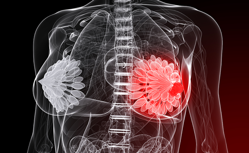
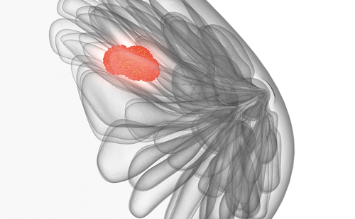
Breast cancer, the most common cancer and the second leading cause of death among women worldwide, remains an important public health problem. More than one million women are newly diagnosed with breast cancer each year, and approximately 500,000 women die annually from this malignancy. Advances in breast cancer therapy have led to improved survival in the last 10 years. However, early breast cancer detection continues to be a clinical challenge.1 Although mammography was approved by the US Food and Drug Administration (FDA) as the ‘gold standard’ tool for breast cancer screening, the limitations of this screening procedure are well known.2 Moreover, the World Health Organization (WHO) has recognized that mammography is not a viable option for many countries because it is an expensive screening tool that requires a large infrastructure and trained healthcare professionals.3 The search for a detection tool for early-stage breast cancer is the subject of extensive research. Efforts to develop an early breast cancer detection method have included digital mammography, computer-aided detection, ultrasound, magnetic resonance imaging (MRI), positron emission tomography (PET), electrical impedance imaging, and infrared (IR) imaging, alternately named thermography or breast thermography. Despite these imaging technologies receiving FDA approval as diagnostic adjuncts to mammography, currently there is no diagnostic tool capable of significantly reducing breast cancer mortality that benefits women at all ages, is non-invasive, has high-quality assurance, has high sensitivity and specificity, is inexpensive, and is easily trainable. Moreover, most imaging modalities, although promising, are too expensive for routine use. Advances in IR imaging and reduced equipment costs show promise as a feasible technology that could meet ideal characteristics for an early breast cancer detection tool. The incorporation of cameras with high-resolution digital imaging, high-speed image capture, image manipulation software, high-speed computers, and computer-aided detection are new technologies suitable for breast cancer screening. Diakides and Bronzino have provided a comprehensive review of recent advances in IR imaging.4 Low- and middle-income countries require new strategies to increase early breast cancer detection and access to care. In this article we review the role and feasibility of IR imaging for early breast cancer detection in rural communities in southern Mexico.
Definition of Infrared Imaging
Clinical thermography is a non-invasive, non-contact, and non-ionizing radiation imaging technique that detects, records, and forms an image of the temperature distribution on the surface of the body. IR imaging is not related to morphology; it is a functional imaging technology. The physics rationale is that all objects with a temperature above absolute zero (-273°K) emit IR radiation from their surface. Because the emissivity of human skin is high, it can be converted into temperature values. IR imaging equipment has been designed to provide both qualitative and quantitative information on temperature patterns. In the electromagnetic spectrum, the IR are located between visible and microwave spectral regions covering wavelengths that range between 0.75 and 1,000μm. The full IR spectral region is subdivided into: near IR (NIR), which covers wavelengths between 0.75 and 1.4μm; short-wave IR (SWIR), from 1.4 to 3.0μm; mid-wave IR (MWIR), between 3.0 and 8.0μm; long-wave IR (LWIR), from 8 to 12μm; and far IR (FIR), beyond 12μm. Despite the new generation of IR cameras detecting the full spectral region, for medical IR imaging purposes LWIR (or body IR rays) is the most important IR spectral region. Thermal IR (TIR) refers to the region that covers wavelengths beyond 1.4μm where the IR emission is heat or thermal radiation and covers the SWIR, MIWR, LWIR, and FIR regions. IR rays cannot be detected by the human eye; however, IR radiation can be detected using IR cameras and detectors.4–9
Clinical Infrared Imaging Equipment
First-generation IR cameras were large and heavy, and were expensive because they required the use of a cooling system in the form of a nitrogen or compressed-air cooling bottle. Scanning mirrors were incorporated to generate the picture. Advances in detector technology and microelectronics, favored the incorporation of uncooled thermal detectors and 2D imaging device types as a focal plane array (FPA) that is sensitive in the IR spectrum, avoiding the use of mirrors and improving image quality. Currently, the thermal sensitivity of the uncooled cameras is 0.05 or 0.02ºC and the FPA detector detects wavelengths from 3–5 or 8–12 to 3.0μm. Cancer cells can be imaged as hotspots in the IR images due to their high metabolic rates and angiogenesis, generating a higher temperature than the normal cells around them. Because of the advances in detector technology it has been reported that the average breast cancer tumor size undetected by new IR cameras is 1.28cm, versus 1.66cm by mammography.10 Moreover, new IR cameras are much smaller, lighter, more reliable, and less expensive. The new generation of IR cameras has benefited from important advances in computer and information technology. IR cameras coupled with high-speed computers, high-speed image capture systems, and high-resolution digital imaging generate much smoother and clearer pictures.4,9
Methods of Acquiring Infrared Images
The most common methods of acquiring IR images to detect breast cancer are classic static thermal imaging and active or dynamic IR imaging. Static thermography monitors and measures temporal changes in the temperature of selected areas. It has been the most frequently used method in clinical studies of breast thermography. Dynamic thermal assessment (DTA), another term for dynamic thermal imaging (DTI), monitors physiological processes and measures temperature distribution over certain areas.9 DTA could provide more information about angiogenesis and metabolic changes associated with breast cancer. Although promising, DTA has not yet been sufficiently evaluated in clinical trials. Other innovative IR imaging methods such as active dynamic thermal (ADT) IR imaging or thermographic non-destructive testing (TNDT), thermal tomography or thermal texture mapping, thermal multispectral imaging, and 3D IR are still under intensive research and development.4–6,9
Image Processing and Analysis
Advanced image processing, image manipulation software, and computer-aided detection are feasible tools that play an important role in the analysis and standardization of IR imaging. The incorporation of both image processing techniques and smart processing approaches, also known as artificial intelligence, will help to reduce the false-positive diagnosis rate and avoid bias associated with physician analysis of IR images. Asymmetry analysis, artificial neural network classification, smart brain-like neural network algorithms, and automatic target recognition are technologies under development and evaluation.9 Since thermograms are recorded and stored in digital format, the organization of larger IR image databases is a feasible option for standardization of IR imaging. The Digital Imaging and Communications in Medicine (DICOM) medical imaging standard emerges as a feasible option for a standardized thermogram format for storage and communication of thermograms.11
Infrared Imaging in Medicine
IR imaging has been evaluated in medicine since the late 1950s when military developments in IR surveillance were transferred to medicine. After the first report by Lawson in 1956,12 IR imaging was mainly used in early breast cancer detection, but years later was reported to have clinical utility in different medical applications, such as diseases of the skeletal and neuromuscular systems, rheumatology, detection and diagnosis of deep venous thrombosis, diabetic microangiopathy, pain management and control, skin temperature and metabolism during exercise, coronary bypass operations, and orthopaedic and urological surgery.4,13 DTI has been applied to evaluate the response of soft-tissue sarcomas to chemotherapy and monitor angiogenesis activity in patients with Kaposi’s sarcomas.14,15 IR functional imaging has also recently been evaluated as a mass screening tool of fever for infectious disease outbreak.16
Infrared Imaging and Early Breast Cancer Detection—Early Experience
Between 1956 and 1963 pioneer studies reported by Lawson demonstrated that breast cancer increases regional skin temperature and can be accurately measured by IR imaging.12,17,18 In 1963, Lawson and Chughtai also suggested that the temperature changes observed could be explained by both increased vascular flow and increased metabolism.19 Despite the biological mechanism explaining these temperature changes remaining unclear, it was suggested that the elevation in temperature could be due to either hypervascularity as a result of tumor angiogenesis or nitric-oxide-mediated vasodilatation resulting from nitric oxide release from breast tumor cells. The role of angiogenesis in cancer,20 and particularly in early breast cancer, was established several years later.21–24 Today, it is also known that nitric oxide is a potent neurotransmitter that induces vasodilatation. All three isoforms of nitric oxide synthase (NOS)—inducible (iNOS), endothelial (eNOS), and neuronal (nNOS)—have been detected in tumor cells and could be involved in promoting or inhibiting cancer;29–31 NOS has also been identified in breast tumor cells.25–28 Nitric oxide produced by tumor cells leads to vasodilatation and that mechanism could explain why breast cancer is associated with local hyperthermia. This hypothesis has been experimentally corroborated by using digital IR imaging.32
In the 1960s and early 1970s, thermography was actively evaluated as a feasible screening tool for early breast cancer detection.33,34 Approximately 3,000 thermography clinics were operating in the US during this time.7,8 However, the negative results reported in 1977 by Feig35 and in 1979 by the Breast Cancer Detection and Demonstration Project36 decreased interest in breast thermography. In these studies, which evaluated the roles of physical examination, mammography, and first-generation thermal IR imaging in breast cancer detection, thermography showed high false-positive rates and low sensitivity. Although thermal IR imaging was approved by the FDA in 1982 for use as an adjunctive breast cancer screening procedure,36 this diagnostic approach has been abandoned.
Infrared Imaging and Early Breast Cancer Detection in the 21st Century
To date, thermography has been considered an experimental and unproven tool for early breast cancer detection because of a lack of well-designed and controlled clinical trials evaluating its role for either screening or diagnosis. IR imaging has dramatically improved and interest in this technology has been renewed. However, in the last decade the new generation of IR cameras, coupled with the advantages of computer and information technology, have not been evaluated in studies designed as clinical trials, except one reported by Parisky et al.37 In that study, 769 patients diagnosed as having mammographically suspicious lesions were evaluated by computerized IR imaging analysis, as was a subgroup of 448 patients who had micro-calcifications but were assessed as false-negative by IR imaging. The index of suspicion for the whole group and the group with microcalcifications was, respectively: sensitivity 97 and 99%; specificity 14 and 18%; negative predictive value 95 and 99%; and positive predictive value 24 and 27%. The role of IR imaging as either a screening or a diagnostic tool for breast cancer was reviewed by Kerr,38 but there were no significant studies found. It is important to note that between 1985 and 2004 the only study that evaluated IR imaging for breast cancer screening did not show evidence of benefit.39 However, the study by Parisky et al.37 and the retrospective case–control study reported in 1998 by Keyserlingk et al.40 documented that IR imaging provides an adjuvant benefit. In the last five years, clinical trials evaluating the role of IR imaging in breast cancer have not been reported. Currently, there is no evidence to support the role of IR imaging for screening or as an adjuvant diagnostic tool. However, the limitations of mammography as an early breast cancer detection tool necessitate evaluation of the new generation of IR cameras in well-designed and controlled clinical trials.
Experience of the Center for the Study and Prevention of Cancer with New-generation Infrared Imaging Screening for Breast Cancer in Rural Communities
Breast Cancer in Mexico
Breast cancer is the second highest cause of death from cancer among Mexican women. The median age at diagnosis is 51 years, and 45.5% of all breast carcinomas develop in patients <50 years of age.41 Up to 90% of women are diagnosed as having advanced disease. However, only 7% of women >50 years of age report having had a mammogram. Although rural women are generally considered to be at lower risk for breast cancer than women living in the main cities, the incidence of this malignancy has been rising rapidly in rural populations. A low frequency in the practice of mammography, ranging between 12 and 18%, has been reported.42,43 These findings can be explained by lack of availability of medical resources, including insufficient mammography equipment and trained healthcare professionals. With the aim of improving early breast cancer detection in this population, the Center for the Study and Prevention of Cancer (CEPREC) has designed educational, preventive, and early detection programs. As part of this public health strategy, we have evaluated the feasibility of new-generation IR imaging as a screening tool for breast cancer.44
Patients and Methods
From November 2006 to December 2008, women attending cancer screening programs at our clinic were invited to participate in this study and offered clinical breast examination (CBE) followed by breast thermography and mammography, and biopsy if indicated. CBE and breast thermography were correlated with mammography and histological diagnosis.
Infrared Camera Characteristics
IR imaging was obtained using an IR camera (DL-700; 320*240 UFPA, Zhejiang Dali Technology Co., Ltd). The main characteristics are: detector type FPA; uncooled microbolometer; resolution 320×240; spectral 240; spectral range 8–14μm; thermal time constant 4ms; lens field of view 20.4–15.4°; focus range 1m–∞; and thermal sensitivity 0.08–30oC.
Patient Position and Image Capture
In order to increase accuracy and precision, IR imaging was performed in a controlled environment. Temperature was maintained between 18 and 23oC (within }0.1oC) and the humidity was about 60}5%. After clinical breast examination, patient preparation included an acclimatization period in the imaging room of at least 15 minutes. Then, with the purpose of facilitating anatomical presentation of the breasts, the patient placed her hands on top of her head. The bilateral frontal breast, the right and left oblique views, and right and left single breast close-up views, if indicated, were included in the series.45
Image Processing and Analysis
In a first step, images captured from the IR camera were obtained in rpt format and then converted into bitmap (BMP) file format, as shown in Figure 1 (bilateral frontal view), Figure 2 (right oblique view), and Figure 3 (left oblique view). On the basis of these images, a preliminary IR imaging diagnosis was established according to thermobiological criteria (TH):45 TH1, normal uniform non-vascular; TH2, normal uniform vascular; TH3, equivocal; TH4, abnormal; or TH5, severely abnormal. The results reported in this study are based on this classification. An ongoing second step of this study is the image processing of TH3, TH4, and TH5 thermograms using image manipulation software developed by us and based on image filtering that incorporates the full IR spectrum and an algorithm. Preliminary image processing with this software allows us to detect hotspots (see Figure 4) that cannot be observed in the conventional thermogram (see Figure 1).
Results
Overall, 911 women (100%) with a median age of 44 years were evaluated. Five hundred and three (55%) were 40 years of age or older. The median age of this group was 58 years (range 40–83). Two hundred and eighty-seven (57%) women were 40–49 years of age, 154 (31%) were between 50 and 59 years of age, and 62 (12%) were over 60 years of age. Cancer was diagnosed in 14 women (2.7%); a TH5 diagnosis was previously established in 13 (92.85%) of these 14 women. Cancer was diagnosed in three (1.18%) of 253 women 30–39 years of age; the three women (100%) had a TH5 diagnosis. Overall, cancer was diagnosed in 17 (2.2%) of 756 women >30 years of age. Sixteen of 17 women (94%) were previously diagnosed as TH5 using IR imaging. Cancer was not found in the group of women under 30 years of age.
Conclusions
In this study, breast cancer was diagnosed in 2.2% of women over 30 years of age. Ninety-four percent of women diagnosed as having cancer were previously diagnosed using new IR imaging technology. These findings are encouraging and require independent confirmation, assessing larger patient numbers in the context of well-designed and controlled clinical trials. IR imaging emerges as a potential screening tool for early detection of breast cancer. However, the development of international co-operation among physicians and IR imaging centers is required. ■


