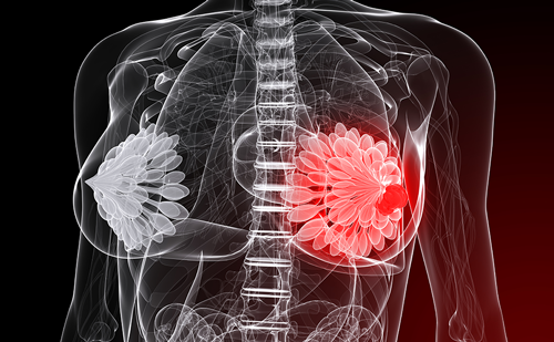
Sentinel lymph node (SLN) biopsy has revolutionized breast cancer management and has been generally accepted as a mainstay in lymph node evaluation for breast cancer patients. Still, there are many controversies that surround this technique, in terms of both the surgical procedure involved and how the SLNs are evaluated pathologically. The ramifications of isolated tumor cells (ITCs) and micrometastases and the need for axillary node dissection in all SLN-positive patients remain subjects of debate. The literature with regardsto these issues is reviewed herein and the current state of the science of SLN biopsy presented.
Sentinel Lymph Node Biopsy Technique
Giuliano et al. utilized a peri-areolar blue dye injection to identify the sentinal lymph nodes in the axilla.1 Generally, the blue dye travels quickly to the first draining lymph nodes in the axilla. Therefore, the dye is injected just after induction of anaesthesia intraoperatively. A small incision is made in the axilla and blue lymphatic channels are traced to blue SLNs, which are then sent for pathologic evaluation. While this technique does not allow for pre-operative identification of the lymph node basin involved, the vast majority of primary breast cancers drain to the ipsilateral axilla and SLNs may be found in this location.
Krag et al. used an alternative technique of injecting a radioactive tracer into the breast and a hand-held gamma probe intraoperatively to identify the SLNs.2 The radioactive tracer tends to take longer to reach the axillary SLNs and therefore is generally injected about an hour prior to the biopsy. Some have found, however, that taking into consideration the half-life of the tracer, one may inject the tracer the day prior to surgery with equivalent success.3 This technique allows for pre-operative imaging with lymphoscintigraphy to map drainage patterns of the breast and identify where the SLNs may be found. While this technique is useful in melanoma, where the drainage patterns of a tumor may be variable, lymphoscintigraphy has been shown to be of little benefit in breast cancer.4 One exception to this, however, is for mapping of internal mammary lymph nodes. While some surgeons will use pre-operative imaging to identify patients in whom an internal mammary SLN biopsy may be performed, many will not use this information to change their management.5 Furthermore, most patients with internal mammary drainage will also have axillary SLNs identified and the rate of finding a positive axillary SLN appears to be the same regardless of drainage pattern.5 Therefore, unless the finding of positive internal mammary SLNs will change management, the utility of lymphoscintigraphy may be limited.
However, patients who present with recurrent breast cancer who have had previous axillary surgery may have alternate drainage patterns and may benefit from lymphoscintigraphy.6
While using either blue dye or radioactive tracer allows for successful SLN mapping, using a combination approach with both agents has been shown to be associated with a higher SLN identification rate.7 In a metaanalysis, Kim et al. found that the SLN identification rates of studies using blue dye alone (n=18), radiocolloid alone (n=16), or both (n=34) were 83.1, 89.2, and 91.9 %, respectively (p=0.007).8 Similarly, the falsenegative rates were 10.9, 8.8, and 7.0 % for each of these groups, respectively (p=0.047).8 Traditionally, a SLN is defined as:
- the hottest node;
- any blue node;
- any node at the end of a blue lymphatic channel;
- any node that has ex vivo counts greater than 10 % of the hottest node; and
- any palpably suspicious node.
Despite removing all of the nodes that meet these criteria, on average only two SLNs are identified. Some have argued that given that the first positive SLN is generally identified within the first three SLNs removed, further SLNs need not be removed.9 Larger studies, however, found that to abort the SLN biopsy procedure at three nodes when other nodes that met the criteria of being SLNs remained would miss the first positive SLN in a subset of patients.10,11
Intraoperative Pathologic Evaluation
As SLN biopsy evolved as a technique, progress in intraoperative pathologic evaluation was also made. While there is some debate as to whether intraoperative assessment is advantageous,12 this continues to be the norm. It allows one to proceed immediately to axillary lymph node dissection (ALND) in cases of a positive SLN without having to return the patient to the operating room for a second procedure. Conventional intraoperative pathologic evaluation of SLNs consists of frozen section, touch imprint cytology, and/or scrape cytology. The sensitivity of these tests varies according to various studies (see Table 1). More recently, intraoperative molecular assays based on polymerase chain reaction (PCR) technology have been evaluated and found to have a fairly high sensitivity (see Table 2). Caution should be exercised, however, in the interpretation of pathology results of SLNs. As SLNs are scrutinized more vigorously than routine axillary lymph nodes, smaller foci of metastases may be identified. Using immunohistochemistry, serial sectioning, and/or molecular techniques, studies have found that metastases will be found in 4–33 % of nodenegative patients (see Table 3). The sixth edition of the American Joint Commission on Cancer Staging System has defined metastases measuring less than 0.2 mm as ITCs.13 While a number of studies have debated the significance of ITCs, these are considered node-negative. Patients who have metastases measuring 0.2–2.0 mm, however, are classified as having micrometastases and are considered node-positive.
Studies have found non-SLN metastases in 0–9 % of patients with ITCs and in 22–33 % of patients with micrometastases in their SLNs (see Table 4). A meta-analysis by Cserni et al. found that, overall, patients with ITCs or micrometastases had a 20 % chance of having non-SLN metastases (95 % confidence interval [CI] 16–25 %).14
The outcome of patients with ITCs and micrometastases has been the subject of a number of studies. In a recent study by de Boer et al., patients with ITCs and those with micrometastases had significantly reduced five-year disease-free survival rates compared with truly nodenegative patients (77.2 versus 85.7 % [p<0.001] and 75.9 versus 85.7 % [p=0.002], respectively).15
In another multicenter study of 1,259 patients who underwent an SLN biopsy between 1996 and 2005, with a mean follow-up of 4.9 years, the distant recurrence rates of patients with negative SLNs, those with ITCs, micrometastases ,and macrometastases were 6, 8, 14, and 21 % respectively (p<0.001).16 The hazard of distant recurrence was 1.5 times higher in patients with ITCs than their truly SLN-negative counterparts (CI 1.0–2.2; p=0.02).16
Other studies, such as that of Kahn et al., found no difference in disease-free interval or disease-specific survival between nodenegative patients and those with ITCs or micrometastases with a median follow up of eight years.17 Similarly, a study by Imoto et al. found no difference in six-year recurrence-free survival between patients with ITCs and micrometastases and those who were SLN-negative (94 versus 88 %; p=0.39).18
The true biologic significance of minimal disease in SLNs therefore remains controversial. The ACOSOG Z-0010 and the NSABP B-32 trials, which recently completed accrual, will shed light on the biologic significance of ITCs and micrometastases. For now, however, guidelines are to treat ITCs as node-negative and therefore avoid completion ALND in these patients. It recommends micrometastases be treated as node-positive and that completion ALND in these patients should be carried out.Prediction Models
Given that the 25-year follow-up data from the landmark NSABP B-04 trial showed no difference in survival for node-positive patients who received ALND compared with those who did not,19 the need for completion ALND in SLN-positive patients may be questioned. While ALND certainly may improve staging and local control, a number of studies have found that many patients with SLN metastases will have no further disease in the axilla (see Table 5). As such, a number of authors have tried to develop clinical prediction models to identify which SLNpositive patients are at low likelihood of having non-SLN metastases and who may therefore be spared the morbidity of an ALND. Several of these models are compared in a study by Coutant et al.20 Overall, they found that the Tenon score and the Memorial Sloan Kettering nomogram outperformed the other models.
Perhaps the best known of these is the Memorial Sloan Kettering nomogram, developed by Kimberly Van Zee and colleagues.21 This model predicts the probability of non-SLN metastases based on:
- pathologic tumor size;
- grade;
- lymphovascular invasion;
- multifocality;
- estrogen receptor status;
- the number of positive and negative SLNs; and
- the method used to identify SLN metastases (immunohistochemistry, routine hematoxylin and eosin staining, serial sectioning with hematoxylin and eosin or frozen section).
While a number of authors have validated this nomogram,22,23 some have found that it could not predict non-SLN status, particularly in patients at low likelihood of having non-SLN disease.24–26 This has led to some debate as to the utility of this model to guide clinical management. Furthermore, this model requires information that is available only postoperatively and so has limited utility in the intraoperative setting.
The Tenon Score was developed by Barranger et al. on the basis of 71 SLN-positive patients who underwent completion ALND.27 Of these, 52 (73 %) had no further non-SLN metastases. A score was developed using three factors: if the ratio of involved SLN to total number of SLNs removed was 1 or more, 2 points were assigned, a ratio between 0.5 and 1 was given 1 point, and a ratio below 0.5 scored zero points.
Macrometastasis in the SLN was given two points, and its absence was given zero points. Tumor size scored zero points if ≤10mm, 1.5 points for tumors measuring between 11 and 20mm, and 3 points if >20 mm. Patients with scores of 3.5 or less had a 97.3 % chance of having no further non-SLN metastases.
A clinical prediction rule, not evaluated by Coutant et al., was developed at the University of Louisville using pre-operative and intraoperative factors alone. Using a multivariate analysis of a multi-institutional series of 1,253 SLN-positive patients, a simple integer point-based system was developed to identify a subset of patients at low likelihood of having further non-SLN metastases. With one point being given if there was more than one positive SLN, an additional point being given if more than 50% of SLNs harvested were positive, and up to four points being given for tumor size (T1a=1, T1b or T1c=2, T2=3, T3=4), patients with only one point had a 95% probability of having no further axillary disease.28 Others have also found that the risk of non-SLN metastases is negligible in patients with a single positive SLN who had more than four SLNs removed29 or those with small tumors (T1a).30 This simple clinical prediction model has been validated by a multicenter European group,31 but is somewhat limited by the small number of patients who fall into the lowest-risk group. Therefore, regardless of the development of clinical prediction models to predict the absence of non-SLN metastases, most surgeons proceed with ALND based on intraoperative pathologic assessment of SLNs.32 Studies are ongoing to incorporate molecular studies to current clinical prediction models, with the objective to improve the ability to identify patients who will not have any further axillary metastases.
Conclusion
SLN biopsy has become the mainstay of axillary staging of breast cancer patients and involves a multidisciplinary effort between surgeons, pathologists, nuclear medicine physicians and others. As this technique has evolved, however, nuances have emerged in terms of the technique itself and how these nodes should be evaluated pathologically. The greater scrutiny with which the SLNs are evaluated has led to some patients being upstaged. The implications of ITCs and micrometastases on outcomes remain an area of debate. Nonetheless, patients with tumor deposits less than 0.2mm are considered node-negative and will generally not proceed to completion ALND. Those with larger deposits may proceed to immediate ALND based on intraoperative pathologic evaluation. While a proportion of these patients will have no further axillary disease, prediction models to identify these patients continue to be refined. While the concept of SLN biopsy in breast cancer patients is relatively young, it continues to enjoy an ongoing metamorphosis as nuances to this technique evolve.