Neuroendocrine gastroenteropancreatic (GEP) tumors constitute less than 2% of all gastrointestinal (GI) malignancies. The incidence of the largest group of patients, those with small intestinal carcinoid tumors, is two to 2.4 per 100,000 inhabitants.The true incidence is probably underestimated due to sometimes vague clinical presentation and low awareness among physicians. The incidence in autopsy series is significantly higher at 8.4/100,000 inhabitants.1,2
Neuroendocrine gastroenteropancreatic (GEP) tumors constitute less than 2% of all gastrointestinal (GI) malignancies. The incidence of the largest group of patients, those with small intestinal carcinoid tumors, is two to 2.4 per 100,000 inhabitants.The true incidence is probably underestimated due to sometimes vague clinical presentation and low awareness among physicians. The incidence in autopsy series is significantly higher at 8.4/100,000 inhabitants.1,2
Although neuroendocrine tumors can appear at all ages, those of the lung, mediastinum, and the GI tract are, in general, age-related with the highest incidence from the fifth decade upwards. Exceptions to this are the carcinoids of the appendix, which occur with the highest incidence below 30 years of age. Some of the neuroendocrine tumors of the GI tract are part of inherited diseases such as multiple endocrine neoplasia type 1 (MEN-1) and neurofibromatosis (NF) type 1, and von Hippel-Lindau’s disease. MEN-1 is associated with parathyroid hyperplasia/hyperparathyroidism (90%), pancreatic endocrine tumours (50–80%), pituitary adenomas (30–40%), and adrenal cortical adenomas (10–15%). A specific deletion on chromosome 11q13, harboring the MEN-1 gene, is the genetic background of the disease.This gene encodes a protein called menin, which acts as a tumour suppressor.3,4
The von Hippel Lindau’s (VHL) syndrome is an autosomal-dominant neoplastic syndrome characterized by hemangio-ablastomas of the central nervous system (CNS), retinal angiomas, renal cell carcinomas, pheochromocytomas, and neuroendocrine pancreatic tumours. The VHL gene has been mapped to chromosome 3p25.3 and the gene is a tumor suppressor gene, implying that loss of function or inactivating mutations of this gene are associated with tumor formation.5,6
The main two groups of neuroendocrine GEP tumors are the so-called carcinoid tumors and endocrine pancreatic tumors.The carcinoid tumors are divided into fore-gut, which are mainly located in the lung, thymus and gastric mucosa, and duodenum, whereas mid-gut carcinoids, the second group, are located in the distal ilium and jejunum. Finally, hind-gut tumors are located in the colon and rectum. Approximately 40–60% of carcinoids are located in the mid-gut area, 25% in the fore-gut area, and the rest in the hind-gut. A specific clinical syndrome related to mid-gut carcinoid tumors is carcinoid syndrome, including flushing, diarrhoea, carcinoid heart disease, bronchial constriction, and high levels of urinary 5-hydroxyindoleacetic acid (5-HIAA). This syndrome is mainly seen in mid-gut carcinoids, but an atypical syndrome can be seen in patients with lung carcinoids, due to histamine production.7,8
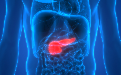
Endocrine pancreatic tumors develop within the pancreas and might be divided into functioning and non-functioning tumors. Functioning tumors constitute approximately 60% and include well-known clinical syndromes such as the Zollinger-Ellison (due to secretion of gastrin), hypoglycemic syndrome (related to insulin/pro-insulin over-production), the Verner– Morrison syndrome (related to high circulating levels of vasoactive intestinal polypeptide), and glucagonomas syndrome (due to secretion of high concentrations of glucagon). The non-functioning tumors are secreting agents, which usually do not cause any distinct clinical syndrome, but merely present as ordinary GI cancers. These tumors are usually large and metastatic at diagnosis. They may secret chromogranin A, pancreatic polypeptide, peripheral hormone peptide YY (PYY) and other hormones, which do not cause any hormone-related clinical symptoms.9–11
Diagnostic Procedures
A correct histopathology is very important in these patients, since the prognosis is significantly better for many of these tumors compared with regular cancers. Patients with non-functioning endocrine pancreatic tumors may sometimes be particularly misdiagnosed with pancreatic cancer, which has a significantly poorer prognosis.12 Biopsy material, preferably surgical or coarse-needle biopsy, specimens are investigated with immunohistochemistry for different neuroendocrine markers, such as antibodies to chromogranin A, neuron-specific enolase (NSE) and synapthophysin. These are routine investigations and can be supplemented by analysis for different hormones in relation to clinical symptoms. If a patient presents with a carcinoid syndrome serotonin should be stained for, and if the patient presents with Zollinger–Ellison syndrome, gastrin should be stained for. Besides that, the proliferation capacity of the tumor is of value for therapeutic decisions and the proliferation marker Ki67 or the MIB-1 antigen is very important. Another method is simple counting the mitotic figures per 10 high power fields in the light microscope.13
In 2000, a new World Health Organization (WHO) classification was established for GEP neuroendocrine tumors. They are now classified according to classical structural criteria, combined with proliferation index (PI) Ki67 into well differentiated endocrine tumors (PI<2%), well differentiated endocrine carcinoma (PI>2% but <15%), poorly differentiated endocrine carcinoma (>15%), mixed exocrine-endocrine tumours, and tumour-like lesions.14
A benign insulin-producing tumor belongs to the group well differentiated endocrine tumor, whereas a patient with a carcinoid syndrome and metastatic mid-gut carcinoid might be classified as a well differentiated endocrine carcinoma in the new classification.
Biochemical Diagnosis
To establish the diagnosis of neuroendocrine tumor, the demonstration of elevated levels of peptides and biogenic amines in the circulating blood is essential. Plasma chromogranin A is a general tumor marker that is increased in nearly all different types of neuroendocrine tumors. No clinical symptoms can be related to the production of chromogranin A. Other general tumor markers are plasma pancreatic polypeptide (PP) and human chorionic gonadotrophin alpha (HCG-α) subunits. For each tumor type with characteristic clinical symptoms, measurement of specific markers such as gastrin, insulin/pro-insulin, vasoactive intestinal polypeptide (VIP), glucagon, and urinary 5-HIAA should be performed.15
Radiological Diagnosis
Computed tomography (CT), magnetic resonance imaging (MRI), and ultrasonography (US) detect <50% of neuroendocrine small gut or pancreatic tumors, but CT and MRI have a higher sensitivity in visualizing liver metastases, larger than 1–2cm.16 Somatostatin receptor scintigraphy (SRS) is based on the presence of somatostatin receptors in 80–90% of neuroendocrine tumors. Tumors expressing somatostatin receptor subtypes 2 and 5 are diagnosed and localized with this method.Tumors lacking these receptors, such as benign insulinomas, can be negative. The method is a whole-body investigation and enables staging of the disease and, hence, can guide the clinicians in the therapeutic decisions. The method should always include single photon emission CT (SPECT).17
Positron emission tomography (PET) is a functional imaging technique that can reflect tumor metabolism. Short-lived positron emitting isotopes such as 18F and 11C are used to label substances of interest. Fluorodeoxyglucose (FDG)-PET is being used as an imaging procedure in common cancer, reflecting increased metabolism of glucose in the tumors. Unfortunately, highly differentiated neuroendocrine tumors do not show increased uptake of FDG.A specific tracer, 5-hydroxytryptophan (5-HTP), a serotonin precursor, has been labelled with 11C and recent studies have shown increased uptake in neuroendocrine tumors. The method is more sensitive than somatostatin receptor scintigraphy (SRS) and CT in detecting small neuroendocrine tumours. In a recent study in neuroendocrine tumors, the sensitivities for PET, SRS and CT were 95%, 87%, and 76%, respectively.18
Endoscopic ultrasound (EUS) combines the techniques of flexible endoscopy with US.Tumors, particularly in the pancreas and duodenum, are identified with a sensitivity and specificity of approximately 80%. Furthermore, it is possible to perform an US-guided fine needle aspiration (FNA) of tumors and regional LNs, which may increase the accuracy.19
Endoscopic investigations, such as bronchoscopy and gastroscopy, are of value in patients with suspected lung or gastric neuroendocrine tumors, respectively. A colonoscopy with biopsies might be needed for the diagnosis of neuroendocrine tumors in the lower GI tract.
Therapy
Surgery is the only way of curing a patient with a neuroendocrine tumor. However, a majority of patients with neuroendocrine tumors of the GI system present with metastatic disease. Cytoreductive procedures have been a major cornerstone in the treatment of malignant neuroendocrine tumors, including debulking and bypassing procedures facilitating the medical treatment. Furthermore, new hormone-blocking agents (somato-statin analogs) have also facilitated the surgical procedures and reduced the risk of carcinoid crisis and other severe events during surgery. Currently, more aggressive surgery has emerged with debulking, laser treatment or radiofrequency ablation (RFA), and embolization of liver metastases. Liver transplantation may be considered in young patients in whom all extrahepatic tumors and metastases are previously removed and a follow-up does not visualize recurrence of extrahepatic tumor tissue.The long-term effect of liver transplantation has to be shown since a majority of the patients present recurrent disease within months to years after the transplantation.20–22
Embolization of Liver Metastases
Selective embolization alone or in combination with intra-arterial chemotherapy (chemoembolization) is a widely performed procedure to reduce clinical symptoms as well as liver metastases. Selective embolization of peripheral arteries induce temporary ischemia and the procedure can be performed repeatedly. The objective response rates have varied between 30% and 70% of significant tumor reduction. This procedure can be performed at any point during the clinical course to facilitate the concomitant medical treatment.23
Medical Therapy
Medical treatment includes somatostatin analogs, alpha interferon (IFN-α) and cytotoxic drugs.24 Somatostatin analogs have been in clinical use for the treatment of neuroendocrine tumors since the mid 1980s. Somatostatin analogs are synthetic somatostatin derivates, with a structure and activities similar to those of native hormone somatostatin, containing 14 amino acids, but with a significantly longer half-life and duration of action than the native substance. Somatostatin analogs are binding to somatostatin receptors—currently, there are five subtypes of somatostatin receptors identified. The currently available somatostatin analogs are binding to receptor 2 and 5 (SST2 and SST5), with high affinity and with lower affinity to receptor subtype 3 (SST3). The somatostatin analogs can exert cytostatic actions via cell-cycle arrest, inhibition of angiogenesis, and effects on immune system. Somatostatin analogs can induce apoptosis, particularly at high doses.25–27 Octreotide and lanreotide are clinically available somatostatin analogs. Both of these exist in long-acting formulations, Sandostatin® LAR® and Somatuline Autogel®, respectively. The dose of octreotide LAR varies from 10–30mg up to 60mg every four weeks. The dose of slow-release lanreotide ranges from 60–120mg, every four weeks. For octreotide there is also a short-acting octreotide with standard doses at 100μg to 500μg three times daily. With somatostatin analogs, symptomatic improvement is achieved in more than 60% of patients, biochemical responses in up to 70% of the patients, but significant tumor response is only found in approximately 5% of patients. In progressive disease, tumor stabilization has been observed in 30–50% of patients, with a duration of stabilization range from two to 60 months. Single patients have been stabilized for more than 10 years.28–31 Recently, a new somatostatin analog, SOM230, has entered into clinical trials. This analog is binding to receptor 1, 2, 3, and 5. Data from a phase II study show an approximate 30% response rate.32
Somatostatin analog treatment is the primary medical treatment for patients with symptoms related to neuroendocrine GEP tumors. For patients with insulinomas and gastrinomas, these agents can be used as second- or third-line medical treatment. They can also be considered for asymptomatic patients with progressive disease, excluding aggressive tumors. Currently, it is controversial as to whether somatostatin analogs should also be considered for asymptomatic patients with stable disease. The most relevant adverse effect is the development of gallstones, because of inhibition of cholecystokinin release, which post-prandially induces emptying of the gallbladder. Up to 60% of patients under long-term treatment may develop sludge in the gallbladder, but less than 10% develop clinically significant gall-stones. Other rare adverse events include pain at injection site, hypoglycemia or hyperglycemia, rash, alopecia, and fluid retention.28
IFN-α
The cytokine IFN-α was introduced in the therapy of mid-gut carcinoids in the early 1980s. Mechanism of action is a direct effect on the tumor cells by blocking the cell cycle in the G1/S-phase, inhibiting protein and hormone synthesis and anti-angiogenesis through inhibition of angiogenic factors, such as basic fibroblast growth factor (b-FGF) and vascular endothelial growth factor (VEGF) and their receptors. It also has an indirect effect through stimulation of the immune system, particularly T-cells, natural killer (NK) cells, macrophages and monocytes.24,33,34 Currently, recombinant IFN-αs are also available in long-acting forms, pegylated interferons such as Peg-Introna® and Pegasys®. The dose should be individually titrated in each patient.The leukocyte count could be of some guidance, aiming at a reduction of the blood leukocyte count to approximately 3.0×10⁹/L.The dose of regular IFN-α should usually be three to five million units three to five times per week subcutaneously, and the precise dose of pegylated IFN-α has still to be established in forthcoming studies although subcutaneous doses of 80–150μg per week could be feasible. The average response rates according to WHO criteria are:
• symptomatic responses in 40–60% of patients;
• biochemical responses in 30–60%; and
• tumor reduction in 10–15%.
Tumor stabilisation is noted in 40–60% of patients with mid-gut carcinoids, lasting for long periods of time (more than 36 months). Single patients have been stabilized for 10 to15 years.24,34
IFN-α can be considered a primary or secondary medical treatment for low-proliferating neuro-endocrine GEP tumors with or without clinical syndrome, either alone or in combination with somatostatin analogs. It could be attempted as a second-line treatment after cytotoxic treatment alone or in combination with somatostatin analogs. Approved indications vary between different countries.
Combinations of IFN-α with somatostatin analogs have generated additive, and also possibly synergistic, effects in the treatment of classical carcinoids and neuroendocrine pancreatic tumors.35,36 IFN-α is known to upregulate the expression of somatostatin receptors, and somatostatin analogs might reduce the side effects of IFN-α. The adverse effect of IFN-α is related to its multiple intracellular actions. Most patients experience flu-like symptoms and fever during the first week of treatment. The most severe and dose-limiting toxicity is the chronic fatigue syndrome seen in 30–75% of patients, mental depression in 5–10% of patients, and neurological disorders in another 5–10% of patients. Other side effects are anemia, leukopenia, thrombocytopenia, and increased liver enzymes in 10–30% of patients. Severe side effects are myositis, vasculatis, and systemic lupus erythematosus (SLE) syndrome in single patients, which necessitate withdrawal of the treatment. Other auto-immune reactions, such as auto-immune thyroiditis, can be managed by standard endocrine procedures. Auto-immune hepatitis, psoriasis, SLE, rheumatoid arthritis (RA), and dementia are contraindications for IFN-α treatment.34
Chemotherapy
Cytotoxic treatment has been the gold standard for most neuroendocrine tumors over the last three decades. However, the increasing awareness that low-proliferating neuroendocrine tumors respond very poorly to cytotoxic treatment has stimulated the development of new biological treatments for slowly progressing neuroendocrine tumors. Cytotoxic treatment is still to be considered a first-line treatment for high-proliferating tumors, i.e. metastatic disease with a proliferation index above 10%. Single-agent cytotoxic treatment has been of limited value in most trials producing response rates of less than 30%. The combination of Streptozotocin plus 5-fluorouracil (5- FU) and doxorubicin has shown response rates of more than 50% in malignant neuroendocrine pancreatic tumors. Poorly differentiated neuroendocrine tumors, particularly fore-gut (pulmonary and thymic) and small-cell colorectal neuroendocrine tumors, might respond to a combination of cisplatinum/paraplatin plus etoposide. These tumors present a high-proliferation capacity and are considered to be poorly differentiated neuroendocrine carcinoma with responses up to 60%.24,37,38
A combination of cytotoxic treatment with somatostatin analogs is recommended in patients with clinical syndromes related to hormone over-production. The addition of somatostatin analogs improves the clinical symptoms and also prevents the development of severe attacks related to release of vasoactive substances during the development of necrosis of neuroendocrine tumors.
The adverse effects of the most common combination of streptozotocin plus 5-FU or doxorubicin include nausea, vomiting, and renal toxicity, and also alopecia with doxorubicin.The combination of cisplatinum plus etoposide is accompanied by significant toxicity, including alopecia, nausea, vomiting, bone marrow and renal toxicity, and neuropathy.24
Radiotherapy
Neuroendocrine tumors are considered to be relatively radio-resistant. Conventional external beam radiotherapy (EBRT) is recommended only for bone and brain metastases. Tumor-targeted radioactive treatments with 111Indium-DOTA-Octreotide or 90Yttrium-DOTA-Octreotide and 177Lutetium- DOTA-Octreotate has generated significant interest over the last few years.The reported response rates with standard WHO criteria have been approximately 20–25% significant tumor reduction with biochemical and clinical responses in 40–50%. The precise role of tumor-targeted radioactive treatment is not yet defined, but future randomized trials will give more information on how to use this kind of treatment. Recently, treatment with 111Yttrium micro-spheres (Sirtex) have been introduced for the treatment of liver metastases of neuroendocrine tumors. Few patients have been treated to date, but single cases have shown significant tumor reductions.39,40
New Agents
There is a rapid development of new agents in the field of neuroendocrine GEP tumors. It is well known that a significant number of neuroendocrine tumors express tyrosine kinase receptors (TKRs), such as platelet derived growth factor-alpha receptors (PDGFR) (α and β), c-kit, EGF-receptors and VEGF-receptors. These receptors might be target for tyrosin kinase receptors (TKR) treatment in the future. Many neuroendocrine tumors are highly vascularized; therefore, the blood supply might be the target for new anticancer treatment of neuroendocrine tumors. A number of new anti-angiogenic substances will be attempted in these tumors. Agents such as imatinib SU11248 and bevacizumab have demonstrated anti-tumor effects in early phase I and II studies in neuroendocrine tumors.41,42 The treatment will be individualized in the future and custom-made for each patient, based on information from molecular genetics and tumor biology of the single tumors. ■


