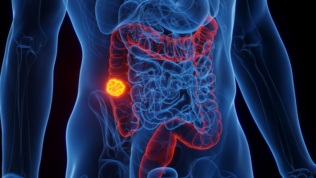
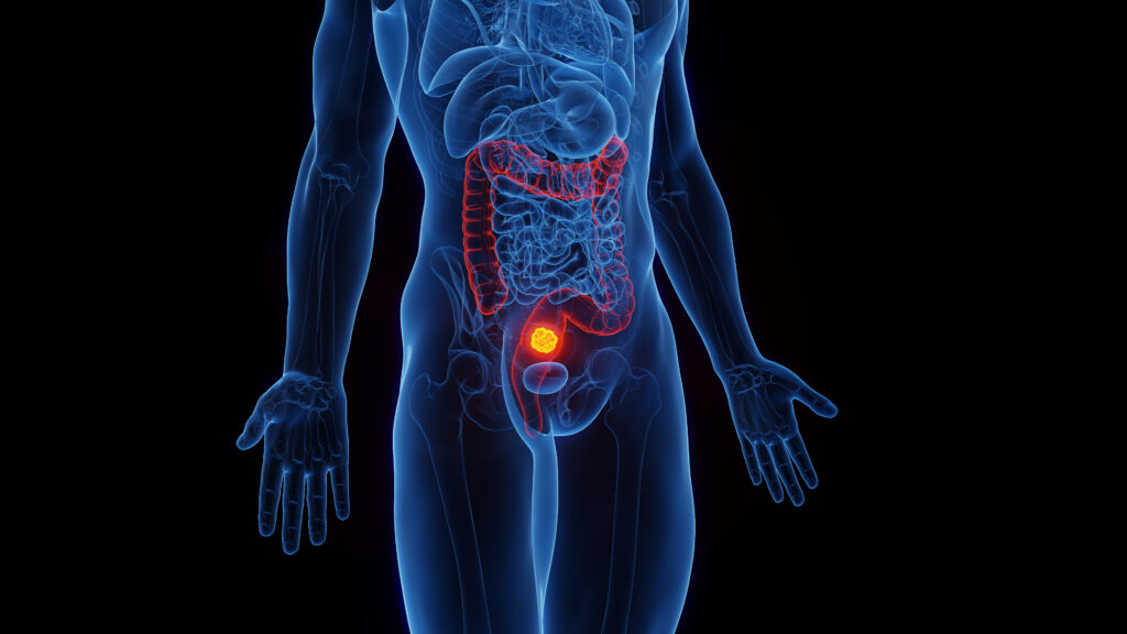
Prior to 2000, the outlook for patients with gastrointestinal stromal tumors (GISTs), the most common gastrointestinal (GI) sarcoma, was poor. This life-threatening disease is highly resistant to traditional management with chemotherapy and radiation, and patients with metastatic GISTs had only one viable treatment option: surgical resection. Even with surgery, many patients suffered relapse and experienced an average survival of 12–18 months.1,2
Prior to 2000, the outlook for patients with gastrointestinal stromal tumors (GISTs), the most common gastrointestinal (GI) sarcoma, was poor. This life-threatening disease is highly resistant to traditional management with chemotherapy and radiation, and patients with metastatic GISTs had only one viable treatment option: surgical resection. Even with surgery, many patients suffered relapse and experienced an average survival of 12–18 months.1,2
However, our better understanding of the disease at the beginning of the new millennium has ushered in dramatic changes in the management of GISTs. The discovery of the KIT receptor tyrosine kinase as the biological driver of GISTs and the introduction of a specific inhibitor of KIT, imatinib mesylate (Gleevec®, Novartis), has revolutionized the treatment of GISTs.3,4 Imatinib mesylate, a protein-tyrosine kinase (TK) inhibitor, also has specificity against the tyrosine kinases ABL and platelet-derived and growth factor receptor (PDGFR).3 Significantly, mutations in KIT are found in about 80–85% of GISTs. Of the GISTs without KIT mutations (fewer than 15%), approximately half have mutations in the gene for PDGFR-alpha (PDGFRA).3–9
The insights gained into the molecular pathogenesis of GISTs have led not only to rapid advances in knowledge about the disease, but also to rapid improvements in the management of the disease. For patients with unresectable or metastatic GISTs, imatinib has provided a much-needed treatment option, providing clinical benefits such as improved median progression-free survival times. Furthermore, imatinib has significant potential in the neoadjuvant and adjuvant settings to improve surgical outcomes for primary, recurrent, and metastatic GISTs.
Pathogenesis of Gastrointestinal Stromal Tumors
GISTs are considered to be the most common mesenchymal tumors, despite accounting for only 1–3% of all malignancies arising from the GI tract.4,10–13 Categorized as epithelioid, spindle cell, or mixed epithelioid neoplasms, GISTs are believed to originate from the interstitial cells of Cajal (ICC) or a cell in that lineage. The ICC forms the neuromuscular network that controls gut motility and shares surface markers including CD117 (KIT protein) and CD34.14,15 However, some GISTs may originate outside of the GI tract in the omentum and mesentery, a location not described for the ICC. This suggests that GISTs may originate from multipotent mesenchymal stem cells.16
The pathogenesis of GISTs stems largely from gain-of-function mutations in the c-kit oncogene that result in constitutive activation of the KIT receptor tyrosine kinase in GISTs.3,4,7,9,17,18 The CD117 antibody is used in immunohistochemistry testing for KIT detection, as a diagnostic marker of GISTs.10,12,14
Imatinib mesylate functions by inhibiting the constitutive activation of the KIT and PDGFRA tyrosine kinases and interrupting the cell-proliferative actions in the signaling pathway. Studies have shown imatinib to be effective in inhibiting tumor growth and inducing regression. Clinical studies in patients with GISTs with nine to 63 months of follow-up observation have shown that under imatinib treatment 4–5% of patients achieve complete remission, while 47–67% of patients experience at least a 50% decrease in tumor size. Stable disease is apparent in 18–32% of patients. The end-points of complete response, partial response, or stable disease after imatinib therapy have been achieved in approximately 90% of advanced GIST patients in all studies to date.19–25 A phase II study with a median follow-up of 4.5 years shows that imatinib therapy was associated with two-year progression-free survival (PFS) of 41–46% and two-year overall survival (OS) of 72–76%.25
Beyond Surgery—The Advent of Imatinib
The standard and most effective approach to treating primary GISTs is through complete tumor resection, which, if necessary, can warrant the removal of adjacent organs.2,26,27 Although surgery is effective, the vast majority of GISTs recur. DeMatteo et al. have reported a 40% recurrence rate within two years,2 and other researchers have shown that the fiveyear survival rate for patients following complete resection of GISTs ranges between 48 and 65%.28–32
Patients with advanced GISTs and those in whom resection is not an acceptable option require alternative therapy. Chemotherapy and radiation are generally ineffective against GISTs, and present additional toxicity risks to the patient.33 Imatinib is approved for the treatment of unresectable or metastatic malignant KIT-positive GISTs. Available in tablet form, imatinib is administered orally daily (QD), usually at a dose of 400mg but sometimes at 600mg. Results from a European phase III trial have indicated that the moderately well-tolerated dose of 400mg twice daily (BID) presented additional benefits to patients with advanced or metastatic GIST characterized by c-KIT expression compared with 400mg QD doses in progression-free survival.22
Changing the Standard of Care
The gain-of-function mutations in c-kit vary in location, occurring most frequently in exon 11 and less so in exons 9, 13, 14, and 17.4,18,34–36 These underlying mutations confer different responses according to the drug agent; patients with GISTs expressing mutations in c-kit exon 11 respond significantly better to imatinib and experience a longer median survival and decreased likelihood of progression than those with GISTs expressing wild-type or exon 9 mutants.6,34,37 Patients with exon 9 mutations are much more responsive to 800mg than to 400mg imatinib, and these are the patients for whom a dose increase to 400mg BID is recommended by most experts.37 The TK inhibitor sunitinib malate (Sutent, Pfizer) is indicated for treatment of patients progressing on imatinib and, similar to imatinib, elicits different sensitivities among the c-kit mutants. Patients with KIT exon 9 mutations are likely to exhibit more active responses to sunitinib than patients with mutations in exon 11.38 By simultaneously inhibiting multiple receptor tyrosine kinases, including KIT, all PDGFRs, and vascular endothelial growth factor receptors (VEGFRs), sunitinib is able to cause tumor regression while significantly prolonging time to progression in imatinib-resistant patients with GISTs, with PFS of 16% for at least 26 weeks.39
Of all patients treated with imatinib, a small percentage (10–12%) have primary resistance to the drug and are unresponsive to treatment, and response rates have been reported to vary depending on the location of exonic KIT mutation.5 A study on imatinib resistance by Demetri et al. suggested that the tumors were becoming resistant through different mechanisms.40
Given that 57–71% of c-kit mutations occur in exon 11,36,41,42 the implications of mutation-based success, in addition to emerging cases of drug resistance, point toward personalized therapy in GISTs. Treatment of GISTs depends on a number of factors, including the general health of the patient and the size and position of the tumor; as such, management of GISTs should be evaluated to encompass the genetic basis of the tumor.
Imatinib has been shown to have both cytotoxic and cytostatic effects in GISTs. Tumor cells have been replaced by myxoid degeneration after as little as four weeks of imatinib treatment, a feature indicative of cell death. The remaining cells lacked expression of the proliferation marker Ki-67 and appeared dormant; some researchers suggest that discontinuation of imatinib therapy in patients with such residual tumor cells would allow these GIST cells to resume cell division afterwards.43 Laboratory studies have also shown an induction of apoptosis in GIST cells by imatinib through an undetermined mechanism.44,45
According to the Response Evaluation Criteria in Solid Tumors (RECIST) guidelines for clinical trials, drug efficacy and response should be assessed with respect to the overall tumor size at baseline.46 Following the RECIST criteria, insufficient shrinkage under the drug therapy would be interpreted as no response. However, the benefits of a drug such as imatinib may not be immediately observable since meaningful tumor shrinkage frequently does not meet the arbitrary cut-off points of RECIST. RECIST has been shown to be an inaccurate early measure of tumor response under imatinib treatment.47–50 Paradoxically, some tumors have been reported to enlarge under imatinib treatment; if associated with decreased metabolic activity in the tumor, this is not indicative of disease progression. Rather, these tumors may very well be responding favorably to therapy while also undergoing intratumoral hemorrhaging or myxoid degeneration.51
The optimal measure of tumor response to imatinib has yet to be defined, but should be expanded beyond reduction of tumor size and disease stabilization and could include decreased tumor radiodensity and metabolic activity.52 Imatinib has shown that not all effective therapies cause tumors to decrease in size; emphasis must therefore be placed on greater understanding of cancer pathways, including how to diagnose cancer at earlier stages and determining the optimal time of treatment during cancer progression.
The current US package insert (PI) recommends that imatinib be used only in those patients with KIT-positive GISTs that are unresectable, metastatic, or recurring. Realistically, however, what is commonly carried out in modern clinical practice and what is recommended by most sarcoma experts does not always adhere strictly to the product PI. The benefits apparent with imatinib therapy in patients with metastatic or unresectable GISTs have suggested possibilities for treatment of patients who are, or have potential to become, candidates for surgery.
New treatment guidelines based on a growing pool of clinical evidence recommend imatinib as first-line therapy for patients with marginally resectable tumors, with surgery and continued imatinib administration post-operatively.19 Imatinib can or may reduce the rates of recurrence and metastases while prolonging disease-free survival.19 The potential benefits of combining surgery and imatinib therapy in GIST management include, but are not limited to, the increase in patient population eligible for surgical resection through decreasing tumor volume. These neoadjuvant and adjuvant imatinib approaches remain experimental.
Neoadjuvant Therapy
A retrospective study by Andtbacka et al. saw promising results for those patients with advanced primary GISTs who received pre-operative imatinib. Pre-operative imatinib was found to decrease tumor volume in patients, leading to a higher rate of complete surgical resection in locally advanced primary GISTs.53 In another case study, a patient with a previously unresectable GIST was treated with imatinib. After six months, a metabolic response and significant size reduction rendered the tumor resectable.54
Panelists at the 2004 GIST Consensus Conference agreed that there was a lack of data in support of imatinib as a neoadjuvant therapy, stating that decreases in tumor size do not necessarily affect surgery.52 However, neoadjuvant therapy with pre-operative imatinib is used to facilitate surgery by reducing tumor size and increasing resectability in some institutions. Reduction of tumor size and tumor debulking through imatinib use can also prevent life-threatening complications in patients with large GISTs, such as hemorrhage or tumor rupture.55 A number of studies have shown that most patients with unresectable GISTs benefit from pre-operative imatinib treatment.19,21,56 What remains to be defined, however, is which surgical patients can benefit most from neoadjuvant and adjuvant administration of imatinib, and at what point of maximal tumor response surgical intervention should be implemented.
Given the frequency of KIT mutations that occur in exon 11 and the responsiveness to imatinib, it may be worthwhile to introduce pre-operative genotyping in the neoadjuvant setting. In doing so, one can anticipate the level of response to imatinib and use this as an aid to the development of a treatment regimen tailored to the individual.
Adjuvant Therapy
During the 2004 GIST Consensus Conference, the majority of panelists agreed that adjuvant imatinib therapy remains investigational.52 However, imatinib treatment should be considered for GIST patients post-operatively to prevent recurrence or spread in the case of incomplete resection.
DeMatteo has recently reported an 82% reduction in the risk of GIST recurrence in patients with completely resected high-risk GIST treated with one year of adjuvant imatinib.57 Data from a pilot study with an average three-year follow-up of adjuvant imatinib saw dramatic improvements in recurrence-free survival. Only one of 23 patients (4%) treated with adjuvant imatinib developed recurrent disease compared with 32 of the 48 patients (67%) in the historic control arm, where no adjuvant treatment was administered. The lack of recurrent disease in the treatment group two years after diagnosis of having high-risk GIST is significant compared with the high degree of relapse in the absence of any adjuvant therapy that usually occurs within two years of diagnosis.58
Recent findings from DeMatteo et al. have suggested a protective role for imatinib in decreasing the likelihood of disease recurrence,59 but the effects on overall survival are far from clear. Researchers from The French Sarcoma Group showed that stopping treatment in patients who were benefiting from imatinib resulted in a large percentage of progression, and many experts now advocate the continuation of imatinib therapy until signs of progression are evident.60 Furthermore, adjuvant imatinib may potentially counteract the appearance of metastases or tumor recurrence, and it is these benefits of adjuvant imatinib that require further study.55 Although promising as an adjuvant therapy, optimal doses and duration of adjuvant treatment have yet to be established.
Summary
The introduction of imatinib has been revolutionary in the management of GISTs, sarcomas that are difficult to treat with radiation, prone to recurrence after surgery and, until recently, resistant to available systemic treatments. Surgery is the first-line treatment of choice for patients with primary GISTs, but as a sole modality is associated with frequent local and distant recurrences. Imatinib appears to improve surgical outcomes in most patients with GISTs in terms of reduction in pre-operative tumor volume, abrogation of GIST metabolism, and potential eradication of micro-metastases.
The current knowledge amassed about GISTs and their response to imatinib treatment may have implications across oncology. With the realization that tumors can behave differently upon exposure to novel anticancer drugs, there is an unmet need for greater understanding of tumor responses and a reassessment of current therapies in use for cancer. Hand in hand with this need is a developing shift toward increasingly personalized management of GISTs. Surgery is performed as monotherapy less frequently; moreover, imatinib is being used in the adjuvant and neoadjuvant setting. Until the optimal treatment strategies and definitive courses of action are established for managing local and advanced stages of GISTs, treatment decisions remain limited to the available evidence and clinical experiences regarding imatinib. In order to better manage GISTs, clinical practice guidelines and actual clinical experience should be integrated, with an ultimate move toward personalized therapy tailored to each individual patient.
Although not yet formally recognized by some of the cancer organizations, the benefits of imatinib in some of these situations have been shown in multiple trials and case studies. The success of predicting and formulating personalized adjuvant and neoadjuvant GIST treatment remains to be seen. Hopefully, the current trials examining adjuvant and neoadjuvant imatinib therapy can definitively state the effects of imatinib on operability, surgical morbidity, disease progression, and disease-free survival for overall amelioration of GIST treatment.


