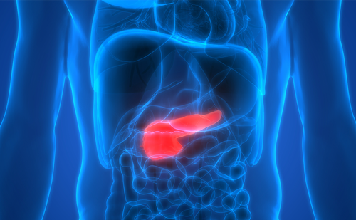
Pancreatic adenocarcinoma (commonly referred to as pancreas or pancreatic cancer) is a devastating disease. Outcome is dismal, irrespective of treatment modality and stage of disease. It is the fourth leading cause of death in the US and is second to colorectal cancer as a cause of gastrointestinal cancer deaths. Most patients with pancreatic cancer will die from their disease. The disease is asymptomatic in its early stages, hence most people present late with unresectable disease.
Pancreatic adenocarcinoma (commonly referred to as pancreas or pancreatic cancer) is a devastating disease. Outcome is dismal, irrespective of treatment modality and stage of disease. It is the fourth leading cause of death in the US and is second to colorectal cancer as a cause of gastrointestinal cancer deaths. Most patients with pancreatic cancer will die from their disease. The disease is asymptomatic in its early stages, hence most people present late with unresectable disease.
Pancreatic cancer is largely a disease of older adults. Despite this, the majority of trials have included mostly younger patients, making conclusions about the clinical management of the older pancreatic cancer patient difficult to derive from the clinical trial literature. This article is a summary of the major contributions to the literature, where possible, with an emphasis on topics or findings exclusive to the older adult.
Epidemiology and Etiology
In the US in 2008, pancreatic exocrine cancer had an annual incidence of 33,700 and an annual mortality of 33,200.1 The prognosis is uniformly poor across all stages, with death within eight to 12 months for patients with locally advanced disease and less than six months for those with metastatic disease. Patients with familial pancreatic cancer may present before 45 years of age, but most patients are over 65 years of age at diagnosis. The male-to-female ratio is equal (1:1).2 The outcome of pancreatic cancer appears to be improving over time in the US. A recent report using the Surveillance, Epidemiology, and End Results (SEER) registries showed that 396 Medicare patients underwent surgery for curative intent between 1991 and 1996 and the three-year mortality rate was 34%.3 The mortality rates are higher in African-Americans than in any other ethnic group, and are also worse the older the patient is at diagnosis.
The risk factors for pancreatic cancer are largely unknown. There are data to suggest a familial aggregation in that 5–10% of patients with pancreatic cancer have a first-degree relative with this disease.4–9 There are hereditary cancer syndromes that are linked to pancreatic cancer. The germline mutation of breast cancer 1, early onset (BRCA1) and breast cancer 2, early onset (BRCA2), especially BRCA2, confers an increased lifetime accumulative risk of 5% for developing pancreatic cancer. The BRCA1 and BRCA2 germline mutation is also associated with hereditary breast cancer and ovarian cancer.10,11 Patients with Peutz-Jegher’s syndrome (PJS) due to mutation in the STK11 gene and transmitted in an autosomal dominant pattern have a 36% lifetime risk for developing pancreatic cancer.12,13 Familial atypical multiple mole and melanoma (FAMMM) syndrome is due to germline mutation in the CDKN2A gene with a lifetime risk of 19% for developing pancreatic cancer.7,14,15 There are data to suggest increased risk for pancreatic cancer in patients with hereditary ataxia-telangiectasia, familial polyposis coli, and Lynch syndrome.16–18 The findings of these genetic factors accounts for only <20% of all cases of familial aggregation.
Both hereditary and non-hereditary chronic pancreatitis are associated with an increased risk for pancreatic cancer.19 Hereditary pancreatitis is caused by mutation of the trypsinogen gene (PRSS1).20 It is an autosomal dominant condition characterized by recurrent abdominal pain in early childhood leading to chronic pancreatitis and a 54% chance of pancreatic cancer by 75 years of age.21
The ABO blood group has been shown by two independent studies to be associated with pancreatic cancer. Both the Nurses’ Health Study and the Health Professional Follow-up Study looked at the relationship between pancreatic cancer risk and ABO blood group by comparing blood group O with blood groups A, AB, and B. This demonstrated that 17% of the 316 pancreatic cancer cases had non-O blood type.22
There is an association between diabetes and pancreatic cancer.23–31 Data indicate that the diabetes may be an early manifestation of pancreatic cancer as opposed to a predisposing factor.32–34 It is postulated that the tumor may secrete islet amyloid polypeptide (a hormone that reduces insulin sensitivity), producing an insulin-resistant state.35 Tobacco exposure is associated with pancreatic cancer.36–45 Approximately 30% of pancreatic cancer46–48 is due to cigarette smoking. The risk increases with the number of cigarettes smoked and the duration of tobacco use.42,43 Data in terms of the relationship between pancreatic cancer and coffee and alcohol consumption, diet, and non-steroidal anti-inflammatory drug (NSAID) use are conflicting.49–60 Other possible risk factors include partial gastrectomy after 15–20 years has a two- to five-fold increased risk for pancreatic cancer.61 Furthermore, an increased risk observed in patients who had cholecystectomy.28 There is also an association between the CagA strain of Helicobacter pylori62 and hepatitis B.63
Clinical Presentation
Upper abdominal pain is the presenting symptom for 80–85% of patients with locally advanced or metastatic pancreatic cancer.64 The pain is usually a dull ache radiating through to the back and is exacerbated by eating. Jaundice, pale stools, and dark urine suggesting biliary obstruction are usually present with tumor involving the head of the pancreas. Painless jaundice is a common presentation in <50% patients with locally advanced or metastatic disease.64 Weight loss, anorexia, fatigue, early satiety, nausea, vomiting, and diarrhea are common. The onset of atypical diabetes in the older adult may pre-date the diagnosis of pancreatic cancer by up to one year. In a Mayo Clinic study, adults with atypical new-onset diabetes were eight times more likely to be diagnosed with pancreatic cancer within three years than the general population.65 Migratory superficial thrombophlebitis (Trousseau’s syndrome) may be present. It is not specific for pancreatic cancer but is associated with mucinous adenocarcinomas. The tumor induces a pro-coagulant resulting in a hypercoagulable state unresponsive to coumadin. Heparin is the treatment of choice.66 A subcutaneous area of fat necrosis (pancreatic panniculitis) may be present.
Pattern of Spread
Pancreatic cancer spreads locally to involve the superficial mesenteric artery (SMA), superficial mesenteric vein (SMV), portal vein, and celiac branches. Lymphatic metastases involve the peri-pancreatic and portal lymph nodes locally and the celiac, SMA, and the root of the distal mesentery. Peritoneal ‘drop’ metastases are fairly common at presentation with nodules around the umbilicus, termed Sister Mary Joseph nodules, peritoneal carcinomatosis, and microscopic peritoneal cytology. Drop metastases may even involve the ovary in females. Distant metastases usually involve the liver and lung, but may involve any organ, including brain and bone.
Diagnosis and Staging
Imaging
The diagnosis of pancreatic cancer is usually based on images derived from one or more modalities. Contrast-enhanced helical computed tomography (CT) (CT angiography) with phase studies provide the most reliable staging information for pancreatic cancer. CT angiography provides useful information about the involvement of major blood vessels, which determines resectability.67,68 Endoscopic retrograde cholangiopancreatography (ERCP) is useful if CT or ultrasound (US) do not show a pancreatic mass and acute pancreatitis is in the differential diagnosis. It has a sensitivity and specificity of 90–95% for the diagnosis of pancreatic cancer. The yield for detecting malignancy via fine-needle aspiration (FNA) at the time of ERCP is lower than endoscopic US (EUS)-FNA. EUS is operator-dependent. It is most useful for the diagnosis of small tumors and may be useful in evaluating nodal or vascular involvement except for SMA and SMV.69,70 Magnetic resonance cholangiopancreatography (MRCP) is better than CT for defining the anatomy of the pancreaticobiliary tree, evaluating the bile ducts both above and below a stricture, and identifying intrahepatic mass lesions.71,72 There is no evidence to support that positron emission tomography (PET) scan or integrated PET/CT provides more reliable information than CT angiography for the diagnoses and staging of pancreatic cancer.73,74 The level of standard uptake variable (SUV) is not reliable for prognosis for pancreatic cancer, although it may be for other cancers.
Serum Tumor Markers
The serum tumor marker of proven significance in pancreatic cancer is the mucinous glycoprotein carbohydrate antigen 19-9 (CA 19-9).75 It is a sialylated Lewis blood group antigen A and occurs in high concentrations in the serum of patients with pancreatic cancer.76 It is not made by the red cells but becomes adsorbed to the red cell surface after it is produced. The CA 19-9 may become elevated in conditions other than pancreatic cancer such as cholangitis, pancreatitis, and biliary obstruction. Approximately 5% of the population is deficient in the enzyme gylcosyl transferase, which is necessary for the expression of Lewis blood group antigen and CA 19-9; therefore, Ca 19-9 will not be a useful tumor marker in these patients.76,77 The expert panel at the American Society of Clinical Oncology (ASCO) stated that CA 19-9 should not be used in screening for pancreatic cancer as it can be falsely elevated (false-positive) or falsely normal (false-negative); rather it should be used as a monitoring tool for patients with resected disease or who are on chemotherapy.78
Biopsy
Percutaneous biopsy under CT or US guidance to a pancreatic mass is performed only if the patient is unresectable or unfit for surgery. The concern with use of percutaneous FNA biopsy is that of seeding of malignant cells along the needle track or in the peritoneum in a potentially resectable patient. Although there are no data to support this concern, it is still quite common.79 EUS-FNA biopsy is less likely to cause peritoneal seeding of the tumor as the biopsy is taken through the bowel.80 It has a sensitivity of 85–90% and specificity of almost 100% for the diagnosis of pancreatic cancer.81 However, patients who are good surgical candidates with a resectable pancreatic mass that is highly suspicious for cancer may proceed to surgery without a biopsy.
Staging Laparoscopy
Laparoscopic evaluation allows the head of the pancreas, peri-pancreatic, peri-aortic, and celiac axis lymph nodes to be visualized and occult peritoneal metastasis to be identified. If there is no evidence of local invasion, laparoscopic ultrasonography is used to evaluate for sub-centimeter lesions on the liver that are not visible with CT or MRI and for vascular invasion.82 Staging laparoscopy is beneficial for reducing the inoperable cases that are discovered at laparotomy.82–84
Peritoneal Washings
Peritoneal washings are usually obtained at staging laparoscopy. However, positive peritoneal cytology from washings in the absence of metastatic disease or unresectable pancreatic mass does not determine prognosis or resectability as the findings have not been conclusive.85–87
Resectable Localized Tumors
A tumor is resectable with potential cure when there is no evidence of distant metastases or involvement of organs and lymph nodes outside the peri-pancreatic area. In addition, the tumor does not encompass or invade the SMA, SMV, portal vein, or celiac, axis, or hepatic arteries. The patient may be considered to have borderline resectable disease if there is focal involvement of the SMV or portal vein and abutment of the tumor on these vessels. Older patients appear to be able to tolerate pancreatectomy compared with younger patients without a substantial increase in peri-operative deaths or complications,88 but all surgical procedures convey some risk on the older adult, which must be weighed against the relative benefits of the procedure.89
Treatment
Surgical resection is the only chance of cure for this disease. Even those with complete (R0) pancreaticoduodenectomy resection (Whipple’s procedure) still have a poor prognosis with a five-year survival of 25–30% for node-negative disease and 10% for node-positive disease.90–94 The use of systemic chemotherapy, radiation (RT), or both before or after surgery to improve the chance of cure in patients with resectable disease is controversial.
Adjuvant and Neoadjuvant Therapy for Pancreatic Cancer
The risk for locoregional recurrence even with R0 resection is high, as >50% of patients will recur without evidence of distant metastases.95 Therefore, the use of chemotherapy, radiotherapy (RT), or a combination of chemotherapy and RT has been investigated in several studies to determine whether there is improvement in the surgical failure rate in these potentially curable patients.
The earliest study was conducted in the late 1970s and published in 1985 by the Gastrointestinal Tumor Study Group (GITSG). Patients were randomly assigned to observation or concurrent chemoradiation with bolus fluorouracil (5FU) and split-course RT then bolus 5FU for one year. The chemoradiation arm showed improved median overall survival of 20 versus 11 months and five-year survival of 18 versus 8%. This study has a very small sample size: only 43 patients were accrued over eight years.96 After the study closed, an additional 32 patients were registered to the combined chemoradiation arm, and a subsequent report that included the original 43 patients confirmed the survival benefit. However, this study did not discuss the individual contributions of chemotherapy versus chemoradiation.97
The European Organization for Research and Treatment of Cancer (EORTC) carried out a similar study randomizing patients to observation or RT 40Gy in split courses plus bolus 5FU 25mg/kg on days one to five in weeks one and five.98 The EORTC results show a benefit of chemoradiation for patients with pancreatic cancer (12.6 versus 17.1 months; p=0.099) but not for patients with peri-ampullary tumors. The median age of the patients in this study was only 60 years, but included patients up to 80 years of age. End-points were not further evaluated by age.
This was followed by the European Study Group for Pancreatic Cancer (ESPAC)-1 study sponsored by European investigators with a 2×2 factorial design. The patients were randomized to observation, chemotherapy, chemoradiotherapy, or chemoradiotherapy then chemotherapy. The chemotherapy was 5FU/leucovorin (LV) Mayo clinic regimen (5FU 425mg/m2 and LV 20mg/m2 intravenous [IV] daily for five days every 28 days for six months). The chemoradiation was 20Gy radiation in split doses with three days of concurrent bolus 5FU. The results showed that the patients who received chemoradiation did worse and those who received chemotherapy did better.99 The conclusion states that bolus 5FU improved survival in resected pancreatic cancer patients, chemoradiation reduces survival when it is given before chemotherapy, and chemoradiation did not affect local recurrence. This study has many problems.
In the chemotherapy arm, one-third of the patients did not complete chemotherapy and another 17% did not receive chemotherapy at all. The 2×2 design was underpowered to statistically evaluate the four treatment groups. The radiation dose was spilt and sub-optimal. There was no control on the final dose of radiation received: it was left to the discretion of the treating physician. The ESPAC-1 trial is difficult to interpret and does not provide clear evidence for the benefit of chemoradiation. In addition, no patients were over 67 years of age, making it even more difficult to apply the findings to an older adult with pancreatic cancer.
The multinational European Charité Onkologie (CONKO)-001 trial randomized patients to gemcitabine versus observation after surgery. The patients had R0 and R1 resections and were stratified according to resection margins, tumor size, and nodal status. The primary end-point was disease-free survival (DFS). The gemcitabine arm shows a statistically significantly increased median DFS (13.4 versus 6.9 months).100 Although overall survival was different, it was not statistically significant. In an update at the ASCO 2008 meeting, there was statistically significant improvement in median overall survival (22.8 versus 20.2 months; p=0.005) and five-year survival (21 versus 9%).101 Therefore, adjuvant gemcitabine significantly improves both DFS and overall survival compared with observation in pancreatic cancer patients with R0 or R1 resections, node-positive or node-negative disease, and all tumor sizes. This study provides the evidence for use of adjuvant gemcitabine in pancreatic cancer. This trial included patients up to 82 years of age, although the median was a relatively young 61–62 years of age depending on the arm. Although some subgroup analyses of DFS were performed, age was not one of the variables analyzed.
The John Hopkins series is one of the largest prospective non-randomized data sets, providing evidence to support adjuvant chemoradiation in pancreatic cancer. The series included 616 patients who had pancreaticoduodenectomy followed by observation or 5FU chemoradiotherapy from 1993 to 2005 with an age range of 34–92 years. Patients who received adjuvant chemoradiation did better (median survival 21.2 versus 14.4 months, two-year survival 44 versus 32%, and five-year survival 20 versus 15%) compared with observation.102 In general, those who received chemoradiation were younger than those who were on observation (63.9 versus 68.8 years of age; p<0.001). The age trend was toward a shortened overall survival, which is not surprising given the frequent comorbid illnesses observed with age. However, there was a substantial overall survival benefit for adjuvant chemoradiation for patients over than 65 years of age (14.6 versus 21 months), which was roughly similar to the benefit seen for younger patients (12.2 versus 21.2 months).
The Radiation Therapy Oncology Group (RTOG) 97-04 study compared gemcitabine followed by 5FU-based chemoradiation followed by gemcitabine with 5FU chemotherapy followed by 5FU-based chemoradiation followed by 5FU. They showed that patients with pancreatic head tumors responded better in the gemcitabine arm (median survival 18.8 versus 16.7 months, three-year survival 31 versus 21%). There was no difference between the treatments in patients with body/tail tumors.103 Although people up to 84 years of age were enrolled, this study included no additional information about the influence of age on treatment-related outcomes.
Many studies looked at pre-operative chemoradiotherapy using concurrent 5FU-104–110 or gemcitabine-111–113 based therapy for potentially resectable pancreatic cancer. The results suggest improved results compared with surgery alone. As expected, there are many additional side effects associated with a neoadjuvant treatment approach. This has not yet become a widely accepted approach, outside of a clinical trial in the US.
In summary, the management of patients with resected pancreatic cancer varies between Europe and the US. The CONKO-001 trial provided strong evidence in support of adjuvant gemcitabine that improves DFS and overall survival irrespective of R resection status, nodal status, or tumor size.100 RTOG 97-04 provides evidence for gemcitabine chemotherapy before and after 5FU-based chemoradiation in patients with pancreatic head tumors.103 The ESPAC-1 trial has some data to support 5FU/LV chemotherapy.99 A neoadjuvant approach is also evidence-based. Data are lacking on the question of which regimen is best specifically in older adults.
Therapy for Locally Advanced Pancreatic Cancer
The early studies114–117 supported the use of 5FU chemotherapy (200mg/m2 daily) with concurrent external-beam radiation compared with RT alone or chemotherapy alone for locally advanced pancreatic cancer. Compared with best supportive care,118 5FU chemoradiation increases median survival (13.2 versus 6.4 months). There is a growing body of evidence119–121 supporting the substitution of capecitabine as a radiosensitizer for 5FU. The ECOG 4201 trial randomized patients with locally advanced pancreatic cancer to gemcitabine followed by gemcitabine or gemcitabine with radiation followed by gemcitabine.122 The study was closed prematurely due to poor accrual. Although the sample size was small (approximately 25% of the intended sample size), there was a statistically significant improvement in median overall survival with gemcitabine and radiation (11 versus 9.2 months; p=0.034).
However, there was more toxicity in the gemcitabine + RT arm, and response rate (RR) and progression-free survival (PFS) were no different. The E4201 data suggest a role for radiation with concurrent gemcitabine in the treatment of locally advanced pancreatic cancer. A therapeutic strategy will be to give initial chemotherapy to identify patients with a poor tumor biology, thus sparing them the radiation should their tumor progress while on chemotherapy.
The data for the use of gemcitabine-based chemoradiation compared with 5FU chemoradiation reveal no better outcomes but increased grade 3 and 4 toxicity with gemcitabine as a radiosensitizer. The recommendation from a panel of experts is that 5FU is the reference chemotherapy for concurrent RT for this disease.123 Another proposed therapeutic option is to give an initial course of gemcitabine-based chemotherapy followed by conventional 5FU-based chemoradiation for those without evidence of disease progression. The Groupe Cooperateur Multidisciplinaire en Oncologie (GERCOR)124 and the MD Anderson report125 retrospectively looked at patients with locally advanced pancreatic cancer who received initial chemotherapy then 5FU-based chemoradiation compared with chemotherapy alone. There was an improvement in median overall survival and PFS in patients in the combined modality treatment group.
Treatment for Metastatic Pancreatic Cancer
Gemcitabine
After many decades with very little to offer metastatic pancreatic cancer patients, in 1996 the US Food and Drug Administration (FDA) approved the use of gemcitabine for stage IV pancreatic cancer. In the initial pivotal phase II trial in 1996,126 gemcitabine showed a low response rate (11%). However, clinical benefit was observed in patients who lacked objective response. Clinical benefit was defined as improvement in pain, performance status, or weight. The follow-up trial used this summary score of ‘clinical benefit’ and overall survival as the primary end-points.127 There was a 5% response rate and an improvement in median survival by six weeks, a median time to progression of two months, and a one-year survival of 18% (versus 2%). The drug was approved due to a clinical benefit rate of 22%. In subsequent phase III trials, single-agent gemcitabine has consistently shown a median survival of five to six months and one-year survival of approximately 20%. A Japanese study of single-agent gemcitabine therapy in metastatic pancreatic cancer looked at the influence on patients over 60 years of age and found no association between older age and toxicity, survival, or PFS, suggesting that older patients tolerate the therapy equally well and have equivalent benefit to younger patients.128 Another retrospective analysis of patients over 70 years of age treated on clinical trial protocols with gemcitabine from Belgium showed that overall survival, time to progression, and response to chemotherapy were similar in younger and older patients.129 A Japanese group published their experience with empirical dose reductions for older patients, which suggested they compromised efficacy, arguing that full-dose gemcitabine should be used when treating older adults.130 The data suggest that older patients enjoy the same relative benefit from gemcitabine as younger patients.
Gemcitabine and Erlotinib
The National Cancer Institute of Canada (NCIC) conducted a phase III randomized, double-blind, placebo-controlled study, the NCIC PA3 Trial, comparing gemcitabine with or without erlotinib in 569 patients with locally advanced or metastatic pancreatic cancer.131 The optimal dose of erlotinib was not known; therefore, the initial group of patients received erlotinib at 150mg, but in the end most of the 521 patients received 100mg of erlotinib. Combined therapy was associated with improvement in median PFS (3.75 versus 3.55 months, hazard ratio [HR] 0.77; p=0.004), median overall survival (6.2 versus 5.9 months, HR 0.82; p=0.038) and one-year survival (23 versus 17%). Subgroup analysis by patient age has not yet been published. The absolute gain in survival with erlotinib-gemcitabine combination is a modest 14 days, leading many to question the clinical relevance of the statistically significant prolongation in survival. The toxicities with erlotinib include rash and diarrhea, and there is a suggestion in the literature that older patients suffer more frequent side effects from erlotinib as a single agent for lung cancer.132 The recommended dose for patients with pancreatic cancer is 100mg erlotinib with gemcitabine for the treatment of locally advanced or metastatic pancreatic adenocarcinoma, which is lower than the dose used in lung cancer. There is no role for erlotinib alone for pancreatic cancer outside of a clinical trial.
Gemcitabine and Capecitabine
The gemcitabine plus capecitabine (GEMCAP) study is a randomized trial comparing gemcitabine with or without capecitabine.133 The preliminary data were last presented in 2005. At that time, 70% of their patients had died and half the toxicity data were not available. There was an increase in response rate (14.2 versus 7.1%; p=0.008), median survival (7.4 versus six months), and one-year survival (21 versus 19%). The author concluded that gemcitabine–capecitabine should be the standard for pancreatic cancer; however, the data set is not fully matured. Additionally, there have been three other phase III trials with gemcitabine plus fluoropyrimidine that were negative. Therefore, the data set needs to be completed before conclusions are made.
Fixed-dose Rate Gemcitabine
It has been hypothesized that giving gemcitabine over longer infusion times appears to have a pharmacokinetic advantage. Gemcitabine is a prodrug that must be phosphorylated to its active metabolites, gemcitabine diphosphate, and triphosphate. The activation of gemcitabine to gemcitabine triphosphate is saturated at infusion rates of 10mg/m2/minute.134,135 Therefore, gemcitabine given at a ‘fixed-dose’ rate infusion (administered at a fixed rate per minute over a prolonged period of time) optimizes the accumulation of intracellular gemcitabine triphosphate and the maximum tolerated dose is higher.136,137 The ECOG 6201 trial compared 30 minutes gemcitabine versus fixed-dose rate (FDR) gemcitabine versus FDR gemcitabine and oxaliplatin;138 the results were disappointing. The response rate and one-year survival were not statistically significant. A subsequent observational study of FDR gemcitabine specifically in older adults with advanced disease found a response rate of 10%, and 39% of patients had grade 3 higher toxicity, with an overall survival of 10 months roughly approximating the experience in younger patients.139
Second-line Therapy
Patients who are refractory to gemcitabine-based regimens should be offered second-line therapy provided they have a good performance status. There is no second-line standard regimen. The CONKO-003, a randomized phase II trial, compared oxaliplatin plus infusional 5FU and LV versus infusional 5FU and LV alone in patients refractory to gemcitabine. This showed that the oxaliplatin-containing regimen was associated with significantly longer median PFS (13 versus nine weeks) and median overall survival (26 versus 13 weeks).140 At our institution, folinic acid (leucovorin), fluorouracil (5FU), oxaliplatin (eloxatin) (FOLFOX) and folinic acid (leucovorin), fluorouracil (5FU), irinotecan (Camptosar) (FOlFIRI) regimens are used for second-line therapy,141,142 or patients are encouraged to enroll in clinical trials.
In summary, gemcitabine chemotherapy is given primarily for its clinical benefits in improving quality of life. Gemcitabine–erlotinib combination modestly improves survival and the rate of stable disease. We await the remaining data for gemcitabine–capecitabine to determine a similar benefit. Many other phase III trials, including recent studies with FDR gemcitabine, oxaliplatin, bevacizumab, and cetuximab, have not shown a survival benefit over single-agent gemcitabine. Patients with a good performance status should be offered second-line therapy with platinum and fluoropyrimidine. Thus far, there is a suggestion that older patients have similar benefits in terms of survival and disease control without excessive amounts of toxicity, although further research is needed.
Conclusions
Pancreatic cancer is a disease that most commonly occurs in older adults, and age alone is a prominent risk factor for developing the disease. The backbone of treatment remains surgery for resectable disease and gemcitabine for unresectable or advanced disease. Older adults appear have an acceptable risk–benefit ratio for pancreatectomy and also seem to benefit to a similar degree from chemotherapy, although the research performed in this area is largely retrospective in nature.
Future research should prospectively seek to define the benefits and risks of treatments in older adults. Clinicians should seek to place their older adults with pancreatic cancer on clinical trial protocols if they meet the trial eligibility criteria. Large phase III trials that have already been performed should publish the results of older versus younger patients to assist the understanding of the relative benefits of the treatments for older adults. ■