Progress in Diagnosis
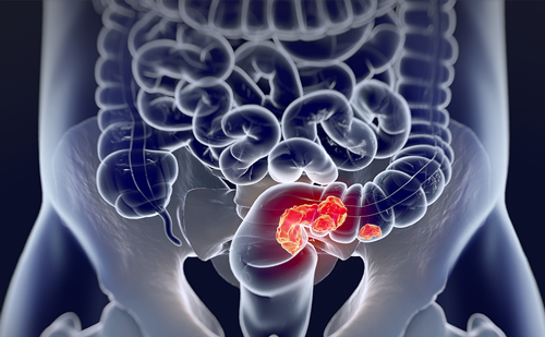
The two major symptoms in patients with peritoneal mesothelioma are abdominal pain and abdominal distention. The patient may also show constitutional symptoms, such as weight loss and fever. In women, diffuse malignant peritoneal mesothelioma (DMPM) is frequently diagnosed as an incidental finding. This fact may be responsible for the improved survival as a result of earlier detection of the disease in women.1
The signs of this disease are ascites, an abdominal or pelvic mass, a new onset abdominal wall hernia, guarding and rebound tenderness especially in the lower abdomen, and a pleural effusion (see Table 1).2 Unfortunately, a definitive diagnosis still eludes the physician in a significant number of patients. The cytologic examination of fluid removed by paracentesis is negative in approximately 90 % of patients. Therefore, either laparoscopy with tissue biopsy or mini-laparotomy with tissue obtained for biopsy may be necessary for a definite diagnosis. Jacquet and colleagues have cautioned that laparoscopy ports or sites for percutaneous tissue sampling under computed tomography (CT)- guidance should be confined to the midline.3 When fluid leaks out along these tracts from the peritoneal space, cancer cells will often be trapped within these tissues. The management of lateral laparoscopy port sites through the rectus muscle or needle tracts through the rectus muscle may be difficult and sometimes impossible to deal with surgically (see Figure 1).
Preoperative Quantitative Prognostic Indicators
Preoperatively, diagnostic tests can be used to select patients who are likely to benefit from the prolonged surgery combined with hyperthermic perioperative chemotherapy (HIPEC). Cerruto and colleagues have shown the prognostic significance of histomorphologic parameters in assessing prognosis.4 They quantitated the nuclear and nucleolar size and showed that it had a profound impact on outcome when patients had a standardized treatment. This histologic assessment of the epithelial type of DMPM is extremely important because 90 % of the patients do not show either sarcomatoid or biphasic histology. The majority of patients have the epithelial type of disease and requires a more sophisticated assessment of prognosis other than the tissue type alone. Currently, patients with sarcomatoid or biphasic DMPM are recommended for systemic chemotherapy as an initial treatment. Cytoreductive surgery with HIPEC has not been effective in this group of patients unless the extent of disease is small.
Patients with nuclear/nucleolar size 1 show a uniform round and small nucleus of 10–20 μm. The nuclear size is similar to that of red blood cells seen in the field. The chromatin pattern is granular and diffuse. Nucleoli are inconspicuous (see Figure 2).
With nuclear/nucleolar size 2, the nuclei remain moderately uniform in size, measuring between 21 and 30 μm. The chromatin pattern is granular and diffuse. The nucleoli are conspicuous and measure less than 3 μm (see Figure 3).
With nuclear/nucleolar size 3, the cells exhibit large and irregular nuclei measuring 31–40 μm. There is eccentric clearing of the chromatin. The nucleoli are prominent, measuring 3–5 μm (see Figure 4).
With nuclear/nucleolar size 4, the nuclei measure greater than 40 μm. They are similar in size to lymphocytes or macrophages seen in the field. There is eccentric clearing of the chromatin. There is a presence of macronuclei greater than 6 μm (see Figure 5).
When the survival of 62 patients treated in a uniform manner with cytoreductive surgery, HIPEC and EPIC paclitaxel were reviewed, there were clear survival differences when patients were placed in prognostic groups 1–4 based on nuclear/nucleolar size. There were no treatment failures with nuclear/nucleolar size 1 patients. Five-year survival was 50 % in those with nuclear/nucleolar size 2 and significantly less in nuclear/nucleolar sizes 3 and 4. These data are shown in Figure 6. It is clear that careful study of a biopsy from the peritoneal mesothelioma is extremely important in the preoperative assessment of prognosis in these patients.
Preoperative Prognostic Assessment using Computed Tomography
Yan and colleagues performed an analysis of the preoperative CT scans of 30 patients with peritoneal mesothelioma who were then treated in a standardized fashion using cytoreductive surgery and perioperative intraperitoneal chemotherapy at a single institution.5 These patients were divided into two groups depending on the extent of disease that remained after the cytoreduction. Those with residual lesion less than or equal to 2.5 mm were placed in the adequate cytoreduction group. Those with lesions greater than 2.5 mm remaining after maximal surgical efforts were placed in a suboptimal cytoreduction group. The preoperative CT scans for each patient were evaluated by a standardized scoring system. Thirty-nine CT parameters were obtained and statistically analyzed to determine their association with the adequacy of the cytoreductive surgery. Following the radiologic assessments, the statisticians were asked to construct a tree-structured diagram to allow the preoperative assessment of adequate cytoreduction. All the different combinations of the CT parameters were statistically tested for their predictive value. The two concerning radiologic features that combined most effectively to identify patients who would have suboptimal cytoreduction were tumor size greater than 5 cm in the epigastric region and class 3 changes within the small bowel and small bowel mesentery (see Figure 7). If a tumor in the epigastric region was less than 5 cm and there were no class 3 small bowel and small bowel mesentery change, 94 % of patients had an optimal cytoreduction. By contrast, if tumor size in the epigastric region was greater than 5 cm and there were class 3 small bowel and small bowel mesentery findings, 100 % of patients had a suboptimal cytoreduction.
The interpretative CT classification of small bowel and its mesentery is an important parameter in preoperative evaluation. Table 2 shows the interpretative CT classification of small bowel and its mesentery that was used by Yan and colleagues. In class 1 disease, the CT shows free intraperitoneal fluid only. The mesentery becomes stranded and stratified as the fluid accumulation outlines the small bowel mesentery. The small bowel vessels are identified within the mesenteric fat (see Figure 8, top). In class 2 classification, involvement of the small bowel mesentery by tumor is evident. The peritoneal lining is thickened and enhanced due to the presence of tumor nodules or tumor plaques. There may be an increased amount of ascitic fluid. The small bowel mesenteric vessels remained identifiable (see Figure 8, middle). With the interpretative CT classification 3, there is an increased amount of solid tumor so that adjacent bowel loops are brought together by the malignant process. The normal configuration of the small bowel and its mesentery is distorted and the structures thickened. The small bowel mesenteric vessels are difficult to define due to the obliteration of the mesenteric fat density (see Figure 8, bottom).
Yan and colleagues showed that CT can effectively identify the presence of peritoneal mesothelioma at crucial anatomic sites and create a list of concerning radiologic features that suggest suboptimal cytoreductive surgery. As will be seen below, this is extremely important because adequate cytoreduction is necessary to achieve a prolonged survival with the combined treatment. Preoperative CT scans are an accurate prognostic radiologic test for patient selection for a comprehensive treatment plan.
Intraoperative Assessment of Prognosis
As the surgeon explores the abdomen of a patient with DMPM, a careful assessment of the distribution and extent of disease must occur. This is quantitatively recorded on a diagram referred to as the peritoneal cancer index (PCI) (see Figure 9). The lesion size between no tumor seen and tumor masses greater than 5 cm or a confluence of disease determines the lesion sizes 0–3. Thirteen abdominopelvic regions numbered 0–12 are used to identify the anatomic sites for the disease. The abdominopelvic regions 0–8 are associated with the abdominal and pelvic surfaces exclusive of the small bowel regions.
The abdominopelvic regions 9, 10, 11, and 12 indicate the upper jejunum, lower jejunum, upper ileum, and lower ileum, respectively. A lesion size is assigned to each one of these abdominopelvic regions 0–12 to arrive at a PCI between 0 and 39.
The PCI has been determined to be a useful prognostic indicator in DMPM patients. Sugarbaker and colleagues showed that a PCI between 0 and 28 was associated with a median survival of 67 months.2 A PCI between 29 and 39 showed a 26-month median survival (p=0.046).
Intraoperative Prognostic Assessment by Cytoreduction Score
The second intraoperative quantitative prognostic indicator is the completeness of cytoreduction (CC) score). This is determined after the surgeon’s best efforts using peritonectomy procedures and visceral resections to clear the abdomen and pelvis of disease. A CC score of 0 indicates no visible evidence of DMPM. A CC-1 score indicates tumor nodules less than 0.25 cm in greatest diameter. No assessment of the number of nodules for CC score quantitation is needed. However, a CC-1 score indicates that the small nodules are individual nodules and not a confluence of disease. A CC-2 score indicates nodules between 0.25 cm and 2.5 cm. For a CC-2 score, the nodules are individual nodules without a confluence of disease on either visceral or parietal peritoneal surfaces. For a CC-3 score, the nodules are greater than 2.5 cm. Alternatively, a layering of smaller tumor nodules so that there is a confluence of disease on peritoneal surfaces would be considered a CC-3 score.
Sugarbaker and colleagues determined the importance of the completeness of the CC score in assessing prognosis. In 41 patients with a score between 0 and 2, there was a 67-month median survival. In patients with a completeness of CC score of 3, there was a 26-month median survival (p=0.003). The completeness of CC score was a dominant prognostic indicator in terms of the success of these treatments.2
It is important to establish that the CC score in patients with peritoneal mesothelioma may not be as definitive as for patients with gastrointestinal malignancy. For gastrointestinal cancer the CC score is a determinant of survival. By contrast, for peritoneal mesothelioma patients, CC-0 (no visible evidence of disease), CC-1 (nodules less than 2.5 mm in diameter), and CC-2 (nodules between 2.5 mm and 2.5 cm in diameter in the absence of a confluence of disease), survival was not clearly separable by statistical analysis. Patients with CC-3 cytoreduction did significantly worse with these treatments. It is quite possible that larger numbers of peritoneal mesothelioma patients would increase the prognostic implications of the CC score.2
Treatment
Currently, the surgery for patients with DMPM is referred to as cytoreductive surgery and it involves a series of peritonectomy procedures combined with visceral resections in an attempt to clear, with a goal of complete visible removal of all disease. The peritonectomy procedures and visceral resections are listed in Table 3.
The treatment plan that has evolved in these patients over the last 2 decades is shown in Figure 10. The first step in treatment is to establish a diagnosis. With the biopsy, the prognostic significance of histomorphologic parameters is determined. A CT scan of chest, abdomen, and pelvis is then used and the concerning radiologic features are summarized. If it is thought that the histology and preoperative CT suggests a high likelihood of complete or near complete (CC-0, CC-1, or CC-2) cytoreduction, the patient is recommended for surgery using the peritonectomy procedures and visceral resections outlined above.
At the completion of the cytoreduction, HIPEC with doxorubicin and cisplatin is used. Also, there is an intravenous administration of ifosfamide by continuous infusion. At the time of surgery, a Tenckhoff catheter is positioned to maintain a high inflow of cancer chemotherapy. Four drains are positioned so rapid outflow of peritonealfluid can occur. A heat pump recirculates the chemotherapy solution to maintain the temperature within the abdomen and pelvis at 42–43°C (see Figure 11).
The inflow catheters and outflow drains are retained within the abdomen and pelvis postoperatively. Through the Tenckhoff catheter and four closed-suction drains the patient is treated with 20 mg/m2 of paclitaxel for 5 days. This calls for a total of 100 mg/m2 of paclitaxel given in the early postoperative period. A second intraperitoneal access device is the intraperitoneal port, which is used for intraperitoneal pemetrexed combined with intravenous cisplatin every 4 weeks for 6 cycles. The standardized orders for the HIPEC and EPIC are shown in Tables 4 and 5. The standardized orders for long-term intraperitoneal chemotherapy through an intraperitoneal port are shown in Table 6.
Results of the Evolution of Treatment Strategies for Peritoneal Mesothelioma Over 2 Decades
The evolution of treatments that has occurred at the Center for Gastrointestinal Malignancies, MedStar Washington Hospital Center is as follows. In group 1 patients, the treatments were limited to cytoreduction plus HIPEC doxorubicin and cisplatin (n=42). The group 2 patients had cytoreduction plus HIPEC cisplatin and doxorubicin combined with EPIC paclitaxel (n=59). In group 3, the patients had cytoreduction plus HIPEC doxorubicin and cisplatin, EPIC paclitaxel, and combined intraperitoneal pemetrexed and systemic cisplatin for 6 months (n=29). These data clearly show the marked improvement in survival of these patients when the long-term regional chemotherapy treatments were used.
The 10-year survival for group 1 is 24 %, group 2 38 %, and group 3 73 %. These survival estimates were significant with a p=0.0276. These results need to be interpreted with great caution. It must be clearly stated that patient groups were not compared in the context of controlled trials. Also, improvements in the learning curve, especially improved patient selection, may have contributed (at least in part) to better prognosis. The study has been carried out over 20 years.
In conclusion, peritoneal dissemination of peritoneal mesothelioma is the only major pathway for spread of this disease. This regional dissemination suggests that advanced treatments within the abdomen and pelvis may be associated with benefit. Patients can now be selected for benefit based on preoperative quantitative prognostic indicators using histomorphologic pathologic parameters and a carefully performed CT scan of the chest, abdomen, and pelvis. Clearly, these two preoperative assessments are of great value in excluding patients who will not profit from the aggressive management plan that does carry a mortality of 1 % and serious morbidity of 12 %. Following the best efforts at cytoreduction, regional chemotherapy treatments using HIPEC (cisplatin, doxorubicin, and ifosfamide) combined with EPIC (paclitaxel) are used. Long-term intraperitoneal or combined intraperitoneal and systemic treatments are associated with a marked improvement in survival suggesting that this long-term treatment should be added to the standard of care for these patients. It is suggested that this long-term local-regional treatment strategy be substituted for systemic chemotherapy in patients with this disease process.