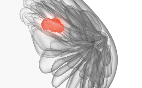
In the past few decades, a major shift in the local management of breast cancer has occurred: mastectomy was replaced by breast-conserving surgery (BCS) followed by post-operative whole breast radiotherapy (WBRT).1,2 Solid evidence from randomised trials supports that the combined treatment has equivalent results to mastectomy in terms of local control and survival rates.1,2 This is a paradigm of a changing standard of care in favour of a new treatment that provides the same clinical results in terms of cure rates, decreased side effects, organsparing and the prospect of a better quality of life.
The role of radiotherapy after breast cancer surgery is to minimise the local relapse risk. Post-operative radiotherapy includes the irradiation of the whole remaining breast with about 50Gy delivered daily for five to six weeks with an additional local boost dose (10–16Gy) to the tumour bed, when appropriate.3 Recently, a new concept of accelerated partial breast irradiation (APBI), which consists of the irradiation of a limited volume of mammary gland immediately surrounding the tumour bed, has been investigated as an alternative to WBRT.
Rationale for and Potential Advantages of Partial Breast Irradiation
The rationale for the use of PBI was based on clinical4–7 and pathological8,9 observations of long-term studies reporting that the majority of breast tumour recurrences occur in proximity to the lumpectomy cavity. In addition, breast cancer relapses outside of the original tumour bed appear to occur with the same frequency following lumpectomy regardless of whether or not adjuvant whole breast irradiation is delivered.6,10 Consequently, WBRT may not be necessary since most ipsilateral breast tumour relapses occur in the vicinity of the primary tumour and radiotherapy does not seem to prevent other quadrant relapses.
Furthermore, PBI offers increased convenience due to a shorter duration of radiation therapy (five to seven days versus six weeks). This significant shortening of the treatment time is extremely important in order to overcome socioeconomic barriers that negatively affect patient compliance with radiotherapy. Indeed, a high rate of breast cancer patients tend to choose mastectomy instead of lumpectomy plus radiotherapy due to limited financial means, and/or long travel distances to the radiation facilities, and/or lack of time and/or due to their advanced age.11–13
Moreover, in countries with limited radiotherapy institutions, patients treated with BCS may wait a prolonged time before beginning radiotherapy. Consequently, there is a delay in the initiation of systemic adjuvant chemotherapy, which may affect overall survival.14
Another important potential advantage of PBI could be the avoidance of side effects, especially cardiac and pulmonary toxicities. In PBI less radiation is delivered throughout normal breast tissue and other organs since treatment targets are more narrowly focused. Finally, the reduction of overall treatment time needed for radiotherapy in PBI might save costs compared with WBRT.15
Partial Breast Irradiation Techniques
PBI can be carried out with four principally different techniques: interstitial brachytherapy with multiple catheters, intracavitary brachytherapy (Mammosite system, Cytyc, Marlborough, MA), intraoperative radiotherapy and external 3D conformal radiotherapy. Each of these techniques has unique advantages and limitations and is in a different stage of development and acceptance.
Interstitial brachytherapy with catheters is the PBI technique with the longest reported period of follow-up.16 In this approach, catheters are placed at 1–1.5cm intervals through the breast tissue surrounding the lumpectomy cavity. The number of catheters is determined by the shape and the size of the target. Interstitial brachytherapy has been used with all possible dose rates, including low- and high-dose rates. This technique permits individual conformation of the irradiated volume precisely adapted to the anatomical conditions.17 However, this approach results in significant heterogeneity of dose delivered,18–20 the target coverage might be inferior to other PBI techniques20 and the skin dose might be elevated.21
Intracavitary brachytherapy (MammoSite) has been developed to be a less operator-dependent procedure compared with interstitial brachytherapy. The MammoSite is a balloon catheter, which consists of a double-lumen catheter with an inflatable balloon at the distal tip. The balloon is inserted in the lumpectomy cavity, either during or following breast-conserving treatment, and is then filled with saline and contrast material such that the surrounding tissue is stretched tightly around it. Treatment is delivered immediately in the lumpectomy cavity using a high-dose source, which is inserted into the centre of the balloon. The method is simple, with a short learning curve.22 Conversely, the limitations of the technique are that the target volume is standardised without allowing individual conformation and the therapeutic range is only 10mm.17 Additionally, the distance between the balloon and the skin appears to be the most important factor for achieving optimal cosmetic results.23,24 Moreover, catheters can be a source of discomfort and potentially promote bleeding, infections and late damage such as fibrosis and telangiectasia.
Intraoperative radiotherapy (IORT) consists of single-fraction treatment targeted at the tumour bed during the surgical procedure, immediately after the removal of the tumour mass. Two modalities of IORT have been described using either electron, as developed in Milan,25 or photon beams (based on ‘soft’ X-rays of 50kV) developed by the University College of London.26 As this irradiation is performed during the same surgical procedure, there is no need for future hospitalisation and transportation of patients. Moreover, the application of a high-dose precisely targeted to a limited volume can be performed while sparing the surrounding tissues.17 The major flaw of this technique is the lack of definite pathological data regarding resection margins, histological features and axillary nodal status at the time of radiation therapy.
A recent consensus statement from the American Society for Radiation Oncology (ASTRO) concluded that there are insufficient clinical and dosimetric data to determine the optimal technique for APBI delivery.16
Current Randomised Evidence
To date, five randomised trials have been published comparing PBI and WBRT in patients with early breast cancer. Four of them28–30,32 reported data on locoregional recurrences and overall survival, while the fifth study31 was a preliminary acute toxicity analysis of an ongoing randomized trial. A meta-analysis33 of three completed randomised studies28–30 has been recently published while the fourth study32 was presented after the completion of the meta-analysis and was not included.
The earliest phase III randomised trial on APBI was conducted from 1982 to 1987 at the Christie Hospital in Manchester.29,34 Overall, 713 patients with breast tumours less than 4cm and negative lymph nodes were randomised, after BCS, to receive either WBRT (40Gy in 15 fractions in 21 days) or limited field irradiation only to the tumour bed (40–42.5Gy in 10 fractions in 10 days). In this study, the limited field irradiation was significantly associated with higher local and regional recurrences, while there were no differences in terms of overall survival and distant metastases between the two treatment arms. These disappointing results should be interpreted with caution due to the old radiation technique used in the study, the poor quality control, the inadequate axillary and systemic management and the incomplete pathological examination.35 In addition, a single field size was used for all patients in the limited-field arm irrespective of the tumour dimensions or other characteristics, which could have resulted in several instances of ‘geographical miss’. The lack of appropriate patient selection criteria was highlighted when the results were analysed according to the type of primary tumour and it was found that limited-field radiotherapy was inadequate only for patients with infiltrating lobular cancers or cancers with an extensive intra-ducta component.
The second study was performed during 1986–1990 by the Yorkshire Breast Cancer Group.30 In this study, 174 patients, irrespective of nodal status, were randomised to receive either WBRT or irradiation only in the tumour bed. In this study, a higher risk of locoregional recurrences was demonstrated. However, there were uncertainties on target-volume definition, the irradiation technology used was inadequate, more patients in the tumour-bed-only irradiation arm had axillary node positive disease and the study did not reach its target on participants due to a low accrual rate.
The first well-designed randomised phase III trial was performed by the National Institute of Oncology in Hungary.28,36,37 In this study, 258 patients with T1, N0–1mi, grade 1–2, non-lobular breast cancer undergoing BCS were randomised to either WBRT or APBI (high-dose-rate interstitial brachytherapy or limited-field electron-beam radiotherapy). At the median follow-up of 60 months there were no significant differences in locoregional recurrences or overall survival. Regarding toxicity, there were no significant differences between the two treatment arms in terms of incidence of fat necrosis and late radiation side effects.37 Patients with APBI had a better cosmetic result compared with WBRT.37 The Hungarian study is the only randomised trial that analysed the cosmetic results and the late side-effects of APBI.
A preliminary analysis of an Italian study including 259 patients who were randomised to receive either WBRT or APBI demonstrated that APBI has a very low acute skin toxicity.31 A meta-analysis of the three above-mentioned phase III randomized controlled studies comparing partial breast irradiation with whole breast-radiation therapy was recently presented.33A total of 1,140 patients were included: 575 were randomised to whole breast irradiation and 565 to limited-field or PBI. The meta-analysis revealed no statistically significant difference between the partial and whole breast radiation arms associated with death (odds ratio [OR] 0.912, 95% confidence interval [CI] 0.674–1.234; p=0.550), distant metastasis (OR 0.740, 95% CI 0.506–1.082; p=0.120) or supraclavicular recurrences (pooled OR 1.415, 95% CI 0.278–7.202; p=0.560). APBI therapy resulted in a statistically significantly higher risk for developing local recurrences (pooled OR 2.150, 95% CI 1.396–3.312; p=0.001] and axillary recurrences (pooled OR 3.430, 95% CI 2.058–5.715; p<0.0001). The results of the meta-analysis are encouraging about the future role of APBI since it does not seem to jeopardise overall survival in patients with early breast cancer. The higher locoregional recurrence risk in the APBI arm needs to be considered with caution due to biases of the eligible studies,28–30 as discussed above.
Nevertheless, the limited available randomised data (only three eligible trials), the variety of the PBI techniques used, the poor methodological quality of the two included trials29,30 and the relatively shorter median follow-up (five years) in two trials28,30 compared with the median time needed in order to demonstrate the impact on mortality (seven to eight years)38 are considerable limitations of the meta-analysis.
Recently, the results of the Targeted Intraoperative Radiotherapy (TARGIT-A) trial,32 a large international randomised controlled trial of targeted IORT versus WBRT for breast cancer, were presented at the American Society of Clinical Oncology (ASCO) Annual Meeting 2010.32 In this study, 2,232 patients were randomised to receive either intraoperative targeted radiotherapy or whole breast external beam radiotherapy. With targeted radiation technique the surface of the tumour bed typically received 20Gy that attenuated to 5–7Gy at 1cm depth. The study presents data in terms of local recurrences and toxicity with a median follow-up of four years. There were six local recurrences in the IORT group and five in the external beam radiotherapy group, with no difference between the two arms. In terms of frequency of complications and major toxicities, no differences were observed between the two arms, while radiotherapy-related toxicities were significantly lower in targeted radiotherapy arm. This study represents the largest randomised trial on this topic and provides additional randomised evidence about the safety of PBI and the potential benefits from the use of this technique in women with early breast cancer.
Currently, six ongoing phase III randomised trials in the US and Europe are testing APBI (with all available techniques) against WBRT after BCS.39–44 Data from these trials are expected to provide level I evidence about the application of PBI in clinical practice.
Concerns Regarding Partial Breast Irradiation
Besides the above-mentioned theoretical advantages of APBI, there are also data from studies that raise concerns in terms of the rationale for this new technology. The data from clinical45,46 and histopathological47 studies show that a considerable proportion of local recurrences after BCS occurred away from the primary tumour. Several studies have attempted to identify the pattern of ipsilateral breast tumour relapse after conservative surgery; however, the results are contradictory and not easily comparable, since the definition of same-site relapse has no generally accepted criteria and the extension of surgery varies. This means that a local recurrence close to the surgical cavity after a quadrantectomy corresponds to breast recurrence elsewhere if a lumpectomy had been performed.48 Another concern is raised from detailed histopathological analysis of the entire specimen after mastectomy47,49,50 and magnetic resonance imaging51 studies suggesting that multifocal-same-quadrant or multicentric-other-quadrant foci are relatively common in patients with early-stage breast cancer. The extent of the disease in these patients cannot be encompassed using partial breast irradiation techniques. The clinical significance of multifocal and multicentric foci are uncertain;49 however, the reason these foci did not reach clinical significance may be that patients who suffer from the first local recurrence, which in the vast majority of cases occurs close to primary tumour, undergo mastectomy before a tumour focus becomes clinically apparent. Finally, evidence suggests that rates of ipsilateral recurrence away from the primary site after BCS and WBRT are lower than new breast cancer in the controlateral cancer.46,52 Even if radiotherapy seems to increase the development of new primary cancer in the controlateral breast,53 these data suggest that WBRT could have a protective effect on other areas of the breast.
Conclusion
PBI is a new technology that offers potential advantages compared with WBRT. The most valid concern regarding PBI as a new treatment modality in oncology is its oncological safety. Although limited, current randomised evidence supports that this new technology is a safe treatment modality as it does not seem to jeopardise survival compared with standard WBRT. Nevertheless, this radiation-delivering technique is unlikely to replace WBRT as the ‘gold standard’ treatment for all early breast cancer patients. Ongoing large phase III randomised trials will identify the subgroups of patients who will benefit from PBI. Until then, PBI methods remain investigational and should be performed only in patients enrolled in controlled clinical trials. ■


