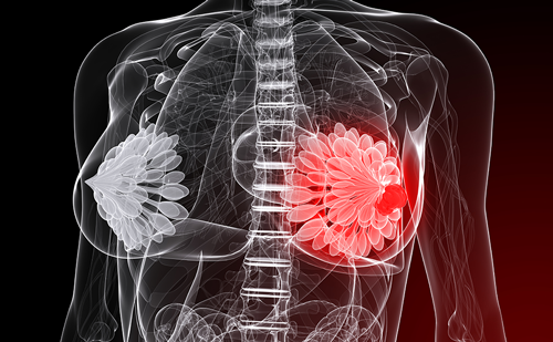
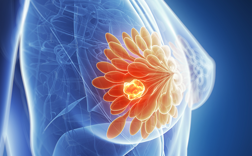
The current standard treatment for early breast cancer includes conservative surgery followed by radiotherapy to the whole breast.1 Longterm follow-up of patients included in the Milan Three Trial (Quart versus Tart) showed that most local relapses occur in the area of the scar tissue,2 suggesting that total irradiation of the breast may not always be necessary.
We started our experience in intra-operative radiotherapy for breast cancer in June 1999 with a dose-finding study to test the feasibility of this new approach during breast-conserving surgery.3 Electron intra-operative therapy (ELIOT) exploits new technologies that make it possible to deliver radiation to a more circumscribed area of the breast. The technique also markedly reduces the time required for a radiotherapy course, improving the quality of life of the patients. From June 1999 to October 2000, we experimented with different dose levels and verified the tolerance of 21Gy prescribed at 90% isodose as a full dose of intra-operative radiotherapy for small-size breast tumours (maximum tumour diameter 2.5cm).4,5
The different dose levels are reported in Table 1. Dose levels of 10 and 15Gy were followed by a reduced course of external fractionated radiotherapy. We obtained excellent results in terms of acute and intermediate tolerance of treatment and we adopted the dose of 21Gy prescribed at 90% isodose as the full dose approach for the randomised trial.6,7
In the pilot study (escalation dose) on 101 patients, after a mean followup of 42 months, 16 patients (16%) developed breast fibrosis (mild in 15 and severe in one), which resolved within 24 months, three patients suffered post-operative haematoma and four developed a lyponecrosis in the treated area. The phase III randomised trial started on 20 November 2000 and enrolled patients older than 48 years affected by unifocal breast carcinoma with a diameter ≤2.5cm. Patients received breastconserving surgery were randomised during the operation for ELIOT 21Gy or external fractionated conventional radiotherapy (50Gy whole breast and 10Gy boost to tumour bed).1 Up to August 2006, we included 1,064 patients in the trial: 535 patients received conventional radiotherapy (60Gy) and 529 received ELIOT (21Gy). We are still recruiting patients and the follow-up of treated patients is ongoing.8
From a surgical point of view, most patients received wide excision with sentinel node biopsy, while a more limited number of patients received wide excision with axillary dissection.9,10 We also treated 1,129 other patients with ELIOT outside the randomised trial by specific request of the patients themselves. From February 2000 to August 2006, we treated 59 cases and the follow-up of these patients is also ongoing. Globally, our experience with 21Gy ELIOT involves 1,759 patients with infiltrating breast carcinomas.11
We are testing ELIOT on selected cases of ductal carcinoma in situ (DCIS) with the premise that DCIS treatment with breast-conserving surgery benefits from post-operative radiotherapy, but whole breast irradiation may appear excessive in very limited size DCIS. Finally, from March 2002 to June 2006 we adopted ELIOT (16Gy) for irradiating the nipple areola complex during nipple-sparing subcutaneous mastectomy for multicentric in situ ductal carcinomas or multicentric infiltrating cancer with negative retro-areolar frozen sections. This is a new technique we developed to relieve the feeling of mutilation during mastectomy, by reducing the risk of local relapse thanks to ELIOT. The preliminary results are encouraging on 666 patients (225 cases of in situ carcinomas) with an excellent cosmetic result.12
Surgical Procedures
Electron Intra-operative Therapy after Quadrantectomy
Patients undergo quadrantectomy according to the Veronesi’s technique with sentinel node biopsy (SNB). (Only patients with positive SNB undergo axillary dissection.) ELIOT requires a special sequence of procedures to facilitate the radiation treatment. Immediately after the removal of the breast quadrant, the remaining parenchyma should be separated from the pectoralis fascia to place an aluminium–lead shielding disc posterior to the parenchyma to protect the thoracic wall, the heart and the lung (see Figure 1). The anatomy of the breast is temporally restored by suturing the gland, taking care to correctly expose the clinical target volume. The treated volume should include the entire surgical scar, plus a safety margin of 1.5–3cm. A metallic ring with non-traumatic hooks is used to hold open the skin and the applicator is positioned (see Figure 2). A wet sterile gauze is positioned between the applicator and the surrounding tissues to absorb the low-energy electrons scattered around the applicator edge. The applicator is connected to the head of the treatment machine (hard docking). The monitor units needed to deliver the prescribed dose using an electron beam of appropriate energy are calculated, and the patient treated. ‘Beam-on’ time is less than two minutes, and the entire procedure lasts about 15–20 minutes. After irradiation, all the materials are removed and cosmetic reconstruction of the breast is performed.9
To perform ELIOT, we currently using two dedicated, mobile linear accelerators – a Novac7 and a Liac – installed in two different operating rooms and delivering electron beams at high dose rate. The two linear accelerators, which can be easily manoeuvred by means of motors acting on the wheels and the articulated arm, deliver electrons at the following different nominal energies: 3, 5, 7 and 9MeV (Novac7) and 4, 6, 8 and 10MeV (Liac). Beam collimation is achieved by a hard-docking system, consisting of round perspex applicators that are 5mm thick. Flat-ended and bevelled (22.5° and 45°) applicators of 4, 5, 6, 8 and 10cm diameter are employed. The nominal source-to-surface distance (SSD) is 100cm for the 10cm applicator and 80cm for all others. For radiation protection, a primary beam stopper (a trolley-mounted 15cm-thick lead shield) and mobile 1.5cm-thick lead shields (100cm long, 150cm high) are used.
Based on our experience, we believe that ELIOT will be rapidly utilised for the treatment of breast carcinoma, with a positive impact on the quality of life of patients and on the treatment organisation. Data from our randomised trial will be critical to establish the effectiveness of ELIOT in preventing local relapse of disease in the breast.