
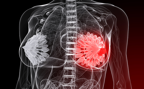
The key to surviving breast cancer, the second most common cancer affecting women, is early detection and treatment. When the cancer is confined to the breast, the five-year survival rate is nearly 100%. Mammography screening is widely regarded as the only proven method for early detection of breast cancer. Large populations can be screened at a relatively low cost. However, mammography does not detect all breast cancers and in some series the false negative rate is between 20% and 30%. Mammography is limited in patients with dense breast tissue and with certain types of tumours. For this reason, different techniques for improving diagnosis of breast cancer, particularly among high-risk women, are being explored.
High-risk women include those who have had a previous breast cancer or other high-risk histologies, those with a strong family history of breast cancer and those who are carriers of either of two familial breast cancer genes, BRCA1 or BRCA2. These genes are responsible for 5% to 10% of all breast cancers. Women with mutations in BRCA1 or BRCA2 genes are at an especially high risk of developing breast cancer when they are relatively young and have dense breast tissue, which lowers the sensitivity of mammography.
Magnetic resonance imaging (MRI) is one of the most promising of the emerging technologies for breast cancer screening in high-risk patients. Of all current modalities, MRI has been shown to have the highest sensitivity for the detection of invasive cancer. Multiple studies comparing the results of breast MRI with pathological outcomes show that breast MRI identifies at least 95% of invasive cancers. MRI is also useful for detecting the presence of ductal carcinoma in situ (DCIS) although sensitivity is variable, ranging from 40% to 100%. About 20% of new breast cancer cases will be DCIS, a potentially curable disease.
To date, no society or professional organisation recommends screening for breast cancer with modalities other than mammography largely because the other modalities, including MRI, are more costly and are not suitable for large populations. However, in 2003, the American Cancer Society updated its guidelines to suggest that certain women, i.e. high-risk patients, could benefit from additional screenings with either ultrasound or MRI.
Once a diagnosis of cancer has been pathologically confirmed, MRI is useful for staging the tumours and assessing tumour response to treatment. Compared with mammography, clinical breast exam or ultrasound – the conventional methods for determining tumour size and extent – MRI is at least as good and in many cases better.
Magnetic resonance has also been shown to be the most accurate means of evaluating breast implants for possible complications, such as rupture, and when contrast is administered, it can evaluate the overlying breast tissue, which may be obscured by the implant. MRI of the breast is best performed with a dedicated breast imaging coil. The patient lies in a prone position (on her abdomen) with her breasts hanging through a cut-out in the table while the images are taken. The examination can usually be completed in less than 45 minutes.
Most breast MRI indications require the use of the contrast agent gadolinium-DPTA. When injected into the patient’s bloodstream, the contrast circulates in the vascular system, and can locate tumours by accumulating in areas of increased vascularity (neovascularity) associated with developing tumours. As with contrast mammography, scans are taken before and after contrast injection. Areas that show high uptake of the contrast are analysed.
Breast malignancies typically show more rapid enhancement and faster wash-out than benign tissues on MRI, but there is overlap in the enhancement curves of benign and malignant lesions. To resolve the question, further imaging or biopsy may be needed. Of the false negative MRIs, most are DCIS but invasive cancers, especially lobular, can sometimes be missed as well.
A number of studies conducted in the US and Europe have shown that MRI may benefit high-risk patients by detecting non-palpable and mammographically occult breast cancer. While the patient groups were small in most of the studies, MRI detected more small cancers (<2cm) and cancers associated with negative axillary nodes than mammography. Mammographic screening has been shown to reduce mortality by detecting these early cancers. Thus, screening high-risk women with MRI may reduce the mortality rate from breast cancer even further. The literature suggests that MRI imaging finds additional sites of breast cancer in 6% to 34% of patients.
Ultrasound also detects some cancers in dense breast tissue that are missed by mammography. It has a lower cost than MRI and is more readily available. However, it has lower sensitivity and specificity than MRI, is operator-dependent and often will not detect DCIS.
A prospective study of 1,909 women with a mean age of 40 years was reported by Kriege et al.1 Forty-five cancers were identified among the 358 very high-risk women who were BRCA1 and BRCA2 carriers. Of the 45 cancers, 18 were seen on mammography; 32 were seen on MRI; eight were seen on mammography only; and 29 seen on MRI only.
Another study at the Institute of Cancer Research in London of 649 women aged 35 to 49 at high risk of breast cancer based on genetic testing or family history was reported at the Annual Society of Clinical Oncology in May. Annual MRI and mammography screening detected a total of 35 breast cancers in the study group. Of them, 19 were detected by MRI only; six by mammography only; and 10 by both MRI and mammography. Again, the benefits of MRI were most evident among women with BRCA1 mutations. Among this subgroup, mammography detected 23% of tumours while MRI detected 92%. MRI is therefore considered to be a complement tomammography and not a substitute.
There is some concern that screening with MRI might produce a high number of false positive results, which could lead to call-backs and unnecessary biopsies. However, this is not what is happening, especially when MRI is reserved for high-risk and carefully selected patients.
Several studies have found that when MRI is performed among high-risk groups, between 15% and 25% of patients require follow-up procedures based on the initial findings. About 40% of patients who undergo biopsy are found to have malignancies. The positive predictive value (PPV) is significant enough to justify MRI screening in high-risk populations.
Once a lesion is identified by MRI, a biopsy can be performed percutaneously under either ultrasound or MR guidance. In some cases, surgical biopsy with pre-operative MR needle localisation may be required. The American Cancer Society’s guidelines for breast cancer screening say, “Breast MRI should be performed in centers with extensive experience in diagnostic MRI and the capacity for MRI-guided biopsy”.
Based on the results at the Faulkner-Sagoff Breast Imaging and Diagnostic Centre of more than 12,000 breast MRIs, the centre’s protocol for high-risk patients is annual mammograms and annual MRI evaluations, alternating at six-month intervals.
MRI is also finding wider use in the clinical management of breast cancer as it is particularly effective for determining the extent of the disease once it has been confirmed by needle biopsy. A number of studies have compared MRI, mammography and ultrasound for estimating tumour size, and MRI showed the highest agreement with histopathology. MRI shows additional disease in up to one-third of patients, which changes management in approximately one-half of these patients. A contralateral cancer is detected by MRI in 4% to 9% of newly diagnosed patients. Because local recurrence rates are low following breast conservation and radiation therapy, and overall survival is equivalent to mastectomy, MRI may not change the outcome, but could potentially reduce the number of surgeries if the extent of disease is better defined on MRI. The information provided by MRI can be used to determine whether neo-adjuvant chemotherapy is indicated and whether breast conservation is appropriate.2
A number of randomised clinical trials, including NSABP B-18, have shown that while neo-adjuvant systemic chemotherapy does not change disease-free and overall survival, it does improve the chance for breast conservation. Consequently, the use of preoperative or neo-adjuvant chemotherapy has been on the rise.
The staging accuracy of MRI makes it an attractive method for monitoring patients during pre-operative chemotherapy. MRI is also useful for staging residual disease following excision with positive margins and after chemotherapy. In comparisons of MRI, mammography, ultrasound and/or clinical examination for residual disease after chemotherapy, MRI proved to have the closest agreement with histology.
MRI was evaluated in a study of 30 patients at the Institute Curie undergoing pre-operative chemotherapy. MRI would have led to major appropriate changes in treatment for six patients, including five with multicentric disease, and would have helped in surgical planning. MRI would have shown that five of 14 patients with single residual foci would have been candidates for breast conserving therapy (BCT) and nine with multiple residual foci and limited reduction in tumour size were less appropriate candidates for BCT.
Tillman et al.3 reviewed 207 women with 212 early-stage breast cancers who underwent breast MR as part of their evaluation. Based on the MR images, management was changed in 30% of the cases. Of those changes, 63% were deemed favourable.
A study by Drew and colleagues4 of patients with locally advanced breast cancer receiving neo-adjuvant chemotherapy found MRI to be 100% sensitive and specific for defining residual tumour after chemotherapy. In comparison, mammography achieved only 90% sensitivity and 57% specificity, while clinical exam was 50% sensitive and 85% specific.
Another study by Partridge et al.5 of 52 patients confirmed the sensitivity of MRI of the breast after chemotherapy. The patients were imaged before and after chemotherapy. Researchers measured the tumour size prior to treatment using MRI and clinical examination. They examined the same area on the post-chemotherapy images looking for any remaining enhancement. MRI found every case of residual disease while clinical assessment found five false negatives among the 52 treated lesions. In a later study of 62 patients reported by Partridge,6 MRI monitoring of change in tumour size with treatment was more significantly predictive of disease recurrence than clinical or pathological assessments.
The role of MRI in assessing response during treatment is not as clear because only a few small studies have been reported. It would be useful if MRI could reliably identify patients whose tumours are not responding to neoadjuvant chemotherapy. Therapy could be changed or discontinued if it were shown to be ineffective. However, it would be harmful if the lack of tumour response was judged prematurely and the treatment changed or discontinued too early. More research is needed in this area.
MRI has also shown a high level of accuracy in identifying chest wall invasion. A prospective study by Morris et al.7 was conducted of 19 patients with breast tumours suspected to involve the pectoralis major muscle. Clinical examination showed 13 of the tumours to be fixed to the chest wall while 12 appeared on mammography to have pectoral muscle involvement. MRI was 100% sensitive and 100% specific for identifying the five tumours that actually involved the pectoralis major muscle. Given its high level of diagnostic accuracy, it is fair to conclude that MRI is an appropriate imaging modality in this setting.
A small subset of patients present with positive axillary nodes, but no evidence of breast cancer on conventional imaging. MRI can detect the primary tumour in almost two-thirds of patients. This can be used to guide breast conserving surgery rather than mastectomy in a large number of patients.
Finally, of the three techniques used to detect implant ruptures (mammography, ultrasonography and MRI), a number of studies suggest that MRI is the most sensitive and specific.
The future of breast MRI is very exciting. MRI offers the potential for improved detection, especially among high-risk populations, as well as improved staging and follow-up treatment. Certainly, caution must be exercised as the technology is implemented into clinical practice. The technology is expensive, but when used appropriately, the benefits to patients justifies its cost. MR also has the advantages of being non-invasive and has no ionising radiation. As MR technology continues to mature, MRI surely will gain wider acceptance as a breast imaging procedure and will play a more central role in a variety of clinical indications. ■
Magnetic Resonance Imaging Gaining Importance in Breast Cancer High-risk Screening and Disease Management
Article
References
- Kriege M, Brekelmans C T M, Boetes C, Besnard P E, Zonderland H M, et al., “Efficacy of MRI and mammography for breast cancer screening in women with a familial or genetic predisposition”, N. Engl. J. Med. (2004);351: pp. 427–437.
- Berg W A, Gutierrez L, NessAiver M S, et al., “Diagnostic accuracy of mammography, clinical examination, US and MR imaging in preoperative assessment of breast cancer”, Radiology (2004);233: pp. 830–849.
- Tillman G F, Orel S G, Schnall M D, Schultz D J, Tan J E, Solin L J, “Effect of breast magnetic resonance imaging on the clinical management of women with early stage breast carcinoma”, J. Clin. Oncol. (2002);20 (16) pp. 3,413–3,423.
- Drew P, Chatterjee S, Turnbull L, et al., “Dynamic contrast enhanced magnetic resonance imaging of the breast is superior to triple assessment for the pre-operative detection of multifocal breast cancer”, Ann. Surg. Oncol. (1999);6: pp. 599–603.
- Partridge S C, Gibbs J E, Lu Y, “Accuracy of MR imaging for revealing residual breast cancer patients who have undergone neoadjuvant chemotherapy”, AJR (2002);179: pp. 1,193–1,199.
- Partridge S C, Gibbs J E, Lu Y, et al., “MRI measurements of breast tumor volume predict response to neoadjuvant chemotherapy and recurrence – Free Survival”, AJR (2005);184: pp. 1,774–1,781.
- Morris E A, Schwartz L H, Drotman M B, et al., “Evaluation of pectoralis major muscle in patients with posterior breast tumors on breast MR images: Early Experience”, Radiology (2000);214: pp. 67–72.
Further Resources

Trending Topic
It is with great pleasure that we present the latest edition of touchREVIEWS in Oncology & Haematology. This issue highlights the remarkable progress and innovation shaping the fields of oncology and haematology, featuring articles that delve into both emerging therapies and the evolving understanding of complex malignancies. We open with an editorial by Mohammad Ammad […]
It is with great pleasure that we present the latest edition of touchREVIEWS in Oncology & Haematology. This issue highlights the remarkable progress and innovation shaping the fields of oncology and haematology, featuring articles that delve into both emerging therapies ...

The incidence rate of breast cancer (BC) is the highest in Pakistan among all Asian countries.1 In 2018 alone, 2.1 million cases were diagnosed, although the exact number is likely much higher due to poor reporting in rural areas and the lack ...
Endocrine therapy (ET) has changed the natural history of hormone receptor-positive (HR+) breast cancer (BC) and is the cornerstone of the treatment of HR+ BC. There are several ETs approved for the treatment of BC, including selective oestrogen receptor modulators (...

Welcome to the latest issue of touchREVIEWS in Oncology & Haematology. We are honoured to present a series of compelling articles that reflect cutting-edge developments and diverse perspectives in this ever-evolving field. This issue includes a series of editorials and ...
Trastuzumab deruxtecan (T-DXd) is a novel human epidermal growth factor receptor 2 (HER2)-targeted antibody–drug conjugate (ADC) designed to effectively deliver a potent topoisomerase I inhibitor (exatecan derivative) to HER2-expressing cancer cells and limit potential systemic toxicity.1 T-DXd has ...
In the latest edition of touchREVIEWS in Oncology & Haematology, we are pleased to present a collection of articles that delve into the latest research and advancements in the field. From innovative therapies and genetic treatments to analyses of digital ...
Breast cancer is the leading cause of cancer death in Hispanic women in the USA. According to the American Cancer Society, as many as one in nine Hispanic women will develop invasive breast cancer during their lifetime and one in 49 ...

Breast cancer is the most common cancer among women worldwide, and more than a fifth of those diagnosed will develop incurable metastatic disease.1 Molecular testing to investigate genetic and genomic variation is essential to identify the most effective treatment plans ...
Trastuzumab deruxtecan (T-DXd) is a highly effective drug for the treatment of human epidermal growth factor receptor 2 (HER2)-positive advanced recurrent breast cancer. It was approved by the US Food and Drug Admninistration even though it had only been through ...
Human epidermal growth factor receptor-2 (HER2)-positive breast cancer accounts for 15% of all breast cancers.1 This breast cancer subtype was historically associated with poor outcomes; however, the development of HER2-directed therapies has dramatically improved outcomes for patients with both ...

Welcome to the latest edition of touchREVIEWS in Oncology & Haematology, which features a wealth of topical and practical content for oncologists and haematologists, as well as being of interest to the wider medical community. We open with an editorial ...
Triple-negative breast cancer (TNBC) is a biologically aggressive form of breast cancer defined by the absence of the oestrogen receptor and progesterone receptor, as well as lack of amplification of the human epidermal growth factor receptor 2 (HER2). TNBC accounts for ...
Log into your Touch Account
Keep track of your clinical interests and newsletter subscriptions.
Sign up with an Email
Or use a .
Register now for FREE access
Already registered? Login below.
