Clinical Staging
Two widely accepted clinical staging systems exist for CLL: the Rai1 and Binet2 systems. The Rai system is mostly used in the US, while the Binet system is more widely used in Europe. A modified Rai system has also been described, which reduces the number of prognostic groups from five to three. These staging systems categorise patients based on clinical features of CLL, and still form the basis of clinical decisions. The use of computed tomography (CT) scans and new biological markers are currently recommended only for evaluation in clinical trials, not in routine practice.3
New Prognostic Factors
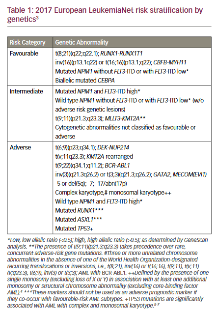
New prognostic factors have also been developed based on the genetic profile or immunophenotypic markers of the B lymphocytes (see Table 1). These include the mutational status of the immunoglobulin heavy-chain variable genes (IGVH),4–7 cytogenetics,8,9 CD38 expression9,10 and levels of the signalling molecule zeta-chain-associated protein kinase 70 (ZAP70)11–15 and serum markers.16–18 These new markers may allow for classification of patients into more refined risk categories than was previously possible using the classic Rai and Binet staging systems. Some are more useful in the context of early stage A disease, when they can aid in prediction of risk of disease progression and need for therapy, while others – such as fluorescence in situ hybridisation (FISH) analysis for the presence of p53 deletion – have particular utility in predicting response to therapy. Since response to therapy is the most important prognostic factor for patient survival, predicting subsets of patients who will or will not respond to a given therapy can prevent unnecessary treatment toxicity and the emergence of clones resistant to treatment and enable selection of targeted therapies. The stratification of CLL patients into prognostic groups using these new markers is increasingly feasible, but before being used in routine clinical practice the methodology of the tests needs be fully standardised and validated. Furthermore, these analyses need to be evaluated in prospective clinical trials. Nevertheless, they have the potential to have an impact on the routine management of CLL in the future. Treatment Decisions – The Current Standard of Care
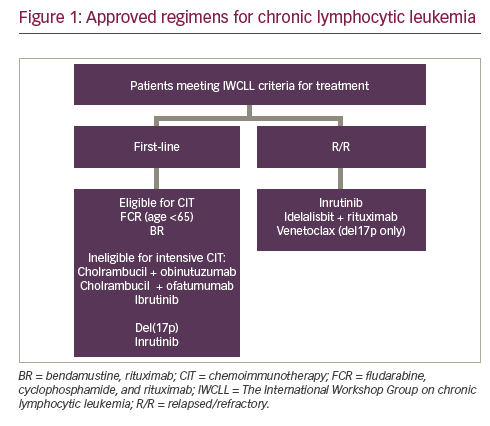
Current treatment modalities for CLL include alkylating agents, purine analogues, monoclonal antibodies and stem cell transplantation. Current international guidelines,3 based on studies from the French Co-operative Group on CLL19 and the Cancer and Leukemia Group B,20 and confirmed by a meta-analysis,21 recommend that asymptomatic newly diagnosed patients with early-stage disease (Rai 0, Binet A) should be monitored without therapy until they have evidence of disease progression.3 Although patients at Binet stage B usually benefit from treatment, some can also be monitored without therapy until there is evidence of progressive or symptomatic disease. Stage C patients require therapy.
In the past, the alkylating agent chlorambucil, with or without corticosteroids, was the most widely applied first-line treatment in patients with active disease, with clinical responses achieved in 40–77% of previously untreated patients.22,23 Alkylator-based combination regimens, including those incorporating anthracyclins, have been compared with chlorambucil alone in prospective randomised studies.24 The results indicated superior response rates (RRs) for patients treated with combination chemotherapy, but no benefit on survival.25 A meta-analysis of 2,022 patients in 10 trials comparing combination therapy, six of which included anthracyclines, with chlorambucil with or without prednisolone showed an identical five-year survival of 48% in both groups.21
The introduction of purine analogues, specifically fludarabine monophosphate, was a major breakthrough that shifted CLL therapeutic strategy from palliation to substantial improvements in RRs and outcome. Fludarabine has been extensively studied in CLL and has been shown to give better complete remission (CR) rates and progression-free survival (PFS) compared with the cyclophosphamide, doxorubicin and prednisone (CAP) regimen and with chlorambucil in randomised trials.26–29 Although all patients eventually relapsed, the conclusion drawn from these studies was that fludarabine was one of the most active single agents in CLL and should form the building block of subsequent therapies. Since then, combinations of fludarabine and cyclophosphamide (FC) and, more recently, fludarabine, cyclophosphamide and mitoxantrone have been investigated. These agents gave high RRs in single-institution studies and showed higher CR rates and longer PFS than single-agent purine analogues.30,31 In a small fraction of patients these combinations eradicated minimal residual disease (MRD). As with many malignancies, eradication of MRD translates into longer PFS and long-term survival.31
In the last two years, three large phase III studies comparing fludarabine alone versus combination FC therapy for CLL patients requiring their first treatment have reported the superior objective response rate (ORR), CR and PFS seen with the FC combination.32–34 These trials included more than 1,000 patients overall, and in one study one-third of the patients were over 70 years of age and had similar RR and toxicity to those in the younger age group.34 This has shifted the standard of care for CLL towards the use of FC combination therapy for younger patients (<65 years of age) and for fitter older patients without significant co-morbidities. Despite these encouraging results, patients with disease refractory to standard chemotherapy have a particularly poor prognosis, and currently there is no accepted standard treatment.Immunotherapy in Chronic Lymphocytic Leukaemia
Over the past two decades significant advances in the development of monoclonal antibodies have improved treatment results in a number of haemopoietic malignancies. In CLL, two antibodies have been studied: rituximab, a chimeric human–mouse anti-CD20, and alemtuzumab (campath-1H), a humanised anti-CD52 antibody.
Rituximab has proven efficacy for patients with B-cell lymphomas, especially when used in combination with chemotherapy. In CLL, when used as a single agent it is less effective, probably because of the lower expression of CD20 in this disease. However, in combination with purine analogues and alkylating therapy in the fludarabine and rituximab (FR) or fludarabine, cyclophosphamide and rituximab (FCR) regimens, results have been very promising.35,36 As first-line therapy, FCR has induced remissions in >90% of patients, with CR seen in 70%.36
Alemtuzumab was the first of this class of drug to receive approval in the US and Europe for use in the treatment of patients with refractory CLL. More recently, it has been approved for the first-line treatment of patients with B-cell CLL (B-CLL) for whom fludarabine-based chemotherapy is not appropriate.
Alemtuzumab in Chronic Lymphocytic Leukaemia
Alemtuzumab is a recombinant monoclonal antibody against CD52, a surface protein highly expressed on most normal and malignant B and T lymphocytes37 but not on haemopoietic stem cells.38 Once bound to CD52, alemtuzumab induces cell death by one or more of the following mechanisms: complement-dependent cellular cytotoxicity, antibody-dependent cellular cytotoxicity and/or induction of apoptosis. In patients with relapsed or refractory CLL after fludarabine and alkylator therapy, alemtuzumab induced a 33% ORR, including some CRs, with additional clinical benefit seen in patients with stable disease.39 A number of other small studies have confirmed the activity of alemtuzumab in patients with fludarabine-refractory CLL. In one of these studies a proportion of patients achieved eradication of MRD, and this was associated with improved survival.40 Importantly, alemtuzumab is particularly active in those patients who have a TP53 deletion detected by FISH and who typically are resistant to conventional fludarabine-based treatment.41–43
Another approach to the use of alemtuzumab is based on the concept that this monoclonal antibody may be effective at purging residual disease in patients who achieve a maximal response after fludarabinebased induction treatment. Results from a preliminary study in nine patients were encouraging, since all but one patient achieved CR and 33% became polymerase chain reaction (PCR)-negative for IGVH. Several studies using different schedules and doses have shown the feasibility of this approach, with improved responses in 50–60% of patients.44–47 One study randomised patients to receive consolidation with alemtuzumab compared with observation and demonstrated improved PFS for the treatment arm.48–49
As yet, there is rather little published data on completed trials of alemtuzumab in combination regimens in either the refractory or first-line setting. Preliminary data suggest that fludarabine-based combinations are effective (ORR ~80% in relapsed CLL) and well tolerated; it remains to be seen how these compare with rituximabbased regimens.
Alemtuzumab as First-line Therapy
Results from a phase II trial of alemtuzumab as first-line therapy for B-CLL showed an ORR of 81%. Moreover, seven patients achieved CRs and 26 partial responses.50 In this study, alemtuzumab was administered subcutaneously for up to 18 weeks.The long-term follow-up report on these patients showed a median time to treatment failure of 28 months in the alemtuzumab group compared with 17 months in the matched historical controls who received various chemotherapy agents as first-line therapy.51


