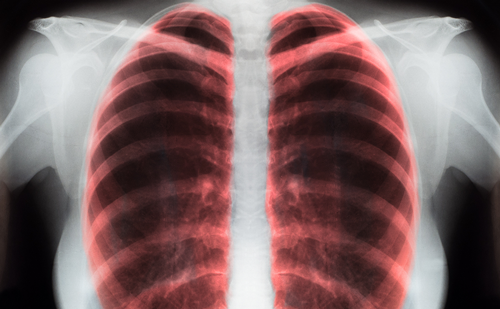
Lung cancer is the leading cause of cancer deaths in the US, with 160,390 deaths estimated in 2007.1 Non-small-cell lung cancers (NSCLC) are the most common type and carry a five-year survival of only 10–15% for all stages.2 Large randomized clinical trials using the bestchemotherapy regimens available have reported similar, but limited, activity, with response rates that have varied from 15 to 22%, one-year survival rates of 31–36%, and median survival of only 10–11 months.3,4 Targeted therapies that can prolong survival are available: epidermal growth factor receptor (EFGR), tyrosine kinase inhibitors (TKI) such as erlotinib, and anti-angiogenic agents, such as bevacizumab.5,6 However, the benefit is restricted to a minority, and most patients who respond to therapy will eventually progress. It is clear that new therapeutic options are needed to improve the survival of these patients. Immunotherapy for lung cancer may provide a new therapeutic option.
Immune Response and Lung Cancer
Historically, NSCLC tumors have not been perceived as good targets for active specific immunotherapy or vaccination because, in general, NSCLC tumors have been considered to be low or non-immunogenic. We also know that NSCLC tumors contain CD4+CD25+ regulatory cells, also known as ‘T regs,’ which suppress cytotoxic lymphocyte (CTL) generation.7 Moreover, suitable target antigens in lung cancer have not yet been identified. For these and other reasons, vaccines targeting lung cancer have not been studied extensively. Therefore, several clinical trials for NSCLC focused primarily on the use of autologous and/or allogeneic whole-tumor-cell-derived vaccines, rather than on specific antigens or peptides.8
Antigen presentation in draining lymph nodes is the first step in triggering an immune response in any tumor. Once CTL clonal expansion and differentiation occur, CTLs are released into the bloodstream and migrate to the tumor tissue by a complex system of chemotactic and adhesion receptors. The initial growth of a tumor has been linked to the failure of immune surveillance of the host. In order for a malignant clone to proliferate and propagate, it has to evade the immune system. Several mechanisms may lead to immune evasion. The first mechanism is the failure to prime the immune system. Tumors such as NSCLC escape recognition by downregulation of major histocompatibility complex (MHC) molecules and secretion of natural killer (NK)-G2D ligands, which is a cell surface receptor expressed on NK cells.9 Several factors have been associated with the lack of adequate priming, including immune suppressive factors secreted by tumors such as: transforming growth factor beta (TGF-β), interleukin (IL)-10, phosphatidyl-serine, prostaglandins, and soluble NKG2D-ligand (which can lead to induction of anergy or tolerance and increased activity of T regs).9–11
A second mechanism of tumor immune evasion is the blockade of the effector phase and lack of efficacy of effector cells against tumor cells. Numerous tumors elicit an immune response as evidenced by the accumulation of tumor-infiltrating lymphocytes (TILs). Although TILs have tumor specificity and the correct phenotype of CTL, many cancer cells may develop defense mechanisms that can neutralize effector cell activity and thereby evade immune destruction. Priming and proliferation of CTLs occur in draining lymph nodes, and CTLs leave the node and enter the bloodstream. Within the tumor vasculature, CTLs must extravasate, enter the tumor tissue (so-called TILs), and locally exert antitumor effector function. As mentioned above, pre-clinical and clinical data suggest that T regs can also play a major role in downregulating immune responses to tumors, including lung cancer, by limiting the expansion of effector clones.11
Lung Cancer Vaccines
Initial attempts at lung cancer immunotherapy were focused on non-specific immune stimulants including thymosin, Calmette-Guerin bacillus (BCG), and other immune adjuvants, especially in patients with small-cell lung cancer (SCLC).12,13 Two larger series of patients with lung cancer treated with BCG revealed improvements in survival compared with historical controls;14,15 however, a randomized study of BCG as adjuvant therapy forSCLC demonstrated no impact on overall survival.16 During the 1990s, nonspecific immunotherapy was attempted using IL-2, other cytokines or inflammatory mediators such as tumor necrosis factor-alpha (TNF-α), or alfainterferon, with little benefit.17,18
Active immunotherapy against lung cancer with the production of cancer vaccines has now been developed. In general, cancer vaccines incorporatetumor antigens and ‘adjuvant’ molecules to facilitate tumor antigen recognition by the immune system. The antigens can be whole autologous or allogeneic cells, specific peptide epitopes, or proteins. In the case of lung cancer, like most other cancers, the most important tumor-associated antigen (TAA) has not been identified. Many immunotherapy approaches involve gene therapy, where a specific gene is transferred to the relevant cell by a vector that is generally a virus such as retrovirus or adenovirus; however, non-viral vaccines such as liposomes and cellular vaccines are also in development.
One approach involves the use of dendritic cells (DCs), which are the most effective antigen-presenting cells (APCs) thus far identified, and play a major role inducing primary and secondary T-cell immuneresponses against cancer in vivo. DCs are capable of stimulating immunologically naïve T cells.19 The cytokine granulocyte-macrophage colony-stimulating factor (GM-CSF) induces differentiation of bonemarrow- derived cells into APCs (macrophages and DCs). A vaccine involving the GM-CSF gene transduced tumor cells is called ‘GVAX.’ In this case, transfection of autologous tumor cells with the GM-CSF gene has shown induction of cancer-specific antitumor immunity requiring both CD4+ and CD8+ cells mediated through DC stimulation.20 Cancer patients may have defects in macrophage and DC antigen presentation to T cells. Antitumor immunity in these trials has been measured by delayed-type hypersensitivity, by antitumor response in tumor samples, and by clinical responses.21,22 There are several published phase II trials with this vaccine. A phase II randomized trial of GM-CSF tumor vaccine (CG8123) with and without low-dose cyclophosphamide in advanced stage NSCLC was presented in Spain in 2005. An interim analysis of the study revealed a median survival of 4.5 months for the vaccine-only treatment arm, and 11.9 months for the cyclophosphamide + vaccine arm. The difference was not statistically significant.23 Another strategy involves transduction of tumor cells with co-stimulatory molecules. In the ‘B7.1’ vaccine developed by us, the co-stimulatory molecule B7.1 (CD80) has been introduced into an allogeneic vaccine to induce T- and NK-cell responses against tumor cells.24 Tumor cells transfected with B7.1 and human leukocyte antigen (HLA) molecules have been shown to stimulate an avid immune response by direct antigen presentation and activation of Tcells, in addition to allowing cross-presentation.25,26 In a phase I protocol performed at the University of Miami, all but one of 19 patients had a measurable CD8 response after three immunizations measured by the release of gamma interferon in ELI spots. The immune response of six surviving patients shows that tumor-vaccine-specific CD8 titers continue to be elevated for at least 38 months. Overall, one patient had a partial response and five patients had stable disease. The median survival for all patients at the time of writing was more than 18 months.24
One important difference between the allogeneic B7.1 vaccine and autologous vaccines such as GVAX is the early activation of NK cells and the innate arm of the immune system due to mismatched MHC. Also, we believe that whole-cell vaccines have a general advantage because of their ability to induce a polyvalent response type against many epitopes, in contrast to a vaccine directed at a single or few epitopes, which may have limited utility due to the evolution of tumor escape mutants. If vaccination is successful and CTLs are generated, the responsible antigens can be identified at a later date. Allogeneic cell-based vaccines are predicated on the assumption that lung tumor antigens are shared between patients and that such antigens can be cross-presented by the APCs of patients. Although there is only limited evidence for shared antigens in lung tumors,26 shared antigens have been observed in other cancers.27 The manufacture of autologous vaccines is difficult due to the fact that a tumor specimen is needed and, frequently, patients have to undergo a second surgical procedure (core biopsy) to acquire autologous tissue, making allogeneic approaches more attractive. In one study, fewer than 60% of the patients scheduled to receive the autologous vaccine GVAX actually received the vaccine due to technical hurdles.
The largest vaccine study—unfortunately unsuccessful—published worldwide was for SCLC with the BEC2 vaccine: there was no difference in the survival of patients who received the vaccine compared with those who received placebo in a multicenter phase III trial.28 It was an interesting approach because BEC2 is an anti-idiotype vaccine that targets the ganglioside GD3 expressed on cells of neuroendocrine origin, including SCLC cells. It targets the binding region of the mouse monoclonal antibody R24, which binds to ganglioside GD3. Hence, BEC2 elicits an immune response against GD3.
Another promising vaccine with a different mechanism of action is the BLP25 vaccine, which is a liposomal preparation of the carcinoma- associated mucin (MUC-1). MUC-1 is expressed on the cell surface of many common adenocarcinomas including lung, breast, pancreas, and others. Cancer-associated mucins are linked to the development of metastases through promotion of the adhesion of malignant cells to the endothelial cell surface. Also, they exhibit uncommon glycosylation patterns, and hence become useful immunogens.29 A randomized phase II trial of MUC-1 peptide vaccine versus best supportive care (BSC) was subsequently developed as a second-line therapy for advanced NSCLC.30 The median survival was 17.2 months in the vaccine arm versus 13 months in the BSC arm (p=0.1802). A multicenter phase III study with this vaccine is ongoing.
Another approach is the development of a vaccine with the MAGE-3 protein, an antigen originally identified in melanoma that is alsoexpressed in some lung tumors. A multicenter phase II randomized trial compared MAGE-3 versus placebo as adjuvant therapy for completelyresected MAGE-3+ stages Ib and II NSCLC. The first results of this study showed that the recurrence rate was 30.3% in the vaccinated group and41.7% in the placebo group (p=0.138); an important difference was seen in stage II patients (30 versus 57%; p=0.55).31
There many more vaccines in development for lung cancer, such as other autologous DC vaccines generated from CD14+ precursors or DCs that are pulsed with apoptotic bodies of an allogeneic NSCLC cell line (1,650 TC), which overexpressed Her-2/neu, CEA, WT1, Mage2, and survivin.32 Also, combinations of telomerase peptides GV1001 and HR2822 have shown immune and clinical responses.33 Also, there is a vaccine made with irradiated genetically altered human lung cancer cell linesengineered to express xenotransplantation antigens by retroviral transfer of the murine α(1,3) galactosyltransferase gene already tested in a phase I trial with several patients achieving disease stabilization.34 Lucanix is a non-viral gene-based allogeneic tumor cell vaccine that demonstrates enhancement of tumor antigen recognition as a result of TGF-β2 inhibition. A randomized dose-variable phase II trial involving stage IIIb/IV NSCLC was presented with clinical responses and a trend in correlation with cell-mediated responses.35
We are developing a new vaccine with Gp96, which is a member of the heat shock proteins (HSP) family. It is an endoplasmic reticulum resident and a chaperone for endogenous peptides and larger protein fragmentsdestined for MHC class I and II loading. Gp96-Ig fusion protein is secreted from transfected cells. In murine studies, tumor-secreted gp96-Ig induces specific CD8 CTL expansion and, when used as a vaccine, mediates tumor rejection and long-lasting tumor immunity by CD8+ cells independent of CD4+ cells.36 Optimal CD8 activation requires IFN-γ, CD28, and B7, but is independent of CD40-L. A phase I clinical trial using an allogeneic Gp96-Ig transfected lung cancer cell line is ongoing at the University of Miami.
The Future of Lung Cancer Vaccines
The development of lung cancer vaccines is challenging. Despite multiple clinical trials including phase I, II, or even III trials, approval of any vaccine for lung cancer has yet to be given, and many logistical and scientific problems for vaccine therapy still exist. For example, the fact that there is not a wellidentified antigen in lung cancer makes the process of developing a lung cancer vaccine difficult. In addition, we still need to find the best strategy togenerate immune responses. Literature exists that proposes B-cell immunosuppression as a way to enhance the Th1 T-cell immune response. Currently, we are looking for ways to enhance the immune response against our b7.1 vaccine.24 One of the options contemplates combination with lowdose chemotherapy or combinations with targeted agents, in both cases with the aim of depleting B-cell immunity, as has been done with GVAX and other vaccines. The availability of funds is important, and the number of lung cancer vaccine trials is minimal compared with the number of ongoing chemotherapeutic trials.
Improvements are needed to better define tumor antigens, select adjuvants, and capitalize on known immune mechanisms to boost the response against tumor cells. Several factors—such as wide variability of the final vaccine product, different sources of APCs used (DCs versus tumor cells), different techniques of preparation, different sources of tumors used (autologous versus allogeneic), the inclusion of cytokineproducing DNA (human or viral), and unknown potent ‘immunogenic’ antigens—have made the selection of optimal approaches and evaluation of outcomes quite difficult. Clearly, challenges facing this new approach include standardization of the technology and the difficulty in correlating immune response to desired clinical outcomes.
Although several pre-clinical trials have suggested antitumor activity, a small number of clinical trials using various vaccine strategies have been developed in lung cancer. These trials have shown some evidence of induction of immune responses and suggested clinical benefit. Responses to immunotherapy may be delayed, and occasionally an increase in tumor size may reflect an inflammatory reaction rather than tumor progression or therapy failure. In this regard, other clinical parameters such as time to progression or survival can be considered for future trials, instead of the classic Response Evaluation Criteria in Solid Tumors (RECIST) criteria that are used to evaluate the solid tumor response, which is based solely on changes in tumor diameter.
Reducing a large tumor burden with immunotherapy alone is unlikely. Vaccination may be best adapted to the adjuvant setting after patients have achieved ‘clinical remission’ with surgery or chemotherapy and radiation, where cancer vaccines may help to control microscopic disease. Also, the role of T regs in suppressing antitumor immune response requires better definition. Depletion of T regs may facilitate improved responses, and selective depletion is rapidly becoming feasible. Future approaches involving the use of immunotherapy include combinations with traditional cytotoxic agents as well as anti-angiogenic agents. Chemokine-mediated tolerance mechanisms and immunogenicity mechanisms may also warrant consideration.
Conclusion
The complicated field of immunotherapy for lung cancer is rapidly evolving. The identification of potential antigenic targets and increasedunderstanding of the immune mechanisms have led to a series of novel strategies for generating antitumor immunity against lung cancer. Despite the difficulties seen in the generation of antitumor response in lung cancer, several agents have already been shown to be capable of priming immune responses in clinical trials, and some have demonstrated promising clinical antitumor activity. These strategies hold promise for improving the survival of patients with lung cancer.
Acknowledgments
This work is partially supported by grants from the James & Esther King Award from the Florida Biomedical Research Program (FBMR)-Florida Department of Health (FDH), and the Flight Attendant Medical Research Institute (FAMRI).


