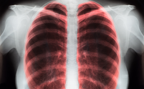
Position Emission Tomography
Positron emission tomography (PET) is one of the most exciting advances in the diagnosis of many cancers, including that of the lung. Briefly, the technique uses the radiolabeled glucose analog F 18 fluorodeoxyglucose. The compound is injected intravenously and is taken up preferentially by metabolically active cells, such as those found in tumors.The patient is placed in a detector similar in appearance to a computed tomography (CT) scanner and the data is recorded. Images are reconstructed and can be viewed in the axial plane (the same as a standard CT scan) or in coronal and sagittal planes. An area of high metabolic activity can be quantified and the number obtained is referred to as the standard uptake value (SUV). In general, areas with SUVs of 2.5 or greater are considered suspicious for tumor.
The clinical utility of PET scanning in patients with known or suspected lung cancer includes the evaluation of the primary lesion, determination of mediastinal lymph node involvement, and detection of distant metastases.
For patients with a new pulmonary nodule or mass, a positive PET scan is highly suspicious for malignancy and mandates tissue confirmation. A negative scan, although not necessarily ruling out malignancy, might encourage a period of observation.The sensitivity and specificity of PET scan for primary lung lesions are 97% and 89%, respectively.1 False positive readings can occur in non-malignant lesions of high metabolic activity, such as tuberculosis or sarcoidosis. Conversely, small tumors (<1.5cm) and tumors of low metabolic activity, such as bronchioalveolar carcinoma and renal cell carcinoma, have low SUVs, and can therefore give false negative results.
The advent of PET scanning has improved the detection of mediastinal lymph node metastases compared with chest CT. In conventional CT, suggestion of mediastinal lymph node involvement is based on size criteria, i.e., a lymph node greater than 1cm in diameter is considered suspicious for metastatic disease. Biopsy is required for confirmation.
In a recent study, PET scanning of the mediastinum had a sensitivity of 81% and a specificity of 96%, similar to that achieved by nodal biopsy via mediastinoscopy.2 In this study, PET scanning also identified positive nodes in areas not accessible to mediastinoscopy and redirected the diagnostic approach. Because of these advances, PET scan has been touted as an alternative to mediastinoscopy. However, biopsy is still advised because of the false negative and false positive rates and the potential consequences of both under-staging and over-staging the patient.
PET scanning has the ability to detect metastatic tumor at distant sites. Unsuspected metastases from lung cancer have been demonstrated in about 15% of patients, thus avoiding a surgical resection.2 Currently, many surgeons are including a PET scan in the preoperative evaluation of lung cancer patients. Efforts to improve sensitivity and specificity of PET scanning have included the correlation of conventional CT images (which provide anatomic information) with PET images (which provide metabolic information). This can be done with socalled fusion software where the images from each test are combined, or with actual hardware that incorporates the CT and PET scanners into one device. Such devices are expected to become more widespread over the next few years.
Video-assisted Thoracoscopic Surgery
Video-assisted thoracoscopic surgery (VATS) involves inserting a television camera into the chest through a small incision. It represents the thoracic equivalent of laparoscopy. Not only can intrathoracic pathology be visualized by minimally invasive means, but a wide variety of procedures can be carried out. Initially, these were limited to biopsies of lung and pleura, or drainage of pleural effusions. However, with improved equipment and experience, virtually any thoracic procedure can be performed with VATS.
VATS is very useful in the evaluation of lung cancer. A new coin lesion or lung mass can be biopsied or excised via small incisions, sparing the patient the morbidity of a major thoracotomy. Mediastinal lymph nodes not accessible by mediastinoscopy can be sampled. Furthermore, finding evidence of unresectability (e.g. pleural implants or tumor invasion of mediastinal structures) can spare the patient a major procedure. Finally, several centers have reported the use of VATS for major pulmonary resections, including lobectomy and pneumonectomy.3,4
Limited Pulmonar y Resections
The standard surgical treatment of carcinoma of the lung is lobectomy (or pneumonectomy). However, this may not be possible in patients with borderline pulmonary function. Also, new primary lung cancers can arise over time, requiring further resection. Since VATS now provides the ability to perform lesser resections through minimal incisions, surgeons have questioned whether such resections (e.g. segmentectomy or wedge excision) in small, peripheral cancers might give similar long-term survival rates while sparing precious pulmonary parenchyma.
To address this issue, a randomized trial was conducted by the Lung Cancer Study Group.5 In a group of 246 patients with small cancers, 122 underwent limited resections (segmentectomy or wedge), while the remaining patients received standard lobectomy. In the limited resection group, the local recurrence rate was tripled and the death rate was increased by 30% compared with the lobectomy group. The evidence suggests that lobectomy or pneumonectomy continues to be the procedure of choice for cure. Lesser resections carry increased risks of local recurrence and reduced survival, and should only be done in poorrisk individuals.
Mediastinal Lymph Node Dissection
During surgery for lung cancer (and many other cancers), evaluation of regional spread (i.e. nodal metastases) is important for accurate staging, subsequent treatment, and, ultimately, prognosis. Surgical opinion varies widely as to whether to perform lymph node sampling or a complete mediastinal lymph node dissection at the time of lobectomy or pneumonectomy. Proponents of a complete dissection note that a recent study has shown that a complete nodal dissection, rather than nodal sampling, appears to confer a survival benefit in certain patient groups.6 It has also been shown that there is a high incidence of occult hilar and mediastinal lymph nodes metastases in small peripheral lung cancers, emphasizing the importance of nodal status in all resections.7 A prospective, randomized controlled trial sponsored by the American College of Surgeons is currently underway to address this question.
It is the author’s current position that mediastinal lymph node dissection should be performed in every case of lobectomy or pneumonectomy. Given the available evidence and the low complication rate, there is every good reason to perform a complete mediastinal lymph node dissection, and no reason to avoid it.Neoadjuvant Therapy
In the past, patients who presented with involvement of mediastinal lymph nodes or local invasion of critical adjacent structures were considered ‘out of bounds’ for surgery (i.e. unresectable), and were offered palliative radiation or chemotherapy. With the realization that surgery offers the best potential for cure, more attention has been given to making resection possible for these patients through the use of pre-operative radiation and/or chemotherapy. Known as ‘neoadjuvant’ therapy, this ‘cart before the horse’ approach has produced some significant improvements in survival in selected patient populations. Most protocols involve both radiotherapy and chemotherapy, as it appears that the results are better overall with both modalities combined than for either one alone.
Patients engaged in neoadjuvant protocols represent a challenge at the time of surgery. The tissue quality has been altered substantially by the pre-operative therapy – scarring, bleeding, and tissue healing are important issues. In particular, the healing of the bronchus following lung resection is critical. Breakdown of bronchial closure can have catastrophic consequences.
Various studies have noted that up to 50% of patients who undergo surgery after chemoradiotherapy will have no living tumor found in the resected specimen.This is referred to as ‘pathologic complete response’ and suggests that the pre-operative therapy has actually destroyed the tumor. A pathologic complete response is a favorable factor and correlates with improved survival.
The study by Stamatis et al. illustrates these concepts.8 In a group of 56 patients with significantly advanced lung cancer treated with a neoadjuvant protocol and surgery, the overall five-year survival was 26%. Historically, survival in these patients has been less than 10%.9 For those patients who were completely resected, the five-year survival was 43% and was 50% in those patients with a pathologic complete response.Although these are impressive results, it is important to note that 39% of the patients did not complete the protocol because of comorbidities or persistence of advanced disease. None of these patients survived five years.
Not surprisingly, patients treated with a neoadjuvant therapy/surgery protocol have higher morbidity and mortality. In another study, Stamatis found that the surgical mortality following chemoradiotherapy was 4.9% but the morbidity was 44%.10 Risk factors for morbidity included increased age, lower functional status, and cardiac disease. Clearly, patient selection is critical to the successful application of a neoadjuvant protocol.
The apparent success of neoadjuvant therapy protocols in improving survival in patients with locally advanced lung cancer has led to the extension of such protocols to patients with less advanced disease. For example, the five-year survival for patients with lung cancer invading the chest wall or hilar lymph nodes is only about 40% to 50%.9 Application of a neoadjuvant protocol may improve these results. As noted, the complication rates are higher and the long-term results are not yet definitive.Therefore, it appears wisest at this point to consider enrolling eligible patients in an ongoing protocol.
Summary
PET scanning has improved the diagnosis of primary lung cancer, as well as the detection of mediastinal lymph node involvement and distant metastases.
VATS is useful for biopsy of lung masses, assessment of mediastinal lymph nodes, local invasion, and pleural involvement, and can even be used in larger pulmonary resections.
Lobectomy/pneumonectomy is the procedure of choice for lung cancer. Lesser pulmonary resections are associated with higher rates of locoregional recurrence and decreased long-term survival, and should be done only in poor-risk patients.
Systematic complete mediastinal lymph node dissection at the time of lung resection improves the accuracy of staging and may improve longterm survival.
Neoadjuvant therapy followed by surgery can improve survival in selected patients with locally advanced lung cancer, and perhaps with less advanced cancer as well.


