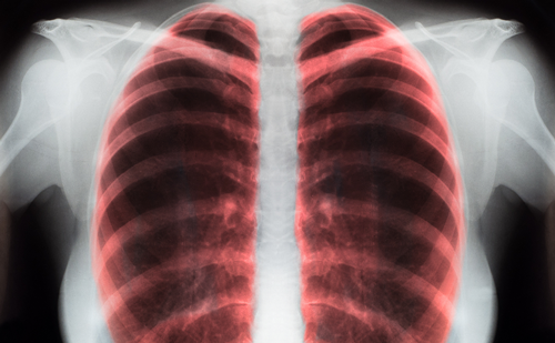
There are several reasons why these disappointing results are not unexpected. First, patients treated with definitive radiation have a lower overall performance status than their surgical counterparts by virtue of the comorbidities that rendered them inoperable. Second, the doses of radiation used to treat these patients are now recognized to yield only a modest rate of tumor control. Understandably, clinicians had legitimate concerns about normal tissue toxicity, and limited doses accordingly. Third, the treatment margins used to encompass a lung tumor were generous, having to account for a) tissue at risk from microscopic tumor extension, b) tumor motion within the respiratory cycle, and c) set-up error.
These limitations were widely recognized and led to several avenues of investigation in an attempt to address them. At Karolinska Hospital in Sweden, Lax and Blomgren began investigations into the stereotactic body frame as a means of targeting tumors more precisely.5 This concept was derived from the stereotactic head frame developed by Lars Leksell at the same institution some 40 years earlier.6 The initial report of this extra-cranial, high-dose-per-fraction technique demonstrated a major response in nearly half of the 31 patients treated, including five of the six patients with lung tumors.5 This represented an advance over conventional 2-D treatment, using computed tomography (CT)-based three-dimensional (3-D) treatment planning, and delivering the treatments using 5–7 non-coplanar beams. This creates a more favorable dose profile in the irradiated normal tissue, sparing the majority of this tissue from doses that may result in clinical complications. With the advent of intensity-modulated radiotherapy (IMRT), these treatments were further refined by the inverse planning method, which involves the assignment of dose parameters to both the tumor and nearby critical structures using a computer-driven iterative process to arrive at an optimized beam arrangement. This is, in essence, the technique that is widely applied today when administering stereotactic body radiotherapy (SBRT) for early-stage NSCLC. Recent clinical reports of patients treated with SBRT for medically inoperable NSCLC demonstrate promising results, with two-year survival rates of 60–80% and local control rates of 80–90%.7–11 While these advances in treatment techniques are promising, they fail to address several fundamental limitations. Foremost among these is the failure of both conventional CT-based treatment planning and port film verification systems to provide an accurate representation of tumor location, given the tumor motion that is inherent in the respiratory cycle. These separate intrinsic errors of 1) target delineation and 2) target verification are both compounded and magnified when delivering highdose, hypofractionated treatments. This is because unintended involvement of critical structures or geographical tumor miss during any single fraction of treatment constitutes a higher proportion of the total treatment than during conventional fractionation. When applied to normal tissue complications, this concept is well illustrated by a phase II trial conducted by Timmerman and colleagues, in which treatment to centrally located tumors (with closer proximity to critical structures such as the trachea, esophagus, and heart) resulted in substantially higher rates of grade 3+ toxicity than treatment to peripheral tumors—46 versus 17% at two years.11 This clearly demonstrates the value of reducing the volume of tissue receiving full dose to the minimum necessary to achieve tumor control. The development of image-guided radiotherapy (IGRT) techniques provides a potential objective basis for this volume reduction.
Target Delineation
Recognizing that CT planning scans provide no meaningful information regarding tumor motion during the breathing cycle, clinicians began investigating methods to generate this information. One of the earlier approaches was the ‘slow CT’ scan, in which the rate of image acquisition is slowed by a factor of 3–5. This allows the scan to capture motion artifact during respiration, which registers as a ‘blurring’ of the tumor. Early evaluation of this technique showed it to be more accurately representative of tumor motion during respiration, and use of the slow CT images during the treatment planning process resulted in improved dosimetric coverage, especially when co-registered with the conventionally acquired CT scan.12,13
Fluorodeoxyglucose positron emission tomography (FDG-PET) has emerged as a powerful diagnostic tool in oncology. Areas of increased glucose metabolism—including most lung tumors—take up this agent avidly, revealing valuable functional information that has the capacity to change the treatment under consideration markedly. In clinical evaluations, FDG-PET has demonstrated the ability both to upstage and downstage lung cancer patients in whom other radiographic studies are equivocal.14,15 It has also been shown to be very useful in the delineation of tumor versus atelectatic lung,16 something which is difficult if not impossible on the basis of CT imaging alone. More recently, evidence is emerging that metabolic response during thoracic radiation therapy measured employing FDG-PET may be a prognostic factor indicating the success or failure of therapy.17
To afford appropriate resolution for diagnostic purposes, PET scans require 35–45 minutes to complete. During this time patients are free-breathing at normal tidal volumes, which creates a motion artifact not unlike that which is observed in a slow CT scan. In the treatment planning process for SBRT, this is a property that can be exploited to generate a de facto ‘motion envelope,’ in which the maximal extent of tumor hysteresis is defined by the attenuated edges of the FDG avid mass. One disadvantage of stand-alone FDG-PET imaging is the lack of useful anatomical information derived from the study. For this reason, FDG-PET is nearly always fused with an imaging study with good anatomical resolution—usually CT. The use of integrated PET/CT scans for treatment planning purposes in lung cancer is well characterized in the literature.18,19 The motion artifact generated during a PET scan can be used in the same fashion as the slow CT images mentioned above, facilitating generation of a gross tumor volume (GTV) that encompasses the tumor throughout the respiratory cycle. One limitation of this technique is that the edges of this motion envelope possess decreased signal intensity, a consequence of time averaging of the signal intensity in areas where the tumor is present for only a fraction of image acquisition. This can lead to some uncertainty regarding the true extent of tumor hysteresis.19 Four-dimensional CT imaging (4-D-CT) represents a further refinement of imaging for treatment planning purposes. First described by Keall and colleagues,20 4-D-CT also creates a motion envelope, but this time uses CT images captured at multiple phases in the respiratory cycle. Fiducials are placed on the patient’s chest and registered by a fixed radio–camera system. The motion of the fiducials is used to register the patient’s respiratory cycle, which is then divided into 10 equal temporal components called bins. CT images are then placed into a bin corresponding to the phase in the respiratory cycle during which they were acquired.
The net result is 10 discrete reconstructed image sets of the thoracic cavity, each representing the position of tumor, normal tissues, and organs at a different phase of the respiratory cycle. Clinical evaluation of the 4-D-CT technique indicates that treatment planning using the 4-D image set has the potential to reduce the volume of lung receiving significant doses of radiation.21 This reduction in volume can be further improved by the restriction of respiratory excursion provided by use of a stereotactic body frame22 or body fixation mold.23 At the University of Wisconsin, all patients receiving SBRT undergo 4-D-CT treatment planning scans. This has resulted in uniformly low mean residual lung doses, and a similarly low rate of clinically significant chronic treatment-related side effects (unpublished data).
Target Verification
Once an SBRT plan is generated, the next challenge is the delivery of this treatment with a high degree of accuracy. Conventional portal imaging, which relies upon alignment of bony anatomy between the daily film and the digitally reconstructed radiograph (DRR), is now recognized to be a poor surrogate for tumor localization.24 Conversely, CT-based ontreatment imaging allows excellent visualization of soft tissue structures, and is especially well suited for visualization of thoracic tumors. It is only relatively recently that refinements in both hardware and software have permitted this imaging modality to be utilized efficiently during patient treatments.The first iteration of CT-based on-treatment imaging was a specially designed ‘CT on rails’ unit. Patients would be positioned on the treatment couch, which was then rotated by 180 degrees to allow for the acquisition of a pre-treatment CT scan using a in-room CT scanner moving on horizontal rails. This scan can then be co-registered with the treatment-planning CT, allowing direct visualization of the lung tumor and comparison of its position with the initial scan. Initial evaluations of this technique in prostate cancer treatment allowed good visualization of soft-tissue structures, permitting positioning modifications as small as 3mm.25 This method has since been applied to SBRT by Wulf and colleagues, who found the combination of external references from the stereotactic body frame and internal references from the CT imaging necessary pre-requisites for the accurate delivery of hypofractionated treatment.8 Some potential limitations of this technique are the requirements for a specialized treatment room—which allows for the mobile CT gantry—as well as patient movement on the treatment table to accommodate CT imaging.
Growing interest in improved targeting has led to the development of CT imaging that is integrated into the linear accelerator itself. This on-board imaging (OBI) technology typically consists of a diagnostic CT platform that is oriented orthogonally to the accelerator head. Both kilovoltage (kV) and megavoltage (MV) CT scanning capabilities are available, varying by treatment unit. Both beam energies allow good soft-tissue resolution, and the integration of OBI into the treatment units streamlines the imaging process further. As with the ‘CT on rails’ technology, the daily scan is co-registered with the treatment planning scan, allowing for critical evaluation and adjustment of tumor position.
Several studies have provided data on the daily shifts made throughout a course of SBRT using OBI with both kVCT and MVCT.26,27 One limitation of the current CT-based imaging platforms is that the image rendered provides no information regarding tumor motion within the respiratory cycle, representing only a part of the motion-adjusted GTV commonly used.
The process of re-visualizing the tumor in all phases of the respiratory cycle using 4-D-CT verification scans is an intriguing refinement of the verification process. Although pilot studies of this technique have been reported,28 the process needs to be streamlined to facilitate rapid processing of the large volume of information acquired while the patient remains immobilized in the treatment position. Additionally, the use of immobilization devices that minimize respiratory excursion and allow for precisely reproducible patient set-up are a critical adjunct to the successful planning and delivery of IGRT. The use of devices such as the stereotactic body frame and double vacuum body mold has been validated in the literature,29,30 and should be a necessary component of SBRT treatment.
Conclusion
The use of image-guided SBRT for early-stage medically inoperable NSCLC has generated encouraging clinical results, and its use is becoming more widespread throughout the radiation oncology community. The integration of IGRT into the treatment planning and delivery process should be considered a necessary step to ensure highquality treatment, and to minimize the risk of associated treatmentrelated toxicities.