Radiofrequency (RF) is a sinusoidal current with a frequency of 400 to 500KHz. When delivered through electrodes inserted in tissues, it can heat the tissues through ionic agitation and produce coagulation necrosis when temperature rises to 60°C. RF ablation has achieved impressive results in the treatment of unresectable liver primary cancer and metastases. This has encouraged interventional radiologists to use the same technique to treat cancers at other sites. The characteristics of tissue that surrounds the tumour such as vascularity and electric conductivity affects heat diffusion and ablation outcome.1 Because RF was initially applied to treat liver tumours, RF algorithm for current delivery had to be tailored to the lung in order to obtain a predictable and reproducible volume of destruction.
However, several experimental works have demonstrated that RF ablation can completely destroy a healthy lung or animal tumour models in the lung.2,3
Technique
The RF current is delivered through an electrode inserted in the target tumour under computed tomography (CT) guidance, in the same manner as a biopsy needle. However, where a biopsy involves heating the tumour tissue anywhere (this being enough for sampling), with RF ablation the electrode has to be placed accurately in the centre of the tumour (see Figure 1). Most electrodes used for lung RF ablation use a needle containing expandable electrodes to be deployed after needle insertion (see Figure 1). Size of the expended electrode is chosen according to the volume of tissue targeted for ablation. The goal is to ablate the tumour and safety margins of 1cm all around the tumour. The procedure can be performed under conscious sedation but general anaesthesia provides a higher feasibility. Indeed, under conscious sedation, 3% to 10% of treatment must be interrupted due to pain or intractable coughing.4,5
Usually, lungs are treated one at a time in order to avoid the possibility of life-threatening complications from bilateral adverse events, such as massive haemorrhage or pneumothorax. Several tumours can be treated on one side during the same session. When patients have had previous lung surgery, the risk of pneumothorax is low and bilateral treatment can be attempted.
Tolerance–Complications
Pneumothoraces are found on CT scans immediately after ablation in about 54% of treatment sessions. This is probably due to the needle caliber of RF electrode being around 14 Gauge, and thus larger than that used for a biopsy. However, in 31% of the procedures they are small enough not to require any treatment. The remaining 23% are expelled manually after inserting a small bore needle catheter with side holes while the patient is still lying on the CT table immediately after or during RF ablation. Finally, chest tube drainage was necessary in 9% of RF sessions, and retrieved one or two days later in every case. Post-procedural minor haemoptysis, which lasts from two to seven days, is encountered in roughly 10% of patients without the need for any treatment.
The only report of severe haemorrhage concerns tumours in contact with the hilum.6 The author treated patients with FEV1 down to 0.8L/second and noticed that post-ablation was uneventful for patients with a FEV1 superior to 1.2L. Patient with lower FEV1 required some oxygenotherapy during the initial post-ablation weeks. Mean hospital stay was two nights in patients who did not have complications from treatment.
Imaging
CT images obtained within a few minutes following the end of RF energy delivery show the lung tumour surrounded by ground glass opacity. This opacity enlarges the diameter of the hyper-attenuating area, and this enlargement is even greater on images acquired after 24 and 48 hours, although opacity then decreases in size during follow-up (see Figure 2). In the authors’ experience of 100 tumours, the largest diameter of the ablated zone was still measuring 19mm at one year, while the initial tumour diameter was 17mm. This enhanced the fact that World Health Organization (WHO) or response evaluation criteria in solid tumours (RECIST) criterion cannot be used because the goal of RF ablation is to produce a volume of ablation larger than the initial tumour volume. It is generally considered that an ablation volume that does not increase in size on subsequent imaging is a complete ablation. This method of evaluation has some drawbacks, namely late discovery of incomplete treatment. Indeed, in the author’s experience, after a minimum of one year follow-up (mean=18 months), six incomplete treatments were depicted at four, six, nine and 12 months in one, two, two and one patients, respectively. Positron emission tomography (PET)-CT, which is under investigation in clinical practice in the author’s centre, appears a promising method of providing early evaluation of treatment response (see Figure 2).7
Results
Recent reports on lung RF ablation presented shortterm retrospective results with minimal follow-up ranging from one to six months and mean or median follow-up ranging from 7.1 to 12 months4,5,8,9 with a success rate of more than 90% in tumours smaller than 30mm. Lee et al. observed success rates declining to 38% for tumours between 30 and 50mm and to 8% for tumours larger than 50mm.
The author’s experience is made up of 60 non-surgical patients bearing 100 tumours (15% primary lung tumour and 85% metastases) less than 40mm (m±sd =17±10mm). Metastatic disease was from colorectal cancer (n=23), renal cell carcinoma (n=12), soft tissue sarcoma (n=8) and miscellaneous tumours (n=8). The estimated rate of complete local treatment at 18 months was 93% [IC95% = 86 -97] per tumour. The relative risk [0.49 (IC95%=0.06-4.2)] of incomplete local treatment was no different (p=0.51) between the 22 patients who received chemotherapy for a new distant tumour and the patients who did not. The estimated rate of incomplete local treatment per tumour at 18 months was 5% for tumours measuring 2cm or less in their largest diameter and 13% for tumours larger than 2cm (see Figure 4). This difference was not statistically significant, but a trend was noted (p=0.066). In the 73 tumours with a ratio between the area of ground glass opacity imaged at 24 to 48 hours and the tumour area before treatment of at least four, the rate of incomplete local treatment was 4%, which is significantly lower (p=0.02) than when this ratio was below four with a 19% incomplete local treatment rate. The rate of incomplete local treatment compares favourably with the rate of incomplete surgical resection reported to be 12% in the largest world report [metastases, 1997 #900]. However, a major limitation of the comparison of these results with the surgical literature is the difference in tumour sizes between this study and most of the studies reported in the literature. Indeed, the rate of incomplete resection is linked to the tumour size in many reports – tumours exceeding 1cm recurred more often for Higashiyama et al. [Higashiyama, 2002 #902], while 2cm was the threshold for Yano et al. [Yano, 1995 #906]. Although follow-up is short, overall survival and lung disease-free survival at 18 months were, respectively, 71% and 34% for metastatic patients.
Additionally, good tolerance of the treatment demonstrated no significant changes in lung function and spirometry one month after ablation when compared with pre-ablation spirometry (FEV1 = 2.2±0.74L before treatment and 2.2±0.8L after treatment). This makes it possible to treat patients with several previous pulmonary resections and to propose repeated treatments on demand for patients with new metastases. In the author’s experience, five patients underwent a second RF ablation to treat a metachronous lung tumour three, six, nine, 18 and 21 months after the first RF ablation. One patient underwent two additional RF ablations 20 and 26 months after the first one for metachronous lung metastases. Follow-up of these six patients after the second or third RF ablation is too short (3.5 to nine months) to present any conclusive results regarding repeated RF ablation of lung tumours. However, these RF ablations of metachronous lung metastases allowed patients to be maintained as tumour-free without any other systemic or local therapy.
Moreover, low invasiveness of RF ablation allowed the combination in a single session of RF ablation of lung and liver metastases in six patients with a limited number of tumour deposits.
Conclusion
RF appears to demonstrate a high success rate for ablation of tumours smaller than 4cm. Follow-up imaging has to be improved in order to depict incomplete treatment earlier. RF should be considered as an alternative in non-surgical candidates, even with poor pulmonary function. Further evaluation of this promising technique is needed. ■
Radiofrequency Ablation of Lung Tumours
Article
References
- Ahmed M, Liu Z, Afzal K S et al., “Radiofrequency ablation: effect of surrounding tissue composition on coagulation necrosis in a canine tumor model”, Radiology (2004);230: pp. 761–767.
- Goldberg S N, Gazelle G S, Compton C C et al., “Radiofrequency tissue ablation in the rabbit lung: efficacy and complications”, Acad. Radiol. (1995);2: pp. 776–784.
- Miao Y, Ni Y, Bosmans H et al., “Radiofrequency ablation for eradication of pulmonary tumor in rabbits”, J. Surg. Res. (2001);99: pp. 265–271.
- Yasui K, Kanazawa S, Sano Y et al., “Thoracic tumors treated with CT-guided radiofrequency ablation: initial experience”, Radiology (2004);231: pp. 850–857.
- Lee J M, Jin G Y, Goldberg S N et al., “Percutaneous radiofrequency ablation for inoperable non-small cell lung cancer and metastases: preliminary report”, Radiology (2004);230: pp. 125–134.
- Vaughn C, Mychaskiw G, 2nd, Sewell P et al., “Massive hemorrhage during radiofrequency ablation of a pulmonary neoplasm”, Anesth. Analg. (2002);94: pp. 1,149–1,151.
- Antoch G, Vogt F M, Veit P et al., “Assessment of liver tissue after radiofrequency ablation: findings with different imaging procedures”, J. Nucl. Med. (2005);46: pp. 520–525.
- Steinke K, Glenn D, King J et al., “Percutaneous imaging-guided radiofrequency ablation in patients with colorectal pulmonary metastases: 1-year follow-up”, Ann. Surg. Oncol. (2004);11: pp. 207–212.
- Gadaleta C, Catino A, Ranieri G et al., “Radiofrequency thermal ablation of 69 lung neoplasms Lung radiofrequency ablation with and without bronchial occlusion: experimental study in porcine lungs”, J. Chemother. (2004);16 Suppl 5: pp. 86–89.
Further Resources

Trending Topic
It is with great pleasure that we present the latest edition of touchREVIEWS in Oncology & Haematology. This issue highlights the remarkable progress and innovation shaping the fields of oncology and haematology, featuring articles that delve into both emerging therapies and the evolving understanding of complex malignancies. We open with an editorial by Mohammad Ammad […]
Related Content in Lung Cancer
Highlights Immunotherapy, especially combinatory immunotherapy, has shown promise with prolonged survival for patients with advanced mesothelioma in the first-line setting (see the sections on ‘Systemic treatment and immunotherapy debut’ and ‘Randomized immunotherapy trials of mesothelioma’). Histology-based therapy is important to ...

Welcome to the latest issue of touchREVIEWS in Oncology & Haematology. We are honoured to present a series of compelling articles that reflect cutting-edge developments and diverse perspectives in this ever-evolving field. This issue includes a series of editorials and ...
Unmet clinical needs for patients with advanced epidermal growth factor receptor-mutated non–small-cell lung cancer Epidermal growth factor receptor (EGFR)-tyrosine kinase inhibitors (TKIs) have become the standard first-line therapy for patients with advanced non–small-cell lung cancer (NSCLC) harbouring ...

Accurately detecting lung tumours and their margins is important for disease outcomes.1,2 However, detection is challenging due to the use of minimally invasive surgery and current localization techniques, such as computed tomography (CT)-guided and endobronchial interventions, which add significantly ...
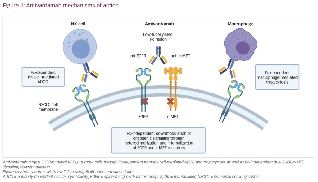
Cancer treatment has expanded rapidly in recent years as advancements in the fields of tumour biology and molecular diagnostics have informed the development of targeted therapies, improving survival in patients with oncogene-addicted cancers with therapeutically relevant molecular lesions. Osimertinib has ...
The treatment of patients with non-small cell lung cancer (NSCLC) has seen significant advances in the past decade, with the availability of multiple targeted therapy agents for oncogenic-driven non-squamous NSCLC and the advent of immunotherapy that has completely revolutionized the ...
Advanced non-small cell lung carcinoma (NSCLC) treatment paradigms have evolved during the past decade. Identification of tumor-specific molecular alteration in cancer driver genes has led to the development of targeted therapies.1–3 Most of the tumors harboring such alterations are sensitive ...

Radiation-induced esophagitis, caused by incidental damage to the mucosal lining of the esophagus during radiation therapy, is a common and clinically important toxicity in patients with lung cancer. Esophagitis generally develops 2–3 weeks after initiation of radiation therapy and presents as ...
Historically, chemotherapy represented the only available treatment for patients with advanced non-small-cell lung cancer (NSCLC) who did not show any target molecular driver, such as epidermal growth factor receptor (EGFR), anaplastic lymphoma kinase (ALK) or the receptor tyrosine kinase ROS1. ...

At initial diagnosis of metastatic disease, central nervous system (CNS) metastases are present in 22–33% of patients with anaplastic lymphoma kinase (ALK)-rearranged (ALK+) non-small cell lung cancer (NSCLC). After crizotinib failure, the incidence can reach up to 70%, compared with 40% in ...
Non-small cell lung cancer (NSCLC) remains a world-wide health issue, accounting for 85% of all lung cancers, of which there were an estimated 2.1 million new cases and 1.76 million deaths in 2018; equivalent to 11.6% of the global cancer burden.1,2 This incidence is expected ...

Welcome to the fall edition of Oncology and Hematology Review (US), which features a wide variety of articles covering topics of interest to oncologists and hematologists, as well as the wider medical community. We begin with some expert interviews, which ...
Latest articles videos and clinical updates - straight to your inbox
Log into your Touch Account
Earn and track your CME credits on the go, save articles for later, and follow the latest congress coverage.
Register now for FREE Access
Register for free to hear about the latest expert-led education, peer-reviewed articles, conference highlights, and innovative CME activities.
Sign up with an Email
Or use a Social Account.
This Functionality is for
Members Only
Explore the latest in medical education and stay current in your field. Create a free account to track your learning.

