Lung cancer is the main cause of cancer deaths for both men and women in the US and worldwide. In the US, there were approximately 232,270 new cases diagnosed and 166,280 deaths due to this disease by the end of 2008.1 Non-small-cell cancer (NSCLC) accounts for approximately 80% of all cases of lung cancer, the rest being smallcell lung cancer (SCLC). Unfortunately, despite efforts by scientists and clinicians aimed at improving survival, delayed diagnosis with subsequent late-stage disease and high rates of recurrence even in patients with early-stage disease ultimately results in suboptimal prognosis and decreased disease-free survival. In fact, overall fiveyear survival rates in lung cancer patients have only modestly improved over the last few decades, with the current five-year survival rate being around 15% in the US and much lower in developing countries.2
Until recently, limited federal resources were dedicated to the study of lung cancer largely due to it being a disease resulting from a lifestyle choice (tobacco smoking) and it typically affecting an older male population. This social prejudice led to stagnation for many decades in understanding the molecular and histopathological basis of lung cancer. However, the staggering lung cancer mortality statistics at a global level have fueled an attitude shift and increased awareness that favors education and basic and translational lung cancer research dedicated toward the development of efficacious diagnostic and therapeutic tools. To this end, molecular genetic studies have recently revealed that histopathologically distinct lung cancers contain an array of genetic and epigenetic anomalies that account for their diverse nature.3 Many of these abnormalities are also found in histologically normal or pre-neoplastic adjacent lung epithelial tissue, thus indicating a multistep process of carcinogenesis encompassing the progressive accumulation of genetic and epigenetic changes that occur concurrent to tobacco smoking, and underlies the initiation, promotion, and progression stages of lung carcinogenesis.4 Clinical translation of these insights into specific molecular mechanisms leading to lung cancer have enabled the development of clinically relevant mouse models of lung carcinogenesis5 and the design of novel targeted therapeutics designed to antagonize chronic activation of epidermal growth factor receptors (EGFRs) with tyrosine kinase inhibitors (TKIs), i.e. gefitinib and erlotinib, which alleviate some symptoms and improve survival in certain subgroups of lung cancer patients.6,7 However, less is known about how to translate underlying mechanisms governing the protumor role of the immune system into meaningful clinical tools.
Over a lifetime, the lungs are constantly exposed to an array of toxic environmental particles that can potentially result in chronic inflammatory responses with life-altering consequences, which is particularly important for tobacco smokers, whose airways are exposed to the carcinogenic agents present in cigarettes. The link between the immune system and cancer is not new, and dates back as far as 1550BCE.8 In the1890s, William Coley, a surgeon in New York, was one of the modern pioneers linking cancer to the immune system. He described how a recurrent facial sarcoma successfully regressed after concurrent infection with erysipelas.9 Unfortunately, his rather innovative hypothesis was not solidly supported by evidence but rather by some anecdotal success, and was met with great skepticism from the medical community. Coley’s views were not further explored for many decades, until it was observed that subsequent to solid-organ transplantation immunosuppressed patients were at increased vulnerability for developing skin and some hematological malignancies, such as lymphoma. These observations partly fueled the immune surveillance theory, simplistically implying that failed antitumor adaptive immunity underlies cancer development.10 More recently, however, fundamental advances in dissecting the nature and bioactivity of leukocytes in lung cancers and other solid tumors indicate that activation of pro-tumor immune response programs may negate antitumor immunity aimed at eradicating neoplastic cells. As ~85% of all lung cancers are thought to have a common etiology in tobacco inhalation, and as chronic inflammatory responses are known to result from this exposure, it seems reasonable to consider novel therapeutic strategies aimed at neutralizing the pro-tumor properties of chronic inflammation while simultaneously bolstering antitumor programs to more effectively eliminate and/or arrest malignant cells. These, together with enhanced national and global strategies to educate people regarding the health risks of smoking and funding of smoking prevention/cessation programs, should be encouraged.11 Thus, in this article we discuss the relevant literature illuminating the role of the immune system in the respiratory tract critical for maintaining physiological homeostasis, as opposed to the role the immune system plays during development of lung cancer. These studies are then used as a backdrop to explore recent advances in immunotherapy of lung cancer and the clinical trials assessing vaccine-based therapies and their delivery in lung cancers. The Immunology Behind Cancer Vaccines
The immune system in mammals is composed of various cell types and mediators that interplay in a complex manner with each other and nonimmune cells in dynamic yet complex networks ensuring the defense against pathogens, while simultaneously maintaining tolerance towards self-antigens. Based on antigen specificity and chronology of activation, the immune system is composed of distinct subsets: innate and adaptive. Innate immune cells, e.g. natural killer (NK) cells, dendritic cells (DC), mast cells (MCs), basophils, macrophages, neutrophils, and eosinophils, are the first lines of defense against organ damage. Macrophages, MCs, and DCs function as sentinel cells pre-stationed in tissues and monitor their microenvironment for signs of noxious insult. If tissue homeostasis is destabilized, sentinels release soluble mediators (cytokines, chemokines, matrix-remodeling proteases, reactive oxygen species, and bioactive mediators) that together induce mobilization and infiltration of additional leukocytes into damaged tissue (i.e. inflammation). Crucial to an effective immune response to any form of tissue damage, including cancer, is induced expression and activation of cytokine and chemokine gradients that direct leukocytes to areas of tissue damage. Cytokines and chemokines are immunomodulatory and chemotactic factors that regulate magnitude, polarization, and resolution of immune responses. Complex interactions between these soluble mediators and their cognate receptors on specific populations of leukocytes not only regulate differentiation of mature leukocytes from hematopoietic progenitors, but also control their bioactivity and effector functions once in tissue.12 Macrophages and MCs also activate vascular and fibroblast responses to orchestrate elimination of invading organisms and initiate local tissue regeneration and remodeling. DCs take up foreign antigens and migrate to lymphoid organs, where they present antigens to adaptive immune cells, thus interfacing between innate and adaptive immunity. NK cells also participate in cellular cross-talk between innate and adaptive cells via their ability to bidirectionally interact with DCs.13–15 Induction of primary adaptive immune responses requires direct interactions with mature antigen-presenting cells (APCs) and a proinflammatory milieu. Adaptive lymphocytes, e.g. β cells, CD4+ (helper), and CD8+ cytotoxic T lymphocytes (CTLs), distinguish themselves from innate cells by expression of somatically generated, diverse antigenspecific receptors formed through random gene rearrangements, allowing a flexible and broader repertoire of responses compared with innate cells expressing germline-encoded receptors. Distinctive CD4+ Tcell subsets, e.g. T helper (Th)-1 or Th2 cells, secrete unique repertoires of cytokines that mediate their responses. Th1 cells produce interleukin (IL)-2 and interferon (IFN)-γ for example, and thereby direct cellmediated immune responses, whereas Th2 cells secrete IL-4 and IL-10 and facilitate local humoral immune responses. Together, activation of innate and adaptive immune response pathways efficiently removes or eliminates invading pathogens, damaged cells, and extracellular matrix (ECM). Once assaulting agents are eliminated, immune cells are critically involved in normalizing cell proliferation and cell-death pathways to enable re-epithelialization, new ECM synthesis, and reestablishment of tissue homeostasis. As immune cells protect against pathogens and promote post-injury tissue remodeling and healing, it seems intuitive that they also participate in protecting against tumor development. Indeed, individuals suffering some types of immune deficiency exhibit increased risk for viral- and/or carcinogen-associated cancers,16 therefore indicating that absence of antiviral immunity affects relative cancer risk. On the other hand, relative risk for common epithelial cancers (breast, prostate, ovarian, and uterine) where cancer etiology is not associated with viral infection or carcinogen exposure is less than 1.0,17 indicating a paradoxical role for immune cells during cancer development where cancer etiology is critical. Lymphocytes and some innate immune cells possess potent anticancer activities that affect growth and/or dissemination of primary tumors. In order to survive, neoplastic cells must evade CTL rejection through subversion of host antitumor immune responses. One plausible explanation for how this occurs is that neoplastic microenvironments favor polarized chronic pro-tumorigenic inflammatory states as opposed to those representing acute antitumor immune states18,19 Clinical data indicate that ‘immune status’ in healthy individuals compared with those harboring tumors is distinct, where in the latter population T cells are functionally impaired.20 In addition, accumulation of chronically activated granulocyte/suppressor cells and regulatory T (Treg) cells can indirectly contribute to cancer via suppression of antitumor adaptive responses, allowing tumor escape from immune surveillance. Furthermore, myeloid suppressor GR1+CD11b+ cells, a subset of innate cells, accumulate in the peripheral blood of some cancer patients21,22 in tumors and lymphoid organs.18,22,23 These induce T-cell dysfunction by direct cell–cell contact and by production of immunosuppressive mediators, and thus actively inhibit antitumor adaptive immunity.22,23 Thus, in the vicinity of a growing neoplasm, the balance between innate and adaptive immunity is often disturbed in favor of cancer progression. Taken together, the accumulated data from human and animal studies support the existence of an immune response involving CD8+ T cells, Th1 cells, and NK cells that protect against tumor development and progression: a system that can be suppressed locally by myeloid suppressor cells and Treg cells. Clinical and experimental data indicate that chronic presence and activation of some innate immune cell types, e.g. neutrophils, macrophages, and MCs, exert a promoting role during cancer development.24 Malignant tissues containing infiltrates of macrophages (breast and lung adenocarcinoma) and MCs (lung adenocarcinoma and melanoma) correlate with an unfavorable clinical prognosis.25–28 In experimental murine models of organ-specific cancer development, genetic elimination of MCs or macrophages minimizes squamous carcinogenesis,29,30 whereas elimination of macrophages during mammary carcinogenesis limits late-stage cancer progression and pulmonary metastasis formation.31 Thus, whereas the historical viewpoint was that host immunity was protective in terms of cancer, it is now clear that subsets of chronically activated innate cells promote growth and/or facilitate survival of neoplastic cells. Innate immune cells directly potentiate cancer risk through the diversity of bioactive mediators they deliver to neoplastic tissues. Leukocytes are loaded with chemokines, cytokines, cytotoxic mediators including reactive oxygen species, serine-, cysteine- and metallo-proteases, membrane-perforating agents, and soluble mediators of cell killing, such as tumor necrosis factor-alpha (TNF-α), ILs, and INFs.32 These molecules are known mediators of acute inflammation and evoke innate cell recruitment and/or activation, tissue remodeling, and angiogenesis, and together create an organ microenvironment favoring cell proliferation, genomic instability, and expansion of neoplastic cells into ectopic tissue, i.e. malignant conversion and cancer development. Clinical and experimental evidence indicates that chronic inflammation is an important epigenetic and/or environmental regulator of epithelial cancer development.24 Initially, it was believed that leukocytic infiltrates in and around developing neoplasms represented an attempt by the host to eradicate neoplastic cells. Indeed, extensive infiltration of T cells in human gastric or colorectal carcinoma is associated with a favorable prognosis,33 whereas malignant tissues containing infiltrates of innate immune cells such as macrophages and/or MCs in human lung and breast adenocarcinoma, gastric carcinoma, and melanoma tend to correlate with an unfavorable clinical prognosis.25–28 Moreover, cohort studies have found increased risk for lung cancer in patients with chronic inflammatory disorders such as rheumatoid arthritis,34 and a modestly increased risk for all cancers, particularly lung cancer and hepatobiliary cancers, in systemic lupus erythematosus patients.34 Chronic inflammation enhances cell proliferation and overall survival of initiated epithelial cells, as well as inducing angiogenic processes in activated stroma.24 During lung carcinogenesis, inflammation has been suggested to play an important role in cancer pathogenesis, particularly in smoking-damaged respiratory epithelium;35,36 however, the mechanisms involved are poorly characterized. Some evidence supports the hypothesis that chronic inflammation contributes to the process of lung carcinogenesis through activation of a number of molecular pathways including differential regulation of nuclear factor kappa B (NF-κB)35,36 and/or cyclo-oxygenase (COX)-2,37 an intermediate early response gene induced by growth factors, oncogenes, and carcinogens. In addition, by studying mouse models of lung carcinogenesis, it has been revealed that activation of Hras alleles in lung epithelia can elicit a primary inflammatory response leading to infiltration of lung parenchyma by various types of innate and adaptive leukocytes that potentiate full malignant progression.38 The functional significance of these infiltrations has not been previously reported (Soto and Coussens, manuscript in preparation). Although there are various mechanisms of vaccine function, in the context of current lung cancer vaccine development we have chosen to emphasize CTL responses, as this is the primary mechanism by which cancer vaccines are believed to act. Following contact between antigens and APCs, a complex chain of events is initiated whose goal is eventual elimination of damaged cells by activated cytotoxic cells including some subsets of CTLs.39,40 As we have already explained, one of the earliest steps in the engagement of a CTL response is the phagocytosis and presentation of antigens by APCs. APCs, e.g. DCs and macrophages, play significant roles in surveying tissues for signs of tissue damage and non-self antigens, or damaged somatic cells, to phagocytose. Following phagocytosis, internalized proteins are digested into short peptide fragments that are shuttled into the extracellular surface of APCs and presented by major histocompatibility complex class II molecules (MHC II). APCs circulate via lymphatics and peripheral blood into lymphoid tissues, where they interact with naïve T lymphocytes.41 Activation of CD8+ CTLs requires the assistance of CD4+ T-helper lymphocytes interacting with APCs. In addition to MHC II interaction with T-cell receptors, this APC–T cell interaction also requires co-stimulatory molecules, such as B7.1 and B7.2, which enhance and modulate subsequent immune responses. In addition to MHC II, a CTL response requires that complementary peptide MHC class I (MHC I) molecules are recognized and targeted by circulating activated CTLs, inducing death of the damaged cell via induction of FAS ligand expression or by granule exocytosis.42 If the immune system is challenged by the same antigen, memory lymphocytes facilitate the readily explosive production of T lymphocytes to fight the culprit pathogen.43 While initial immune responses to emergent neoplastic cells are likely designed to eradicate pre-cancerous cells, molecular and epigenetic alterations in malignant cells often result in an acquired ability to evade antitumor immune responses41,44 and induce a state of immune tolerance to unique tumor antigens associated with cancer progression. Respiratory Tract Maintenance and Immune Surveillance
Mucosal epithelial surfaces of the respiratory tract are constantly challenged to maintain a balance between reacting to harmful and potentially pathogenic antigens, while simultaneously ignoring the myriad non-pathogenic antigens routinely encountered by the respiratory system through breathing. The balanced interactions between innate and adaptive leukocytes regulate cellular and tissue homeostasis and assist healing and repair. If these balanced interactions go awry, they may result in chronic pathologies such as asthma, chronic obstructive pulmonary disease (COPD), and cancer. Additional environmental pollutants contribute to genetic and epigenetic mutations to the proliferating initiated cells.45–47
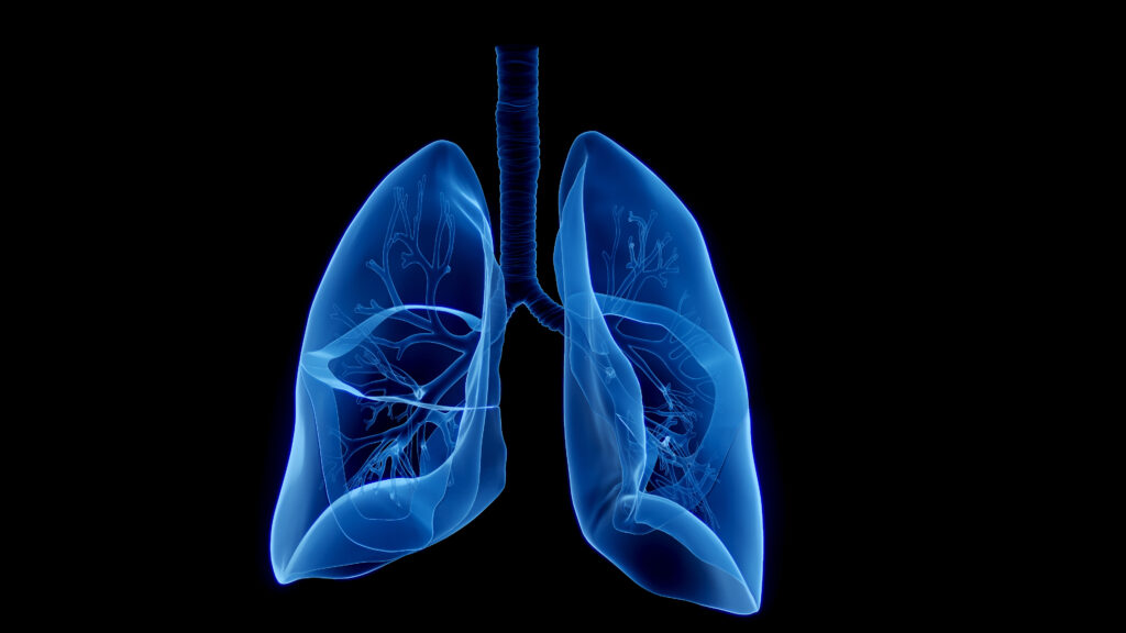
The lungs are functionally divided into two distinct tissue compartments: the conducting airways, covered by the mucosa, and the pulmonary parenchyma, containing thin-walled alveoli involved in gas exchange (see Figure 1). These compartments, with contrasting levels of exposure to antigens, contain distinct resident leukocytes evolutionarily adapted to their differing local microenvironments. In the case of the conducting airways, the lining epithelium of the mucosa is made up of mucus-producing goblet cells and ciliated cells, which in combination are mostly responsible for the process of mucociliary clearance of inhaled antigens. Furthermore, this process is additionally facilitated by locally secreted immunoglobulins (Igs), including IgAs, secreted by B cells. Populations of DCs, myeloid DCs, and plasmacytoid DCs (pDCs), as well as macrophages, take up residence early in life and densely populate the mucosa,48 where resident pDCs are more numerous in the mucosal surface of the airway49 (see Figure 1). Resident airway mucosal DCs (AMDCs)— strategically located in the epithelium and with cellular extensions into the airway lumen—are involved in immunosurveillance by antigen acquisition; however, they are deficient in antigen presentation50, (see Figure 1). Significant numbers of T cells reside within the lamina propria and also intraepithelially in the mucosa, with a mainly effector and/or memory cell functional phenotype. These T cells tend to be CD4+ in the lamina propria and CD8+ within the mucosal epithelium.51 Furthermore, plasma cells mainly producing polymeric IgA and MCs are present in the lamina propria. Some B cells are scattered, but they are uncommon and are thought to play a role in local antigen presentation51(see Figure 1). Bronchial associated lymphoid tissue (BALT) with possible inductive roles is also contained in the airway mucosa. Interestingly, the presence of these discrete lymphoid-cell aggregates varies among species, and their role in humans remains equivocal, as they are most abundant during early life. In experimental rodent models, BALT has been found to provide protective immunity against some viruses.52 Thus, it has been hypothesized that BALT may play a role in respiratory immunological homeostasis in early life compensating for the fact that essential components of the central lymphoid organs are immature. Alveoli are separated by a thin insterstitium that contains lung capillaries that are in close proximity to alveolar spaces and a diverse array of stromal cells. Pulmonary parenchymal immune cells may be located superiorly to the alveolar epithelium or inferiorly in the underlying parenchyma. In physiological conditions, macrophages constitute approximately 90% of the leukocyte alveolar population, with a smaller number of T cells and DCs. In the lung parenchyma, DCs, T cells, B cells, and scattered macrophages co-exist with MCs, but not with plasma cells.
Induction of Cellular Immunity in the Lung
Mucosal surfaces of the upper respiratory airways are chronically exposed to a vast number of environmental antigens, most of which are non-pathogenic in nature. Therefore, in order to protect themselves from unnecessary destructive autoimmunity, the default immune modality is a non-inflammatory, low-level Th2-cell immunity and/or a form of T-cell-mediated immunological tolerance,50,53 although, unequivocally, local DC populations seem to be responsible for the regulation of the protective immunological mechanisms employed by the respiratory airways.50,53–56 Furthermore, the degree of memory T-cell responses that are able to evade these protective mechanisms is controlled by T-cell inhibitory activity of pulmonary parenchymal macrophages.57–59 Moreover, pattern-recognition receptors such as Toll- like receptors (TLRs) expressed by AMDCs effectively help to circumvent the Th2-cell response, with subsequent production of effector memory responses against the culprit stimulating antigens. These antigens are readily transported to regional lymph nodes, the site at which promotion of immunological memory occurs. Subsequently, activated DCs that have migrated from the local lymph nodes acquire the ability to prime naïve T cells and induce immunological responses. These transformed T cells are able to selectively return to the initiating site of immunological stimulus, a phenomenon referred to as tissue-specific homing.60 This activity is possibly due to the ability of activated memory T cells to upregulate tissue-specific adhesion molecules and chemokine receptors that target their migration to non-lymphoid tissue sites while downregulating lymphoid-tissue-homing receptors.60 Although lymphocyte homing is also found in the respiratory system, the mechanisms through which it occurs are not as clearly understood as it is in the case of the gastrointestinal tract and the skin.61–66 Priming of T and B cells in Peyer’s patches and mesenteric lymph nodes preferentially induces the expression of α4β7- integrin and CCchemokine receptor 9 (CCR9); however, peripheral lymph-node primed T cells upregulate cutaneous leukocyte antigen CCR4 and CCR10.61–64 There is increasing research evidence that antigen-presenting DCs process metabolites produced locally to program tissue-specific lymphocyte homing. For example, in gut-associated lymphoid tissue (GALT), resident DCs metabolize vitamin A to retinoic acid, which promotes α4β7-integrin and CCR9 expression by T cells.62 In the skin, local DCs utilize vitamin D3 metabolites to program T cells in regional lymph nodes, which allows their migration to the epidermis.65 In contrast, pulmonary-tissue-specific T-cell homing is less well understood; nevertheless, it is distinctive because it possesses two individual circulatory systems, the first pertaining to the bronchial arteries originating from the systemic circulation and that supplies the bronchial tree, and the second being a low-pressure pulmonary circulatory system through the parenchyma of the lung. In addition, the central airways are lined by mucosa that is an integral part of the mucosal-associated lymphoid tissue (MALT). Depending on the site of induction—whether the intestinal or the respiratory tract—distinct tissue-trafficking patterns for T and B cells are observed in MALTs for various organs despite several shared properties, such as IgA responses. For example, in the respiratory system plasma-cell precursors that are primed in lymphoid tissues that home to the tracheo-bronchial mucosa express low levels of gut-homing molecules CCR9 and α4β7-integrin but high levels of CCR10 and α4β1-integrin.61 Moreover, the airway mucosal endothelial cells constitutively express CC-chemokine ligand 28 (CCL28)and vascular cell-adhesion molecule 1 (VCAM1).61 Respiratory T cells have a different phenotype from gut-homing T cells.67 In the lung parenchyma, leukocytes migrate through pulmonary capillaries. Research in both murine models and humans has shown that memory CD8+ T cells specifically target respiratory viruses that accumulate in the lung parenchyma.68 In non-inflammatory conditions, the pulmonary vasculature expresses intercellular adhesion molecule 1 (ICAM1) and P-selectin; however, it does not express VCAM1. Vascular retention and emergence of effector CD8+ T cells to the normal mouse lung is mediated by the action of lymphocyte function-associated antigen 1 (LFA1)–ICAM1 and CCR5–CCR5-ligand interactions.69 Furthermore, inhibition of P-selectin glycoprotein ligand 1 (PSGL1) resulted in the decrease of T cells in both the alveoli and lung parenchyma.69 Recently, CXC-chemokine receptor 6 (CXCR6) and its ligand were shown to be specifically expressed by human T cells and the lung parenchyma suggesting that they may be involved in tissue-specific lung homing.70 In conclusion, in physiological conditions the immune system, with the effective and tight regulation of its differing subpopulations of immune cells, exerts a status quo of surveillance that is able to differentiate between potentially harmful pathogenic antigens and the other vast majority of non-harmful environmental antigens to which the lungs are constantly exposed. Thus, damage to the lungs through unnecessary immune responses is limited and local respiratory homeostasis is maintained. Rationale for Exploiting the Immune System in Lung Cancer
Traditionally, compared with, for example, melanomas, lung cancers were thought not to contain many tumor-infiltrating lymphocytes, which for some time led to lung cancer being thought of as modestly immunogenic.71 This ethos is changing due to research in animal models and also in human NSCLC specimens demonstrating significant infiltration of leukocytes in cancerous tissues (Yagui-Beltrán et al., unpublished data) Furthermore, CTLs against tumor-specific antigens have been specifically isolated in lung cancer, all indicating that there is a role for immunotherapy in this disease.72,73 Currently, there are several clinical trials evaluating vaccine therapies in the management of lung cancer: L-BLP25 (Stimuvax; Biomira, Alberta, CA),74–76 BEC-2,77,78 belagenpumatucel-L (Lucanix; NovaRx Corporation, San Diego, CA),79 melanoma-associated antigen A3 immunotherapy,80,81 granulocyte macrophage colony-stimulating factor (GM-CSF) transduced allogenic cancer cellular immunotherapy (GVAX; Cell Genesys Inc., South San Francisco, CA),82–85 1E10,86 and PF-35126;76 we will highlight aspects of the most important trials in this article. Recent high-throughput gene expression analyses have led to the identification of novel tumor-associated antigens. Tumor-specific antigens can be obtained autologously from the patient’s malignant lesion subsequent to surgical resection, or can be generated as recombinant proteins or from organ-specific tumor cell lines. Although an important aspect, tumor antigens in isolation may occasionally be insufficient to elicit a clinically significant immune response. A variety of approaches can be utilized to overcome these limitations, including the use of genetically modified tumor cells, viral vectors, DNA-based strategies, and autologous DCs.87 Bacillus Calmette-Guérin (BCG), which has been used non-specifically for immunotherapy of bladder cancer,88 is the most widely used immunogenic adjuvant in lung malignancies.89–91 The individual role of intrapleurally administered BCG in lung cancer has been assessed in several studies with no improvement to survival.89–91 Furthermore, Mycobacterium vaccae (SRL172) with concurrent chemotherapy has been examined as a non-specific immunogenetic adjuvant without success, as shown by a phase III study in NSCLC with no effect on survival.92 Modern techniques to boost immunotherapy comprise mixing adjuvants with tumor cells and/or antigens prior to administration. The strong immunogenic reaction to the adjuvant agents trigger APC responses, with subsequent breakdown of the adjuvant and presentation of the antigens to T cells.93 Biological adjuvants include diphteria toxoid, hemocyanin from marine mollusk Diodora cayenensis, tetanus toxoid, and various plant extracts such as that derived from the soapbark tree Quillaja saponaria. Montanide ISA 51 and aluminum hydroxide are examples of chemical adjuvants. Alternatives to the use of immunogenic adjuvants are the genetic modification of autologous tumor cells or allogenic cancer cell lines to promote the secretion of costimulatory molecules and cytokines in order to minimize immunotolerance.94,95 Moreover, viral vectors expressing an antigen of interest or encoding cytokine of interest utilizes the viral biological apparatus and the innate immune system.96–98 The discovery of TLRs has promoted the utilization of DNA vaccines against cancer.99,100 DNA plasmid vaccines containing unmethylated CpG sites have the power to stimulate TLR 9 on dendritic APCs.101 Similar to the case of viral vectors, DNA vaccines can be designed to contain fusion genes coding for immunogenic molecules of interest.102–104 A more direct method for cancer immunotherapy is the use of DCs directly where peripheral blood is utilized to isolate monocytes with GM-CSF used to induce differentiation into mature DCs.105 These are then exposed to specific tumor antigens and subsequently reinoculated into the patient to induce a therapeutic antigen-specific immune response.106–112 The effector arms of antitumor immunity are CD4+ and CD8+ cells, which can recognize antigen only when it is presented by APCs.107,108 Upon administration, the DCs should migrate to secondary lymphoid organs and induce an antigen-specific immune response.107,111 We will now describe and discuss aspects of various types of lung cancer vaccines and the clinical trials behind their ethos (see Table 1). Antigen-specific Vaccine Clinical Trials
In order for a tumor antigen to be a suitable target for vaccine therapy, it should be expressed homogeneously in the cancer of interest, should differ from those expressed in nearby normal cells, and should be immunogenic and tumorigenic. Specific tumor-associated antigens such as Wilms’ tumor protein (WT1) and some gangliosides, which play a role in cell–cell recognition, cell-matrix adhesion, and cellular differentiation,113,114 have been used in the design of lung cancer vaccines. SCLC expresses ganglioside GD3.115 BEC2 (G2b [IgG2b]) is a mouse antibody that mimics GD3 and promotes an immune response against GD3.78,116 A small pilot study77 showing promising survival in patients receiving the vaccine led to an international phase III clinical trial to assess its role against lung cancer.116,77,78 Unfortunately, and despite the promising results of the phase I study, the use of BEC2–BCG did not result in increased survival or progression-free survival for lung cancer patients.77,78,117 Interestingly, the level of GD3 expression was not evaluated as part of the trial despite the fact that these levels fluctuate among patients,118,119 thus indicating limited prospective determination of a potential benefit for the subgroup of patients overexpressing GD3 impossible.
Fucosyl GM1, another ganglioside, has been used in conjugation with keyhole limpet hemocyanin as a vaccine antigen in adult patients with SCLC with limited or extensive stage who had completed initial treatment with chemotherapy and or radiotherapy at least four but not more than 12 weeks previously. Two phase I studies assessing the therapeutic role of this vaccine found that although most patients developed an immune response, there was no significant clinical end-point.120–122
Mucin1 (MUC1), a transmembrane peptide expressed in epithelial cells, can be anomalously glycated and is overexpressed in many malignancies, including NSCLC,123 and has been linked to chemoresistance and reduced apoptosis.123–125 Therefore, MUC1 has been targeted for the development of immunotherapeutic agents.126
L-BLP25 (Stimuvax; Biomira, Alberta, CA) is a liposome vaccine targeted to the extracellular core peptide of MUC1. The vaccine incorporates an adjuvant (monophosphoryl lipid) and three lipids to enhance delivery of the vaccine to the immune cells.75 Pre-clinical studies showed acceptable toxicity and that the L-BLP25 vaccine was able to generate an antigen-specific T-cell response.74,75 L-BLP25 was evaluated in stage IIIB and IV in a phase IIb clinical study where 171 patients were randomized to best supportive care or MUC1 plus a lipid adjuvant.127 L-BLP25 was administered weekly for eight weeks (at four sites of the body to improve uptake in draining lymph nodes), and there was the option of maintenance therapy, which consisted of vaccination every six weeks starting at week 13. Cyclophosphamide, shown to reduce the activity of suppressor T cells, was additionally administered three days before vaccination.127 Although survival improvement was not statistically significant, a post hoc subgroup analysis determined that the best survival advantage for MUC1 was found in patients with locoregional stage IIIB and lung pathology.127 This has been the basis for the planning of a large international multicenter phase III study to determine whether this vaccine is relevant in the treatment of inoperable stage III NSCLC after treatment with definitive chemotherapy (Stimulating Targeted Antigenic Responses To NSCLC [START] Trial, www.nsclcstudy.com/about-startclinical- research.html). Furthermore, viral vectors, such as the attenuated vaccinia virus modified to express MUC1 and IL-2, are currently under investigation for their potential role in advanced malignancy128 in a cohort of 13 patients, which included four lung cancers and one mesothelioma; disease stabilization was achieved in four patients.128
ALVAC is a canarypox vaccine artificially expressing the carcinoembryonic antigen (CEA), which is expressed in many malignancies, including lung cancer, and the co-stimulatory molecule B7.115 This agent has been tested in a phase I/II study of 18 patients with advanced tumors (three lung cancer patients) bearing CEA; clinically stable disease was obtained in three patients.115
Subsequent to the clinical hype of EGFR-TKIs in the treatment of lung cancer, much effort is being devoted to targeting EGF for immunotherapy. Neisseria meningitides P64k protein was conjugated to a chemical adjuvant and EGF prior to administration to patients.129 Several phase I trials reported that this immunotherapy modality was able to induce specific EGF immunoreaction and, more importantly, this response seems to be clinically relevant translating to improved survival in those who had adequate seroconversion (eight months versus 4.5 months).129
Melanoma associated antigen A3 (MAGE-A3) is a tumor-specific antigen also found in 35% of NSCLC and possibly in a greater percentage in more advanced stages of the disease.130,131 In a recent randomized phase II study of patients with resected MAGE-A3- expressing lesions, this agent was given with an adjuvant and evaluated against placebo in a cohort of 182 patients with stage IB/II NSCLC patients. Although there was a suggestion that this immunotherapy modality promised improved survival, this was not clinically significant and none of the outcome end-points (diseasefree survival, disease-free interval, and overall survival) were reached.81 Despite this, the indication of survival benefit from the phase II study was enough to justify a phase III study where MAGE-A3 vaccine will be administered and evaluated in an adjuvant setting following completion of adjuvant chemotoxic regimes.81
A phase I clinical study evaluated the use of WT1 combined with adjuvant ISA 51 in a total of 26 patients, 10 who had lung neoplasia. A decrease in tumor size was observed in 12 patients out of the 20 developing an immune response.132,133 Further evaluation will reveal whether these benefits are substantial.
Tumor-specific Vaccine Clinical Trials
As discussed above, autologous vaccines are specific to the patient and as such require individual patient tissue for vaccine preparation, with obvious associated time requirements. On the other hand, allogeneic vaccines circumvent these limitations and are created from lung cancer cell lines with consequent compromises due to specificity.82,134 To overcome concerns relating to specificity, several genetic tactics have been utilized.135
GVAX is a vaccine consisting of complete tumor cells with genetic modification to secrete GM-CSF.83,136–138 Initially, autologous tumor cells from metastatic lesions or pleural effusions were transfected with a non-replicating adenoviral vector encoding GMCSF and tested against metastatic NSCLC.83 Vaccines were produced in 37 out of 38 patients recruited into the study, with acceptable toxicity.83 Stable disease was achieved in five patients and two showed prolonged remission extending beyond 40 months.83 A multicenter phase I/II study of 86 patients with advanced stage NSCLC was employed to assess a later version of GVAX: bystander GVAX (consisting of tumor cells and GM-CSF secreting bystander cells).85 A total of 76 patients produced the vaccine, and 49 were subsequently vaccinated.85 A degree of prolonged remission was seen; however, no significant tumor response was observed, despite vaccine-associated immunoresponse.85 GVAX studies in the future are likely to use the earlier version of the vaccine.83 Furthermore, GVAX is currently being evaluated specifically in the context of bronchoalveolar carcinoma (BAC) in a phase II study in patients with stage IIIB/IV BAC (Southwest Oncology Group-SO310/D-0032), the rationale being that the initial data from the phase I studies described above83,84 demonstrated that three out of the four patients who showed remission had BAC.
A phase I study is evaluating the use of modified NSCLC cell lines and AD100, an adenocarcinoma cell line, transfected with the co-stimulatory molecule B7.1.139 Early results are encouraging, with six out of 19 patients showing partial or stable disease and acceptable toxicity.139
Transforming growth factor beta-2 (TGF-β2) has been shown to have many functions in various biological and pathological processes including immunosuppression in cancer.79 (TGF-β2) can suppress DCs and NK cells,79 thus exploiting this as a novel vaccine (belagenpumacel-L) against lung cancer has been investigated. A (TGF-β2) antisense transgene was used to knock down (TGF-β2) expression in allogeneic NSCLC cells lines.79 A phase II study of this vaccine in 75 NSCLC patients showed a dose-related survival difference.79 Patients receiving higher doses had a two-year survival of 52% compared with 20% in those receiving lower doses.79 Phase III studies evaluating Belagenpumacel-L are currently being designed. They will yield useful clinical data that will allow us to fine-tune current available immunotherapy tools for lung cancer. Dencritic Cell Vaccine Trials
There has been a number of phase I and II trials of DC vaccines that have included some patients with malignant lung lesions,140–147 although only a minority of those patients actually had primary lung lesions. Surprisingly, some clinical responses were reported in those patients with advanced-stage lung cancer, and we will highlight the most relevant of those studies. The most common antigen used in lung cancer DC vaccine trials has been CEA.140,141,143–145 Human lymphocyte antigen (HLA)-restricted class I CEA peptides or altered peptides have been exploited as antigens in these studies as effective immunogenic agents.141 In 2001, a study showed that tolerance to CEA could be reversed by immunization with a CEA-derived peptide (made to be a more potent T-cell antigen and loaded onto DCs for delivery as a cellular vaccine).141 Immunization with these antigen-loaded DCs induced CD8 CTLs that recognized tumor cells expressing endogenous CEA.141 After vaccination, two of 12 patients experienced dramatic tumor regression, one patient had a mixed response, and two had stable disease.141 Clinical response correlated with the expansion of CD8 T cells, confirming the role of CD8 T cells in this treatment strategy.141
In a 2004 study, autologous DC vaccines were delivered to 16 NSCLC patients with stage IA to IIIB who had been treated with surgery, chemoradiation, or multimodality therapy.148 The study aimed to evaluate tolerability and immunological responses to DC vaccines in a heterogeneous group of NSCLC patients. DC vaccines were generated from CD14+ precursors pulsed with apoptotic bodies of an allogeneic NSCLC cell line that overexpressed human EGFR 2 (Her2)/neu, CEA, WT1, Mage2, and survivin.148 Individuals were immunized intradermally twice, one month apart. Peripheral blood was drawn serially over 16 weeks, and immune responses were measured by INFγ enzyme-linked immunosorbent spot (ELISPOT).148 Five of 16 patients showed a tumorantigen- independent response, and six of 16 showed an antigenspecific response.148 Immunological responses were independent of stage and prior therapy.148 Favorable and unfavorable clinical outcomes were independent of measured immunological responses, with no unanticipated or any serious adverse events. DC vaccines seem to be well-tolerated with biological activity in a heterogeneous group of NSCLC patients. Establishing an optimal approach will require carefully conducted comparative studies in well-defined NSCLC patient groups.
Conclusion and Future Perspectives
The respiratory tract with a surface area of 70m2 in adults is constantly exposed to the external environment and its associated dwelling particles and pathogens. A single-cell-thick epithelial layer covering the tracheobronco pulmonary respiratory tree allows blood oxygenation throughout life. Therefore, it is obvious that this organ has developed specific tactics over millions of years to maintain homeostasis and prevent the fatality that would ensue upon its failure. Fortunately, recent progress in the understanding of the cellular and molecular events governing this organ during physiology and disease are allowing us to extrapolate and translate knowledge into immunological tools against myriad diseases including lung cancer. There have been many pre-clinical and phase I and II studies evaluating lung cancer vaccines (see Table 1), and some phase III trials are under way.
In order to facilitate these studies and ensure that they yield useful information that can be utilized in a meaningful manner in our quest to develop useful vaccines for lung cancer, a few concepts must be taken into consideration. First, adequate patient selection for clinical trials is important. The majority of trials have concentrated on patients with advanced disease. The rationale supporting this is sound because a higher numbers of events in a short period of time permits the study to be powered to demonstrate survival benefit; however, research in animals has not shown a significant reduction in tumor size,149 which suggests that the optimal candidate for these vaccines is a patient with early-stage disease who has undergone bulky resection but who is at an increased risk for disease recurrence because of microscopic residual disease. This implies better understanding of tumor biology and good biomarkers to identify such individuals (see Figure 2). Second, disease end-points employed in chemotherapy studies are not necessarily extrapolable to cancer vaccines.150 Equally, many studies have shown that immune response does not necessarily correlate to clinical response and, therefore, potentially useful markers of immunity may ultimately be useless. Perhaps it is valid to suggest that the most cost-effective role for cancer vaccines may lie in prevention rather than cure, as is the case in many infectious diseases; however, in cancer the matter of autoimmunity is of crucial importance because these malignant cells originate from the patient. Despite all these logistical limitations, our rapid improvement in understanding the immune system in the context of tumor biology will certainly allow the development of newer perspectives and methods to achieve novel and clinically useful immunotherapeutic agents to target lung cancer. Moreover, within five years it is likely that phase III studies of L-BLP 25, belagenpumatucel-L, and MAGE-3 vaccine will be performed and completed. The results will be pivotal in advancing our understanding of the role of the immune system against malignancy. The clinical experience of vaccines targeting all stages of NSCLC will surely allow immunotherapeutic non-toxic therapeutics to be expanded and optimized for the treatment of a major killing disease. ■