Malnutrition and Cachexia
Viewed as a physical state, malnutrition is a disorder of body composition characterised by macro- and/or micro-nutrient deficiencies resulting from insufficient intake of, increased demand for and/or altered use of nutrients.5 Malnutrition is related to several disease states, as well as situations of famine, and can be reversed by providing nutritional therapy. Cachexia, on the other hand, is a complex syndrome that affects not only cancer patients but also those with AIDS, congestive heart failure and other wasting diseases. This syndrome leads to loss of muscle and fat, which is directly caused by tumour factors or indirectly caused by an aberrant host response to the tumour presence. Several treatments have been tested to overcome this in cancer patients. However, up to now none of them has been 100% successful, perhaps due to the different phenomena involved in this syndrome.
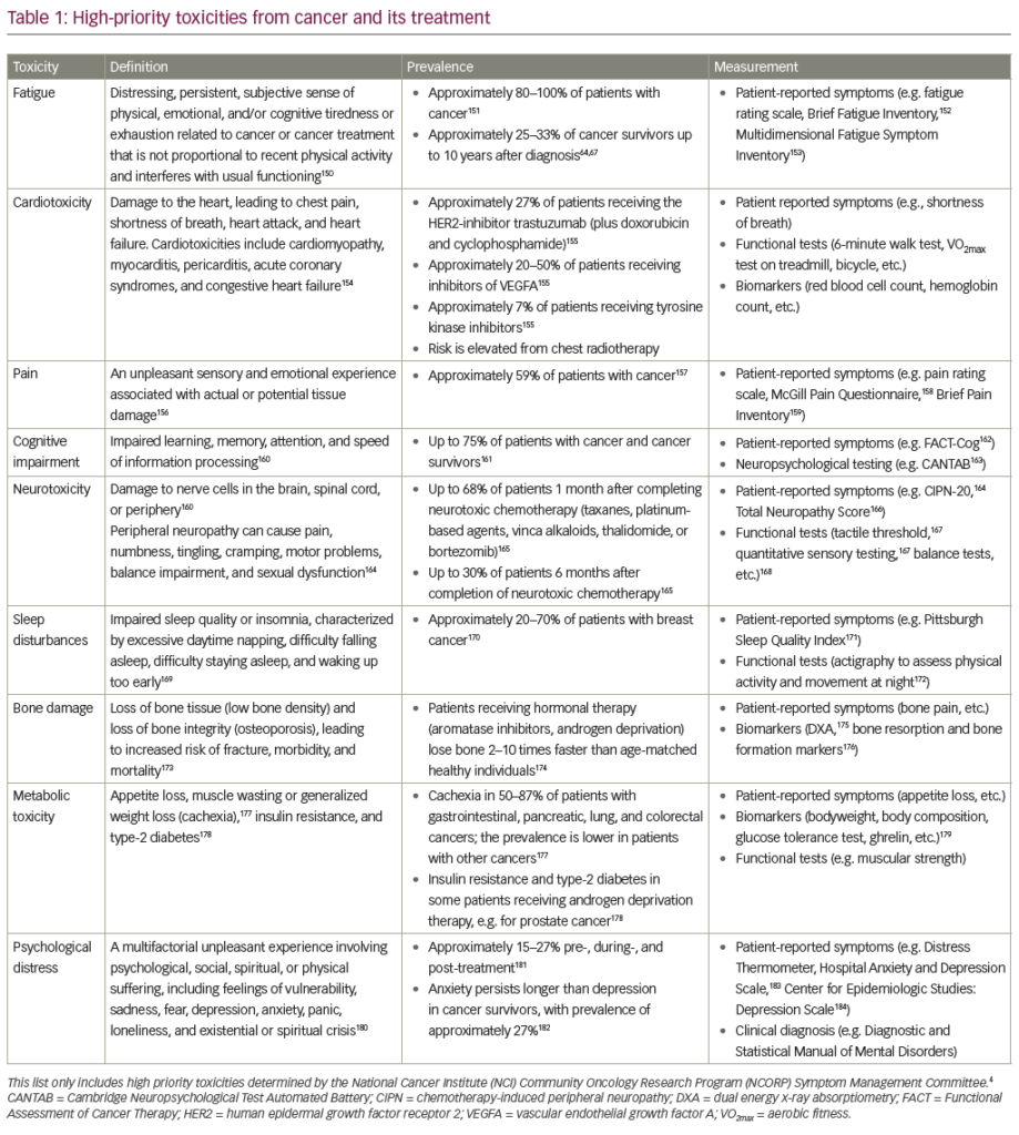
Both malnutrition and cachexia have been related to increased morbidity, mortality, length of hospital stay and costs.4 The functional and metabolic body disorders that justify the previous events are based on the fact that these situations interfere with almost every organ and/or system of the human body. The gut of malnourished patients presents with impaired immune function, digestion and absorption.6 Muscle dysfunction, especially of the thoracic muscles, might explain the high incidence of pneumonias in the malnourished.7 Wound healing is also affected by malnutrition.8 In cancer patients, malnutrition is highly prevalent. A classic study by DeWys et al.9 shows that patients with pancreatic cancer presented with a prevalence of malnutrition of up to 70%, with 38% of them showing losses above 10% of usual weight in the previous six months. Andreyev et al.10 also reported a high prevalence of weight loss among cancer patients with different tumour types. Walsh et al.11 conducted a comprehensive prospective analysis of symptoms in 1,000 patients on initial referral to the palliative medicine programme of the Cleveland Clinic. The median number of symptoms per patient was 11 (range 1–27). Weight loss was reported by over 50% of the patients. Waitzberg et al.3 showed that among 794 with cancer the prevalence of malnutrition was 66.9%, versus 40.9% in the non-cancer group. Severe malnutrition was found in 23.3% of the patients.
Cancer-induced weight loss is directly related to poorer prognosis. Argiles et al.12 showed that two-thirds of patients are cachectic by the time they die, and death is directly caused by cancer-induced weight loss in 22% of the cases, rather than the cancer per se. Other authors have also demonstrated that cancer-induced weight loss negatively impacts on chemotherapy, radiotherapy and surgery. Quality of life is also affected by cancer-induced weight loss. Andreyev et al.,10 who followed 1,555 patients with cancer of the digestive tract, showed that those who had lost weight, and continued to lose weight before and throughout cancer treatment, had a decreased survival time compared with those who had stopped losing weight. DeWys et al.9 also showed that despite the different tumour types, those patients who lost weight presented with overall significant decreased median survival.
Risk Factors for Weight Loss
A variety of factors influence the nutritional status of cancer patients. Decreased ingestion is caused by anorexia, satiety and obstruction. Head and neck plus gastrointestinal tumours often cause obstruction of the gastrointestinal tract. Aside from this, anorexia is common and is related to alterations in taste perception and metabolic disorders. This might be caused by changes in serotonin and leptin receptors. Satiety is influenced by intestinal motility, which is affected by different drugs, such as painkillers, which also lead to abdominal distention. Mucosal atrophy as a consequence of cancer treatment might also influence satiety. Anorexia and appetite are influenced by peripheral stimuli such as pain and gastrointestinal distension, which lead the medial hypothalamic nucleus to signal to decrease ingestion of food. On the other hand, positive stimuli such as good smells and the memory of something tasty will trigger the lateral hypothalamic nucleus to stimulate increased ingestion. These aspects are important when planning nutritional interventions, which should be concentrated and palatable with the possibility of mixing different nutrients to target a wider variety of tastes. It is known that cytokines directly influence the brain either by increasing glucose-sensitive neurons or the corticotropin-releasing hormone, which in turn will suppress appetite. Therefore, efforts should be directed at decreasing or ameliorating cytokine production induced by the presence of the tumour.
Metabolism
Alterations in metabolic pathways may account for energy losses of approximately 300kcal/day in cancer patients. The futile Cori cycle is one of them. Tumours consume large amounts of glucose and convert it to lactate. As the oxygen tension is too low for the Krebs cycle and mitochondrial oxidative phosphorylation to operate, the lactate produced circulates to the liver and is reconverted into glucose in a process known as the Cori cycle. Although the Cori cycle is normally responsible for 20% of glucose turnover, this has been shown to be increased up to 50% in cachectic cancer patients, accounting for 60% of the lactate produced.12 Gluconeogenesis uses six adenosine triphosphate (ATP) molecules for every lactate–glucose cycle and is inefficient for the host, contributing to the increased resting energy expenditure (REE) in cachectic subjects.
| Assessment Method | Advantages | Disadvantages |
| Subjective global assessment | Essentially clinical; inexpensive, good sensitivity and specificity3,4 |
Subjective; demands good training of interviewer |
| Biochemical tests | Good markers of inflammatory response thus good predictors of morbidity and mortality | More expensive, not always available, interference from diseases other than malnutrition |
| Anthropometrics | Inexpensive, objective data | High error factors, comparisons with tables derived from healthy population data, oedema may alter results |
| High-tech body-composition tests | More precise in definition of body composition | Expensive, not available everywhere |
Protein turnover seems to be increased in about 32–35% in cancer patients as a consequence of muscle catabolism; as a result, increased nitrogen excretion is present, leading to negative nitrogen balance.12 This probably explains loss of functional capacity in these patients. Thus, protein repletion is important in cancer patients.
Protein catabolism may be due to three different proteolytic pathways:
• the lysosome system, which is involved in extra-cellular protein degradation and cell receptors;
• the calcium-activated cytosol system, which is involved in tissue damage, necrosis and autolysis; and
• the ubiquitin pathway, an energy-dependent pathway that is the main protein catabolic pathway in cancer patients, leading to increased muscle proteolysis and culminating in cancer-induced weight loss.13
Most of the degraded proteins are myofibril. In order for this to happen a great amount of energy is demanded; this might contribute to the increased resting energy rate in cancer patients. This is a controversial subject, since some authors report that cancer patients do not present with increased resting energy expenditure. It is most likely that cancer patients do have increased resting energy expenditure, but because they diminish their physical activity the overall energy expenditure might not be increased.
In all cases of muscle atrophy, the latter appears to be due to increased activity and expression of the ubiquitin–proteasome proteolytic pathway. In this process, proteins are marked for degradation by a polyubiquitin tag, which is recognised by the 2 MDa complex (26S) proteasome, a large multi-sub-unit proteolytic complex consisting of a central catalytic core (20S proteasome) and two terminal regulatory subcomplexes (19S complex). Degradation of a protein via the ubiquitin–proteasome pathway involves two discrete and successive steps:
• tagging of the substrate by covalent attachment of multiple ubiquitin molecules; and
• degradation of the tagged protein by the 26S proteasome complex with release of free and reusable ubiquitin.In patients with gastric cancer, Bossola et al.14 performed rectus abdominis biopsies and observed that there was twice the amount of messenger ribonucleic acid (mRNA) ubiquitin in those with advanced disease.
Cancer patients who are losing weight show an increased turnover of both glycerol and fatty acids compared with normal subjects or cancer patients without weight loss. There is no evidence for a decreased level of lipoprotein lipase in the adipose tissue of cancer patients, but there is a two-fold increase in the relative level of mRNA for hormone-sensitive lipase, suggesting an upregulation of triacylglycerol hydrolysis.
The tumour is a burden to the host, which is interpreted as a foreign body, triggering a local and systemic immune response that leads to cytokine production and all the previous presented metabolic pathways.
Cancer-induced weight loss shows similarities to tissue injury, infection or inflammation in showing an acute-phase response (APR) in which liver protein synthesis changes from synthesis of albumin to production of acute-phase proteins such as C-reactive protein (CRP), fibrinogen and 1-antitrypsin. The APR is known to be activated by cytokines such as interleukin (IL)-6, IL-8 and tumour necrosis factor, suggesting that they may play a role in cancer-induced weight loss. Lipid- and protein-mobilising factors produced by the tumour also contribute to the inflammatory cascade. The inflammatory status leads to changes in the metabolism of proteins, carbohydrates and lipids as previously seen.
Nutritional Assessment and Therapy
There are several ways of assessing nutritional status. Some of these techniques are sophisticated and expensive compared with others that are less complicated and available in most hospitals. Among all of these, there is yet to be one that is highly sensitive and specific enough to be considered the gold standard.15 Subjective global assessment (SGA)16 and functional assessments; biochemical tests such as albumin, prealbumin and transferrin; anthropometrics such as height, weight, skinfold thickness and arm circumferences; and high-tech bodycomposition tests such as bioelectrical impedance, computed tomography and dual-energy X-ray absorptiometry have been used. Each of these method types have advantages and disadvantages that are outlined in Table 1.Nutritional requirements must be calculated based on either existing formulas such as the Harris-Benedict or the quick formula of 25–30kcal per kilogram per day, considering at least 1.2g of protein per kilogram. Nutritional therapy is prescribed based on the estimated requirements. Ideally, oral nutrition should be indicated because it is more physiological and less expensive. Enteral and parenteral nutrition are reserved for those who cannot eat enough or at all.
As previously seen, cancer patients behave as chronically inflamed patients and therefore regular nutritional therapy, despite offering the patient’s requirements, might not fully reverse nutritional deficiencies. Thus, special nutrients such as eicosapentanoic acid (EPA) should be considered, since it has been demonstrated that EPA can interfere in some cancer pathways.17 The ideal formula to cover the nutritional demands of these patients should contain:
• complex carbohydrates, which would compensate for the insulinresistance status;
• fibre, which would help with intestinal dysmotility and also with insulin resistance;
• increased concentration of high-quality protein to compensate for the increased muscular proteolysis; and • low fat so not to interfere with satiety, but at the same time provide anti-inflammatory properties such as omega-3 (?-3) fatty acids.
The formula should also be concentrated to overcome the satiety that is so frequently seen in these patients and to improve tolerance. It is important to explain to the patient and to the family the different options that can be worked up with the formula, in order to increase compliance. This is a fundamental aspect that should be individualised by the nutritional therapy team.
EPA is a long-chain fatty acid of the ?-3 family. Eicosa refers to the 20 carbon structure, while penta indicates five double bonds and ?-3 indicates that the first double bond follows the third carbon from the methyl end. ?-3 fatty acids aressential dietary components because they play structural roles in membranes and functional roles for enzymes and mediators. The main source for ?-3 fatty acids is oily fish (e.g. sardines, salmon, mackerel and tuna), and typical intake is about 0.25g per day (European/English diet).
The purported mechanism of ?-3 fatty acids in preventing tissue wasting in cancer might be the suppression of the ubiquitin–proteasome system, the inflammatory cytokines and the cancer cachectic factor.18
Conclusions
Cancer patients present with several risk factors for malnutrition, which directly impacts on their outcome. Nutrition interventions should be based on adequate assessment, and special nutrients such as ?-3 fatty acids may have an important role due to the different metabolic pathways enrolled in the mechanism of cancer-induced weight loss


