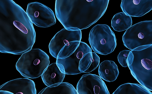
Gaucher disease (OMIM #230800) is an inherited deficiency of lysosomal enzyme acid ß-glucosidase (glucocerbrosidase, GBA1; EC 3.2.1.45) due to mutations in the glucocerebrosidase gene, GBA1.1 Genetic mutations affect the enzyme’s catalytic function, intracellular stability or subcellulartrafficking.2–4 Such enzyme deficiency results in the accumulation of glucocerebroside in lysosomes of macrophages (known as Gaucher cells, Figure 1), which are observed in many organs, primarily in the bone marrow, liver, spleen and lymph node parenchyma. The finding of an association between the GBA mutation in the heterozygous state and Parkinson’s disease indicates that there may be pathogenic roles for GBA mutations beyond enzyme deficiency.5–7
Gaucher cells are typically 20–100 μm in diameter with eccentrically placed nuclei and cytoplasm with characteristic crinkles and striations.8–10However, untreated patients with type 1 Gaucher disease have recently been shown to display a considerable proportion of Gaucher cells with atypical cytomorphology.11
The glucocerebroside concentration in spleens can be increased 10 to 1,000-fold, but high levels may also be present in the bone marrow and liver.12 Glucosylceramide accumulation leads to variable combinations of:13
- Splenomegaly with abdominal discomfort, often the presenting symptom of Gaucher disease.14
- Anaemia with chronic fatigue.15
- Spontaneous bruising and bleeding – due to thrombocytopenia and/or Gaucher disease-associated coagulopathy.16 This is initially a result of enhanced blood cell clearance by the enlarged spleen. At a later disease stage, or in patients who have undergone a splenectomy, replacement of the bone marrow by Gaucher cells adds to cytopenia development.
- Hepatomegaly and abnormal liver function.
- Diverse bone disease manifestations, including chronic bone pain, acute bone crises, defective bone mineralisation, infarction, osteonecrosis, osteolysis and pathological fractures.17 Skeletal disease affects more than 80 % of patients with Gaucher disease and can have a major impact on patient quality of life.18,19
- Impaired neutrophil function and neutropenia may cause an increased susceptibility to infection.20
Gaucher disease is classified into three broad phenotypes:21 Type 1, nonneuropathic disease; Type 2, fulminant neuropathic disease that is fatal in infancy; and Type 3, chronic neuropathic disease, with an expected survival from childhood to mid adulthood.22 An intermediate phenotype between types 2 and 3 has also been recognised.23 The broadest spectrum in phenotypes in terms of age of onset, rate of progression and the organs affected occurs in Type 1 Gaucher disease.24 Clinical phenotype cannot be discerned from the genotype23 although early onset has beenassociated with more rapidly progressive and severe disease.16 Type 1 Gaucher disease accounts for >90 % of all Gaucher disease patients. Gaucher disease occurs at a global prevalence of around 1 in 50,000but is much more common in the Ashkenazi population, in which it is found in around one in 800–850 individuals.15,25–28 Nearly 300 mutations have been identified, including frameshift mutations, point mutations, deletions, insertions and splice site mutations.29 Four mutations N370S (c.1226A>G), L444P (C.1448T>C), 84GG (-c.84dupG) and IVS2+1 (c.27+1G>A) account for approximately 90 % of the disease causing alleles in the Ashkenazi Jewish population. In non-Jewish populations, the same four alleles account for approximately 50–60 % of the diseasecausing alleles. Whereas strong correlations have not been identified between genotypes and the resulting phenotypes, several mutations are associated with a higher risk of neuronopathic forms of Gaucher disease (e.g., homozygosity for L444P or D409H, or the compound heterozygote L444P/D409H).29 By contrast, the N370S mutation is commonly perceived to lead to minimal symptoms, although it could be associated with severe progressive bone disease and cancer risk when homozygous.30 Genotypic data from affected patients show that presence of the single N370S allele is diagnostic of type 1 Gaucher disease. Alleles bearing the 84GG mutation are unable to direct systhesis of any protein and this mutation has therefore never been found in the homozygous state.31 The combination of N370S and 84GG mutation leads to severe disease.32 The most prevalent mutations in Caucasian patients are N370S and L444P.33
The model of Gaucher disease management is underpinned by prompt diagnosis taking place before the development of irreversible complications.34 Enzyme-replacement therapy (ERT) (imiglucerase [Cerezyme®]; Genzyme Corporation, Cambridge, MA, US) ameliorates or reverses many systemic manifestations of Gaucher disease35 and is considered the reference treatment for Gaucher disease type 1 and 3 patients. Other recombinant glucosidases include velaglucerase alfa (Shire HGT, Cambridge, MA, US) and taliglucerase alfa (Protalix Biotherapeutics, Carmiel, Israel and Pfizer, NY, US). However, ERT hasbeen shown to correct only part of the coagulopathies in Gaucher disease and other therapeutic and supportive measures should be considered to treat and/or prevent bleeding.36
Diagnostic Delay
Despite the availability of treatment options, nearly one-quarter of type 1 patients with Gaucher25 disease do not receive timely access to therapy, possibly due to the delays in obtaining a diagnosis after the onset of symptoms.28,37 Concerns about safety of treatments may also account for this delay. Enzyme testing diagnosis of Gaucher disease is unequivocal; however, Gaucher disease is rare with a non-specific and heterogeneous manifestations, and minor overt symptomatology at onset, all of which hinders consideration of the disease in differential diagnosis. Aspiration biopsy of bone marrow is not a reliable diagnostic tool.38
Cytopenias and splenomegaly can lead to suspicion of haematological malignancy, especially in the presence of focal splenic lesions.37 However, concomitant presence of hyperferritinaemia, polyclonal gammopathy, without neutropenia, but together with a more insidious onset may be useful in differentiating from malignancy.22
Early symptoms of Gaucher disease reflect the haematological characteristics such as splenomegaly, anaemia, thrombocytopenia and bleeding.39 Therefore, patients with Gaucher disease are most likely to be referred to a haematologist for initial diagnosis, assessment and ongoing medical care. A global study of 406 haematologists– oncologists found that just 20 % considered Gaucher disease in the differential diagnosis, even in the presence of the classic symptoms of cytopenia, hepatosplenomegaly and bone pain.37 Instead, the most likely diagnosis considered included leukaemia, lymphoma and multiple myeloma. The case series of patients who experienced a delay in diagnosis is summarised in Table 1. Of 136 patients surveyed, the average time from first appearance of Gaucher disease symptoms to diagnosis was 4.1±10.3 years. In particular, patients homozygous for the N370S glucocerebrosidase mutation appear especially vulnerable to diagnostic delays (see Table 1).
In a retrospective review of a single centre cohort of 86 patients, patients most commonly presented with symptoms related to visceral of haematological involvement.22 The almost universal presence of concomitant thrombocytopenia or splenomegaly means that the majority of patients were reviewed by haematologists. Fifty-six per cent of the patients presented primarily with features of thrombocytopenia or splenomegaly. The median time from onset of symptoms to diagnosis was 2 years (range 0.5–26 years) and 19 % experienced delays of ≥5 years. Bone marrow biopsies were performed in the majority of the cohort (68 %), despite the fact that they are not recommended in routine Gaucher disease diagnosis.
Preliminary findings from an observational study of 35 haematological centres in Italy showed that 18 % of all haematological first evaluations are positive for splenomegaly and/or thrombocytopenia and, among them, 11 % did not receive an appropriate diagnosis.40
Diagnostic Algorithms – Overview
A consensus meeting convened in Copenhagen to develop algorithms for Gaucher disease diagnosis and management.28 In the publication containing the algorithms, which expands on ancillary concerns, the authors highlighted that it is advisable to test for Gaucher disease as a first-line investigation in patients of Ashkenazi Jewish ethnicity presenting with splenomegaly and cytopenia. The most common genotype in this ethnic group, N370S homozygosity, is frequently characterised by mild cytopenia and splenomegaly that may be difficult to detect, therefore ancillary information is helpful, including hyperferritinaemia, low high-density lipoprotein (HDL) cholesterol, premature gallstones or osteoporosis and gammopathy (see Figure 2A). In the non-Ashkenazi population where Gaucher disease is markedly less frequent compared with haematological malignancies, it is appropriate to consider Gaucher disease in differential diagnosis when malignancies have been ruled out (see Figure 2B). Bone marrow biopsy is usually undertaken in this setting and it should be routine to search for Gaucher cells as well as for haematological malignancies.
Routine bone marrow examination is not used in diagnosis; however, due to the risk of bleeding complications due to thrombocytopenia and coagulopathy,41 and also the availability of a robust, less-invasive enzymatic test. The diagnostic test for Gaucher disease is the demonstration of low acid ß-glucosidase activity in peripheral blood leukocytes (normal range 2.1–2.5 μmol/l/hour).42
Gaucher Disease and Haematological Malignancies
Physicians need to be aware of the increased risk of haematological malignancy and actively look for signs of the disease.15,39,43 The increased risk may possibly be due to potential chronic stimulation of the immune system associated with glucocerebroside storage in tissue macrophages.43 Elevated glucosyceramide and complex glycosphinogolipids have been proposed to be involved in cancer progression.44 Other possible factors implicated in the pathophysiology of cancer in Gaucher disease include splenectomy, endoplasmic reticulum stress, genetic modifiers, altered iron metabolism and insulin resistance.45
A single-centre cohort study of 131 patients with Gaucher disease of mixed ancestry in Western Europe investigated cancer incidence and mortality compared with controls.46 A 2.5-fold (95 % confidence interval [CI] 1.1–4.7) increased incidence of cancer was reported and a 12.7-fold (95 % CI 2.6–37.0) increased risk of haematological cancers. In particular, the incidence of multiple myeloma was highly elevated with a standardised rate ratio of 51.1 (95 % CI 6.2–184). In a retrospective cohort study, comparing the incidence and type of cancer in 48 patients with Gaucher disease with those of 511 control subjects without Gaucher disease, 10/48 (20.8 %) developed cancer compared with 35/511 (6.8 %) of the control population.47 Data from the International Collaborative Gaucher Group (ICGG) reported an increased risk of multiple myeloma, although the patients with Gaucher disease did not seem to be at a highly increased risk of cancer, at least during early and middle age.43
Gaucher disease, as is the case with multiple myeloma, is associated with biclonal and triclonal gammopathies and monoclonal gammopathy of underdetermined significance (MGUS).48 The risk of progression of MGUS to multiple myeloma or related disorders is about 1 % per year in the general population,49 although it is not known if this rate also applies to patients with Gaucher disease. Immune dysregulation in the bone marrow microenvironment is thought to be involved in the increased risk of MGUS and myeloma in patients with Gaucher disease.50
In a systematic literature review investigating malignancies and monoclonal gammopathy in Gaucher disease, based on the studies included, patients with Gaucher disease have an increased risk of cancer in general (pooled relative risk of 1.7 [95 % CI 1.27–2.31] and in particular, an increased estimated risk of multiple myeloma [25.0–51.1] and haematological malignancies [3.5–12.7]).45
An association between Gaucher disease and myelodysplastic syndrome has been reported but is rare.51 Epidural haematoma in Gaucher disease is a rare manifestation that mimics malignant processes.52
Management and Enzyme Replacement Therapy (see Table 2)
Recommendations for the management of haematological malignancy have been published following proceedings of a European panel of experts in haematology that convened in Paris, France in 2006.39 Bi-annual measurement of serum protein electrophoresis and immunofixation is recommended in those aged <50 years and annual measurement in older patients. There is overlap between some features of malignancy and Gaucher disease, including bone pain, splenomegaly and cytopenias, though the development of these features in otherwise stable patients should be followed by further investigation. The potential role of ERT in the prevention of malignancies warrants investigation. ERT may also improve peripheral blood counts to facilitate chemotherapy.50
The coincidence of Gaucher disease and multiple myeloma does not exclude the patient from receiving ERT.39 There is, however, a paucity of data on the impact of continuing Gaucher-specific treatment during cancer treatment. Therefore, it is advisable to personalise the decision of continuing or interrupting the Gaucher disease treatment according the clinical status of the patient, life expectancy and treatment burden.
Newer treatment strategies for Gaucher disease that are under investigation include substrate reduction therapy, chaperone therapy, gene therapy and receptor-interacting protein kinase-3 inhibitors.50,53
Risk Factors
Age is an important risk factor for cancer in patients with Gaucher disease and may prove more pronounced in people with Gaucher disease than in the general population; the risk of cancer in patients with Gaucher disease compared with the risk in age-matched controls was higher in a cohort with mean age >40 years46 than in cohorts with a mean age <40 years.30,54 Other risk factors include splenectomy, immune dysregulation and impaired proteasomal degradation and of mutant glucocerebrosidase variants and genetic modifiers.
Mortality
The single-centre cohort study of 131 patients with Gaucher disease reported an increased mortality risk of hepatocellular carcinoma (relative mortality 101.4, 95 % CI 12.3–366.1) but no significant increase in mortality for multiple myeloma (mortality risk 7.6, 95 % CI 0.92–202.0) and all cancers (relative risk 3.0, 95 % CI 0.96–6.9).46
Conclusions
Evidence suggests that patients with Gaucher disease may go undiagnosed for years, which leads to severe complications that are preventable or reversible with ERT. Such complications include avascular necrosis, severe bleeding, chronic bone pain, pathological fractures, growth failure, liver pathology and life-threatening sepsis. Gaucher disease, although a rare disorder, has the potential to influence understanding of more common disorders, including Parkinson’s disease and certain malignancies. Most patients with Gaucher disease are initially evaluated by a haematologist– oncologist. Increased risk of multiple myeloma and haematological and non-haematological malignancies has been reported with type 1 Gaucher disease. The precise relationship between Gaucher disease and cancer is still unclear, however, and needs further investigation.